Pests of safflower_Binomics_Identification_Dr.UPR.pdf
Field biology of placozoans distribution diversity
1. Field biology of placozoans (Trichoplax): distribution, diversity,
biotic interactions
Vicki Buchsbaum Pearse1,
* and Oliver Voigt†
*Long Marine Laboratory, Institute of Marine Sciences, University of California, Santa Cruz, CA 95060, USA;
†
Department of Geobiology, University of Go¨ttingen Geoscience Center, D-37077 Go¨ttingen, Germany
Synopsis The goal of this review is to highlight what little is known, and point to the bulk of what is yet to be learned,
about the natural history of placozoans in the field—in order to stimulate a broader search for placozoans and a fuller
exploration of their distribution, diversity, and all other aspects of their enigmatic lives. The documented geographic
distribution of placozoans lies mostly in the nearshore, warm, marine waters of the tropics and subtropics. Although
placozoans have long been viewed as benthic organisms, they can be more readily collected from the water column,
well above the sea bottom. The full life-history of placozoans is unknown, including the nature of this abundant pelagic
phase and all details of sexual reproduction and development. We note observations on the biota associated with
placozoans in field collections, in particular the other regular members of the microcommunity in which placozoans
occur on our collecting plates and on some factors influencing this assemblage. Among the animals found are some
potential predators against which placozoans appear to be defended, although the mechanisms are still to be examined.
Also yet to be uncovered is the full breadth of diversity in this phylum, certainly underrepresented by its single named
species. We report here greatly expanded distributions for known haplotypes and fresh specimens that include a new
haplotype, and we review the evidence that many more almost certainly await discovery. We also describe some methods
for collecting and handling these small, fragile animals.
Introduction
As the simplest of living metazoans, Trichoplax
adhaerens Schulze, 1883, phylum Placozoa, holds
a special fascination for biologists (Fig. 1). Although
its relationships with other animals remain contro-
versial, both morphological and DNA-based phylo-
genetic analyses place it among the diploblasts
and possibly at the very base of animal origins
(e.g., Nielsen et al. 1996; Halanych 2004; Dellaporta
et al. 2006; Signorovitch et al. 2007; Schierwater and
DeSalle 2007). Studies on laboratory-cultured ani-
mals have provided substantial background on
structure and ultrastructure (reviewed by Grell and
Ruthmann 1991) and on cell and molecular biology
(e.g., quantitative DNA measurements: Ruthmann
1977; evidence of a single Hox gene: Schierwater and
Kuhn 1998; documentation of a circular mitochon-
drial (mt) genome: Ender and Schierwater 2003; mt
genomes: Dellaporta et al. 2006; Signorovitch et al.
2007; expression of developmental genes: Martinelli
and Spring 2003; Jakob et al. 2004; Hadrys et al.
2005). Reviews of research on Trichoplax (Syed and
Schierwater 2002; Miller and Ball 2005;
Schierwater 2005) will need frequent updating,
as growing interest in these animals accelerates the
rate of publications. Trichoplax has joined the elite
list of model animals whose entire genome has been
sequenced (now in the process of annotation)
(Joint Genome Institute 2007).
Thus, the rocket science is well launched.
In contrast, almost no one has bothered to glance
down at the launch pad: little is known of the
most basic biology of the living animals in the
field—their biogeography, habitat, behavior,
biotic interactions, or other aspects of their lives.
Grell (e.g., 1981) pioneered the first outlines of
their distribution by extracting placozoans from
samples of seaweeds and other potential
substrates collected from several tropical and sub-
tropical sites. Decades later, we have still barely
begun to document the rich taxonomic diversity that
living placozoans promise (Voigt et al. 2004;
Signorovitch et al. 2006). One goal of this present
review is to stimulate a broader search for
placozoans and fuller exploration of their
distribution and diversity.
From the symposium ‘‘Key Transitions in Animal Evolution’’ presented at the annual meeting of the Society for Integrative and Comparative
Biology, January 3–7, 2007, at Phoenix, Arizona.
1
E-mail: vpearse@ucsc.edu
677
Integrative and Comparative Biology, volume 47, number 5, pp. 677–692
doi:10.1093/icb/icm015
Advanced Access publication June 1, 2007
ß The Author 2007. Published by Oxford University Press on behalf of the Society for Integrative and Comparative Biology. All rights reserved.
For permissions please email: journals.permissions@oxfordjournals.org.
byguestonMarch5,2013http://icb.oxfordjournals.org/Downloadedfrom
2. Since first working with these animals in Hawaii
in 1986, V.B.P. has continued to sample for
placozoans at many locales, in marine laboratory
facilities and in the sea, in order to further document
where they are found. Both authors have contributed
samples, including a new haplotype, to the genetic
and phylogenetic analysis presented here. Additional
information about the field biology of placozoans,
mostly fortuitous, was collected as opportunities
arose. Also described are techniques for collecting
and handling these minute, delicate organisms.
Collection and observation
V.B.P. collected placozoans on standard glass micro-
scope slides (25 Â 76 mm) placed in the sea or in
laboratory seawater systems, usually in racks made
from plastic slide boxes, with most of the top
and bottom cut out to allow water to circulate
through them. The slides were spaced $10–15 mm
apart. The top and bottom of the rack were then tied
together with nylon monofilament line or cable ties,
and the rack was suspended from any handy support
(e.g., a coral branch, mangrove prop, or dock)
(Fig. 2). Racks were oriented so that the surfaces of
the slides were either parallel or perpendicular to the
surface of the water. Some racks were placed directly
on the substrate, with the slides roughly perpendic-
ular to the surface of the water.
Single microscope slides were sometimes sus-
pended in the water (weighted with a small rock by
means of a rubber band slipped around the slide) or
placed directly on the substrate. Being less conspic-
uous, these were less likely to attract tampering.
When the slides were retrieved, typically after
10–15 days, the racks of slides were placed into
securely lidded plastic boxes filled with seawater
in situ, so that the slides would be exposed neither to
air nor to seawater from another location or depth.
Loose slides were placed into racks and treated the
same way. Returned to the laboratory, the slides were
kept and examined in the same water, at room
temperature, within hours or no more than 1–2 days.
To examine a slide for placozoans, it was quickly
transferred to a Petri dish containing enough
seawater to cover the slide. The slide was supported
on small bits of rock, so that, while the upper surface
was being examined, organisms on the lower surface
were protected from rubbing against the bottom of
the dish. Illumination from a mirror or light below
Fig. 1 Placozoan. W. Samoa. (K.J. Marschall) This individual
displays the plasticity and asymmetry of shape typical of larger
placozoans; small ones are roughly circular in outline. Placozoans
are the simplest metazoans known. They look like thin
microscopic pancakes, commonly less than a millimeter across
and only about 20 mm thick. The nearly transparent body consists
of upper and lower epithelia, both flagellated and enclosing
between them a syncytial network of contractile cells. Distributed
throughout the upper epithelium are the so-called shiny spheres,
lipid-filled vesicles; in this photo, they are most visible around the
margin of the animal. The histology is extremely simple, with only
a few cell types and no basal lamina or other extracellular matrix.
The specialized cells, tissues, or organ systems typical of most
animals (digestive, circulatory, excretory, neurosensory, and
muscular) are also absent.
Fig. 2 Racks of glass microscope slides hanging on a coral reef,
Sesoko Island, Okinawa, Japan. Slides suspended in the water
capture more placozoans than do slide racks placed on the
substrate (see section, Benthos versus water column).
678 V. B. Pearse and O. Voigt
byguestonMarch5,2013http://icb.oxfordjournals.org/Downloadedfrom
3. the dish was adjusted to optimal angle and intensity,
and both sides of the slide were scanned system-
atically under a dissecting microscope. Placozoans
were counted; other organisms were recorded as
present.
Factors affecting the likelihood of success in cap-
turing placozoans under various conditions are noted
below (see section Macrohabitat and microhabitat).
Strengths and limitations of collection methods
The first published finding of placozoans on glass
microscope slides suspended in the sea was by
Sudzuki (1977) in Sagami Bay, Japan. This method
was used from the start of studies by VBP in 1986,
et al. have followed suit (e.g., Maruyama 2004;
Signorovitch et al. 2006), because placozoans are
almost invisible on any substrate except clear glass.
Sampling with glass slides is also a relatively benign
process, causing little or no disturbance to the
benthic habitat; there is some by-catch, but it is
minor on an oceanic scale.
Capture of a placozoan on a slide establishes its
presence at a known location and depth, provided
that the slide is kept in seawater from the
original site or in adequately filtered seawater.
However, because settlement may have occurred at
any time after the slide was placed, the time cannot be
known precisely. Likewise, the number of placozoans
observed on a slide depends on where and for how
long a slide is exposed and results from some
combination of settlement and fission, as well as
possible loss of individuals over the period of
exposure and increasing difficulty in seeing placozo-
ans as the slide becomes overgrown with other biota.
Thus, only semi-quantitative sampling has been
achieved.
Assuming that two or more slides bearing
placozoans represent independent settlement events,
reporting the number of positive slides, as well as the
number of placozoans on each, adds information
(e.g., Maruyama 2004). The alternative of estimating
biomass (versus number) is problematic (Pearse
1989). Another method is to collect pieces of rock
or shell and shake them in a plastic bag filled with
seawater (Maruyama 2004). Although faster, this
process further obscures the number of placozoans
present, because many become broken into frag-
ments. However, one is assured of direct sampling of
the benthic phase (provided the seawater is well
filtered), whereas the stage of the life-history that
settles on glass slides, either suspended or placed on
the bottom, is uncertain.
Also, as with any small, easily transported
planktonic organism, the location of the source
population on the benthos is difficult to determine.
Thus, although more than one clade of placozoans
can often be found together in a single sample (Voigt
et al. 2004; Signorovitch et al. 2006), interpretations
of sympatry are complicated by the limitations of the
sampling method. Placozoans swimming or drifting
in the water column settle on the suspended slides.
However, two or more placozoans settling on a
single slide may have originated from benthic
individuals in quite separate and different micro-
habitats. Lacking any definition of ‘‘an ecologically
relevant spatial scale’’ (Signorovitch et al. 2006),
conclusions of sympatry may not apply to the
adjacent benthic habitat, but strictly, only to
sympatry in the water column.
Distribution
Biogeography
Sites where placozoans are known to have been
collected are summarized on a global map (Fig. 3).
The annotated list below includes published records
[P], previously unpublished observations of either
of this review’s authors [A], and personal
Fig. 3 Geographic distribution of reported occurrences of placozoans. The numbers correspond to those of the sites listed under
Biogeography. Previously unpublished reports are shown in black boxes.
Field biology of placozoans (Trichoplax) 679
byguestonMarch5,2013http://icb.oxfordjournals.org/Downloadedfrom
4. communications from other biologists [C]. Reported
haplotypes of the mitochondrial 16S large subunit
ribosomal RNA gene (16S rDNA) are given in bold
(using the numbering of Voigt et al. 2004 and of this
publication).
Mediterranean and Eastern Atlantic
(1) Gulf of Trieste, Adriatic Sea. Schulze 1883;
Stiasny 1903. Schulze’s type locality for the
species Trichoplax adhaerens. [P]
(2) Gulf of Naples, Tyrrhenian Sea. Monticelli
1893, 1896. Monticelli’s type locality for the
species Treptoplax reptans Monticelli 1896,
currently not recognized. [P]
(3) Orbetello Lagoon, Tyrrhenian Sea. Tomassetti
et al. 2005 [P] H2,5
(4) Tunisia. B. Schierwater, H. Hadrys, M. Eitel
2005 [C]
(5) Tenerife, Canary Islands. B. Schierwater, H.
Hadrys 2003 [C]
Roscoff, Brittany, France. Ivanov 1973 [P]
Red Sea and Indian Ocean
(6) Eilat, Red Sea. Grell 1971 [P] H1
(7) La Re´union. N. Gravier-Bonnet 2001 [C]
Pacific
(8) Presumed from Sea of Japan. Ivanov et al. 1980
[P] (Similar to H1)
(9) Oki Islands, Sea of Japan. Y.K. Maruyama [C]
(10) Shimoda, southcentral Honshu, Japan. Sudzuki
1977 [P]
(11) Shirahama, south Honshu, Japan. V. Pearse
1989 [A], Maruyama 2004 [P]
(12) Okinawa, Ryukyu Islands, Japan. V. Pearse 1989
[A], Pearse et al. 1994 [P]
(13) Iriomote, Ryukyu Islands, Japan. V. Pearse 1989
[A]
(14) Hong Kong. V. Pearse 2004 [A]
(15) Zambales, Philippines, from material trans-
ported to Monterey Bay Aquarium, Monterey,
California. V. Pearse 1992 [A]
(16) Guam. Univ. Guam Marine Lab. V. Pearse 1988
[A], Voigt et al. 2004 [P] H8
(17) Palau. V. Pearse 1988 [A]
(18) North of Manado, northeast Sulawesi
Indonesia. V. Pearse 1994 [A]
(19) Madang, Papua New Guinea. V. Pearse 1988 [A]
(20) Lizard Island, Great Barrier Reef, north-
eastern Australia. O. Voigt 2006 [A] LIZ
(similar to H9)
(21) Orpheus Island, Great Barrier Reef, northeast-
ern Australia. V. Pearse 1988 [A]
(22) Heron Island, Great Barrier Reef, northeastern
Australia. A.T. Newberry 1988 [C]
(23) Oahu, Hawaii. Kewalo Marine Lab.
M.G. Hadfield $1978 [C], V. Pearse 1989 [P].
E. Gaidos 2007: H1,2,4,6 [C]
(24) Western Samoa. K.J. Marschall, films, 1970, and
cited in Grell 1980 [P], K.J. Marschall and V.
Pearse 1989 [A]
(25) Moorea, French Polynesia. V. Pearse 1989 [A]
(26) Presumed from southern California (see subse-
quently). V. Pearse 2006 [A] H11
(27) Achotines Laboratory, Azuero Peninsula,
Panama. Voigt et al. 2004 [P] H4
(28) Isla Iguana, Azuero Peninsula, Panama. Voigt et
al. 2004 [P] H6,8
(29) Naos Island Laboratories, Panama. Voigt et al.
2004 [P] H4,7
Caribbean and Western Atlantic
(30) Southeast Atlantic coast of US: North Carolina,
Grell 1980 (R.M. Rieger, personal communica-
tion); South Carolina, Klauser 1982 [P]
(31) Southeast Atlantic coast of US: Florida. V. Pearse
2006 [A]
(32) Bermuda. Grell 1980 [P]. T. Syed, S. Sagasser
2001 [C], Signorovitch et al. 2006 [P] H2,4,9,10
(33) Puerto Morelos, Quintana Roo, Mexico. Grell
and Lo´pez-Ochoterena 1988 [P]
(34) Carrie Bow Cay and Twin Cays, Belize.
Signorovitch et al. 2006. [P] H2-4,6-8
(35) Roatan, Honduras. V. Pearse 1985 [A]
(36) Bocas del Toro, Panama. Voigt et al. 2004;
Signorovitch et al. 2006 [P] H1-4,8
(37) Galeta, Panama. V. Pearse 1997 [A]
(38) Discovery Bay, Jamaica. Signorovitch et al. 2006
[P] H1,8
(39) Grenada. Signorovitch et al. 2006 [P] H6
(40) Cubagua Island/Margarita Island, Venezuela.
Voigt et al. 2004 [P] H4
(41) Sa˜o Sebastia˜o Channel, Sa˜o Paulo State, Brazil.
Morandini et al. 2006 [P]
680 V. B. Pearse and O. Voigt
byguestonMarch5,2013http://icb.oxfordjournals.org/Downloadedfrom
5. Macrohabitat and microhabitat
V.B.P. found placozoans in abundance on coral reefs
and in full-salinity mangroves. Results can be locally
patchy, both positive and negative samples being
obtained from a single site. Nonetheless, consistently
negative samples or smaller yields in certain types of
habitats or conditions point to factors restricting
placozoan distribution. No systematic survey has
been attempted but examples of findings are briefly
summarized here (see also Pearse 1988).
Seasonality/Temperature
Placozoans were found on slides in samples taken at
various tropical sites and all months of the year.
However, no long-term surveys of placozoans at
a single location have been done at tropical latitudes.
At one subtropical site, Sesoko Island, Okinawa,
Japan, where sea surface temperatures vary seasonally
(means 20–288C; Loya et al. 2001), collections were
made at a site close to the Sesoko Marine Laboratory
from March to August. In March and April, when
sea temperatures at Sesoko average $21–228C, all
samples were negative; only during May–August,
when temperatures average $24–288C, were placozo-
ans found on slides. Few sites from temperate
latitudes have been examined; some data suggest
possible seasonality in abundance at Shirahama,
Japan (Maruyama 2004). A 1992 survey by V.B.P.
in Monterey Bay, central California, where annual
sea surface temperatures range $11–188C, failed to
yield any specimens of placozoans. A search was
made specifically for placozoans, using the same
methods, but they also were not found at two sites
illustrating extreme temperature conditions: À1.68C
in McMurdo Sound, Antarctica (Pearse and Pearse
1991) and $38C in the Monterey Canyon, Central
California, $1000–3000 m depth.
Salinity
In laboratory trials at the Christensen Research Inst.
(CRI), Madang, Papua New Guinea, exposure to
seawater of reduced salinity rapidly killed placozoans:
exposure for 1 h to seawater diluted to 75% or 0.5 h
in seawater diluted to 50% proved 100% lethal.
After only 8 min or 1 min, respectively, in the test
solutions, the edges of the animals began to curl,
and the animals detached from the glass and became
motionless and unresponsive to touch. Placed again
into full salinity seawater (32.5ø), some individuals
showed improvement, but all died within 524 h.
This intolerance for seawater of reduced salinity
was evident in field collections, e.g., nearshore,
shallow, previously positive sites near CRI were
negative after rains. In contrast, placozoans have
been found to tolerate elevated salinities in the
lab, up to 40–50ø (A. Signorovitch and L. Buss,
personal communication, 2007).
Currents and wave surge
In a narrow channel with strong tidal flow, samples
were consistently small or negative, relative to
samples from the mangrove pond and fringing reef
that the channel connected (Kranket Island, Madang,
Papua New Guinea). Samples from reef flats with
strong currents likewise failed to yield placozoans
(Guam, Western Samoa), even in places where
placozoans were recovered from nearby protected
inlets (Cook Bay and Opunohu Bay, Moorea,
French Polynesia). See also Associated biota section,
subsequently.
Sandy bottoms
Almost all samples were negative in seagrass beds
(Madang, Papua New Guinea; Apia, Western Samoa)
and other sandy habitats. For example, at Orpheus
Island, Great Barrier Reef, Australia, placozoans were
found on slides placed on a fringing coral reef,
but not at a site only $200 m away where slides were
suspended over a sandy bottom.
Benthos versus water column
Although placozoans have long been viewed as
benthic organisms, they were regularly collected on
glass slides suspended in the water column well
above the bottom (Fig. 2). Moreover, when paired
samples were compared (free slides or slide-racks
placed on the substrate versus others hung in shallow
water at the same site 20–60 cm above the bottom),
those suspended in the water column bore signifi-
cantly higher numbers of placozoans. For example,
for 12 sets of eight slides each in Madang, Papua
New Guinea, the number of placozoans was
significantly greater on slides suspended above the
bottom versus slides on the bottom (CRI dock,
mean Æ SD was 8.1 Æ 3.4 for slides suspended
above the bottom, 0.23 Æ 0.32 for slides on the
bottom, t-test, P ¼ 0.02; Kranket Island, mean Æ SD
was 13.9 Æ 3.1 for slides suspended above the
bottom, 3.4 Æ 2.0 for slides on the bottom, t-test,
P50.01). Comparison of V.B.P.’s midsummer
results in Shirahama, Japan, with those of
Maruyama (2004) likewise indicate a strong effect
of sampling off the bottom versus on the bottom.
V.B.P. found large numbers of placozoans on slide
racks suspended in the water column in July, com-
parable to yields during the fall peaks documented
by Maruyama (2004, his Fig. 3), whereas his
Field biology of placozoans (Trichoplax) 681
byguestonMarch5,2013http://icb.oxfordjournals.org/Downloadedfrom
6. slide racks, always placed on the substrate, captured
very few or no specimens in July.
Substrate orientation
At the CRI dock, Madang, Papua New Guinea,
placozoans were counted on two sets of eight slides
each, hung in the sea for 17 days about 20 cm off
the bottom so that their surfaces were horizontal
(parallel to the water surface) versus vertical (perpen-
dicular to the water surface). The number of
placozoans per slide was not statistically different
(mean Æ SD was 5.9 Æ 3.4 for a set of eight hori-
zontal slides, 5.0 Æ 3.3 for a set of eight vertical
slides, t-test, P ¼ 0.61). In contrast, on horizontal
slides, the number of placozoans was significantly
greater on lower versus upper surfaces (mean Æ SD
was 5.5 Æ 0.96 for lower surfaces, 2.0 Æ 0.93 for
upper surfaces for three sets of slides, t-test,
P ¼ 0.01). Similar results were obtained from a trial
at Sesoko Island, Okinawa, Japan (mean Æ SD was
3.1 Æ 1.6 for lower surfaces, 0.17 Æ 0.13 for upper
surfaces, for four sets of slides, t-test, P ¼ 0.01).
Remarks: biogeography and habitats
Given the limitations of collection methods, we can
nonetheless draw some broad conclusions about the
distribution of placozoans in terms of biogeography
and at other scales. First, they have been widely
documented in tropical and subtropical nearshore
marine habitats, especially coral reefs and mangroves.
V.B.P. seldom recovered them from sandy habitats,
and this was also the most common experience of
Signorovitch et al. (2006), even when hard substrates
positive for placozoans were nearby. Although this
might suggest a measurable limit on the distance that
placozoans can travel between adjacent habitats, the
number of observations is few and cannot be
evaluated without information on the direction of
currents and other confounding influences. Protected
areas yield more placozoans than exposed sites with
strong currents or wave-surge. We would expect to
find fewer placozoans in areas of freshwater runoff,
as near river mouths, as much because of the
quantities of sediment and soft bottoms as because
of their intolerance of reduced salinities.
Although whole biogeographic regions, such as the
Indian Ocean, remain nearly blank on the map of
placozoan distribution, sampling in those waters will
almost certainly yield positive results. In contrast,
our expectations of finding placozoans in deep or
polar waters, or on coasts characterized by cold
upwelling, are minimal. To our knowledge, no one
has looked for placozoans in the open ocean. Where
collections are from laboratory tanks or commercial
or personal tropical aquariums rather than directly
from the sea, some uncertainty about the origins of
the specimens is inevitable because placozoans can
easily be introduced with exotic materials.
Placozoans exhibit an abundant nearshore pelagic
phase and can be efficiently collected from the
water column, well above the sea bottom. In paired
samples, slide racks hung suspended in the water
column yielded more placozoans than did those
placed directly on the substrate. The latter collect
more silt and may be subject to disturbance from
other benthic animals, or perhaps the pelagic phase
settling on the slides is less numerous close to the
bottom than within the water column. Compared to
freestream flow in the middle of the water column,
flow along the bottom is expected to be slower
and therefore to supply fewer settlers per unit time;
arriving settlers, however, probably attach more
easily in slower flow. Thus, no simple conclusion
emerges from the hydrodynamics.
The larger number of placozoans found on the
lower surface of slides might relate to the greater
amount of silt and ultraviolet radiation on the upper
surface. Ultraviolet may be very important in restrict-
ing the microhabitats of these organisms and should
be examined. (Placozoans have been observed to react
strongly when exposed to ultraviolet radiation from
laboratory sources; their sensitivity under field con-
ditions has never been tested). Serpulids and some
other organisms also appeared to settle preferentially
on or migrate to the lower surface, perhaps respond-
ing to the same factors. Alternatively, placozoans
might be responding directly to other biota. A study
of temporal succession in this microcommunity could
provide some answers. No difference was observed
between upper and lower surfaces under laboratory
conditions (e.g., Pearse 1989) and placozoans settled
preferentially on a biofilmed surface, whether upper
or lower. Thus, this differential is likely related to
secondary factors present only in the field rather than
directly to the orientation of the substrate.
Relations to other organisms
Associated biota
At some time or other, small representatives of most
phyla of sessile invertebrates, as well as a great variety
of protists and algae, have been seen on V.B.P.’s glass
slides together with placozoans. Nonetheless, a few
particular organisms often dominate the micro-
community that develops on glass slides and occur
together with placozoans with sufficient regularity
that one may almost predict placozoans by the
682 V. B. Pearse and O. Voigt
byguestonMarch5,2013http://icb.oxfordjournals.org/Downloadedfrom
7. presence of these other associates, in particular
several kinds of sessile ciliates: solitary and colonial
vorticellids as well as folliculinids; spirorbid and
other serpulid polychaetes; and, in smaller numbers,
free-living loxosomatid kamptozoans (entoprocts).
Especially remarkable was the repeated finding of
this same assemblage, though not necessarily repre-
sented by the same species, together with placozoans,
at a variety of sites extending across the tropical
Pacific and into the Caribbean Sea. Occasionally,
the typical assemblage would be present except for
placozoans; even more rarely, placozoans were found
in the absence of their usual associates.
The association may be roughly illustrated by two
semi-quantitative vertical transects (Table 1). At an
exposed, wave-swept coral pinnacle, Tripod Reef,
near CRI Madang, Papua New Guinea, slides were
placed at intervals from near the surface down to
20 m in depth. Along this transect, placozoans were
found only at the three deepest stations, 16–20 m,
largely below the influence of wave-surge, and the
associated microcommunity displayed the same
distribution. In another vertical transect in a quiet
part of Opunohu Bay, Moorea, French Polynesia,
placozoans and the associated microcommunity were
present in comparable abundance throughout the
range in depth sampled, down to 16 m (Table 1).
On a smaller scale, the association between
serpulids and placozoans, was seen even in their
distribution on each single glass slide. For example,
in Madang, Papua New Guinea, data for placozoans
(see section Substrate orientation, earlier) paralleled
that for small spirorbids: on horizontal slides,
the number of spirorbids (diameter 200–500 mm)
was significantly greater on lower versus upper
surfaces (mean/slide Æ SD was 11.4 Æ 4.1 for lower
surfaces, 2.9 Æ 1.6 for upper surfaces for a set of
9 slides, t-test, P ( 0.001).
Diet
Placozoans grew and multiplied on glass slides
bearing a varied assemblage of small organisms that
had settled on slides suspended in the sea; typically,
we were unable to discern what they were eating.
In Madang, Papua New Guinea, some slides that
bore an abundant film of green algae supported
unusually large placozoans. In Western Samoa,
conspicuous feeding tracks were visible in the thin,
red, algal film growing on aquarium glass, and at the
end of each track was a pink placozoan.
Encounters with potential predators and other
organisms
Despite the diversity of protists and animals occur-
ring together with placozoans on the glass slides, and
numerous interactions, not a single instance of
predation was seen, either on or by placozoans.
In several cases, however, potential predators were
observed to recoil after contact with placozoans or
reject them as food. At Orpheus Island, Great Barrier
Reef, Australia, a small snail was watched as it
headed straight for a placozoan, clearing a swath
with its radula as it went. Just before reaching
the placozoan, the snail extended one tentacle
anteriorly, touched the placozoan, recoiled abruptly,
turned, and proceeded in another direction.
Table 1 Two vertical transects, on Tripod Reef, Madang, Papua New Guinea from 3–20 m, and in Opunohu Bay, Moorea, French
Polynesia from 3–16 m. Semi-quantitative estimates of the abundance of placozoans and three other commonly associated organisms
were made from glass slides placed down the reef slope at intervals of several meters. The upper part of Tripod Reef experiences
strong wave-surge, whereas the reef at Opunohu Bay is in a quiet, protected situation
Madang Moorea
Depth (m) Plac Vort Foll Spir Plac Vort Foll Spir
3 À þ À þ þ þþ À À
6 À À À þ þþþ þþ À þ
8 À À À þþ
9 þ þþ þ þ
10 À À À þþ
12 À À À þþ þþ þþ þ þ
14 À þ À þþþ þ þþ þ þ
16 þ þ À þþþ þ þþ þ þ
18 þ þ þ þþþ
20 þ þþþ þ þþþ
Plac ¼ placozoans; Vort ¼ colonial vorticellids (Ciliata); Foll ¼ folliculinids (Ciliata); Spir ¼ spirorbids (Polychaeta). (À) denotes absence; (þ, þþ,
þþþ) denotes increasing abundance.
Field biology of placozoans (Trichoplax) 683
byguestonMarch5,2013http://icb.oxfordjournals.org/Downloadedfrom
8. Similarly, at Madang, Papua New Guinea, a small
rhabdocoel flatworm crawling on a slide among
placozoans was observed contacting a placozoan,
recoiling vigorously, and changing its direction of
movement only to encounter another placozoan and
repeat this behavior several times. A swimming
placozoan caught in the feeding current of a small
sabellid tubeworm was passed down the food-groove
to the mouth, where it was rejected, flicked out into
the water, and then caught again; this process was
repeated six times, until the placozoan finally con-
tacted the substrate, adhered, and crept away, appar-
ently unharmed. Various ciliates, tiny nematodes,
and other small animals sometimes crawled around
or under placozoans without displaying any reaction
or evoking any visible response from the placozoans.
Ciliates were observed to gather around wounded or
degenerating placozoans (Pearse 1989).
In none of these instances did the other organisms
appear to be harmed by the placozoans. In contrast,
in laboratory trials carried out by L. Buss and
colleagues (personal communication, 2007), placo-
zoans that were dropped onto the tentacles of three
species of hydroids caused paralysis. After transfer-
ring the placozoans from the tentacles to the mouth,
the polyp became immobile and unresponsive.
By 24 h after contact with a single placozoan, some
polyps had recovered, but exposing a polyp to placo-
zoans two days in a row was fatal. Only small
placozoans fed to large polyps were ever swallowed;
most placozoans, or fragments of them, were able to
crawl away.
Remarks: relations to other organisms
Associated biota
Studies of the assemblage of microbiota that includes
placozoans share many common problems, a major
one being the difficulty of determining the natural
substrates of these organisms that we see settling
together on glass slides. Attempts to examine prefer-
red placozoan substrates by collecting seaweeds,
corals, and other possible habitats, and placing
glass slides among them, typically failed because
equal numbers of placozoans settled in the no-
substrate controls, an early hint of the presence of
placozoans in the water. Minute, solitary loxosoma-
tids were thought to live exclusively as commensals
on other organisms, almost certainly because they
were observed only incidentally by biologists whose
interest was in their hosts. Using glass slides as
settling plates, however, Iseto (2003 and references
therein) has described numerous new species of free-
living loxosomatids, and in the course of his
sampling has, not coincidentally, also captured
placozoans (T. Iseto, personal communication 2004).
No specific interactions were ever observed by
V.B.P. between placozoans and the members of the
associated microcommunity, and their relationship
must be presumed to consist of no more than
a tendency to settle under a shared set of conditions,
although it remains possible that the settlement
of one positively influences that of another. Their
patterns of abundance, similar among the taxa but
differing between vertical transects at two quite
different sites (Table 1), appear most easily explained
by their common response to differences in wave
action (see section Associated biota, earlier).
Diet
In the laboratory, placozoans grow on cryptomo-
nads, other algal unicells, or heat-killed nauplii of
Artemia (Grell 1983; Grell and Lo´pez-Ochoterena
1988) or on commercial food for aquarium fish
(Maruyama 2004, personal communication). Kept on
clean glass slides versus biofilmed slides, they grew
significantly only on the latter (Pearse 1989). They
have been seen to capture and eat ciliates in a culture
dish (Klauser 1982). Wild placozoans are probably
opportunistic grazers and scavengers on organic
detritus and on algae and bacteria in biofilms
covering a diversity of substrates.
Potential predators
The sole published report of predation on placozoans
involved the opisthobranch Rhodope (Riedl 1959).
K.J. Marschall told V.B.P. of seeing a small nemertean
apparently preying on placozoans in an aquarium in
Western Samoa (personal communication, 1973).
We have never observed any animal to prey upon
healthy specimens; on the contrary, we report several
instances of potential predators reacting to contact
with placozoans as they would to a strongly noxious
substance. We suggest that the most logical site of
such a defensive substance is the so-called shiny
spheres that lie throughout the upper epithelium
(see, e.g., Fig. 2 of Rassat and Ruthmann 1979).
The high lipid content of these osmiophilic vesicles
has been confirmed; such lipid deposits normally
serve for nutrient storage, and in placozoans they do
become more numerous when the animals are well fed
(Rassat and Ruthmann 1979). A more likely and
better protected location for nutrient storage, how-
ever, would be in the lower, nutritive epithelium.
Instead, these large lipid-rich vesicles are situated at
the surface of the animal between the upper epithelial
cells, where they are freely exposed to contact and
appear to be readily expelled to the outside
684 V. B. Pearse and O. Voigt
byguestonMarch5,2013http://icb.oxfordjournals.org/Downloadedfrom
9. (Rassat and Ruthmann 1979, their scanning electron
micrograph Fig. 4, and observations of live animals
by V.B.P)—a most perverse fate for nutrient reserves.
Thus, it seems more plausible that the large invest-
ment represented by the abundant shiny spheres of
placozoans serves for the defense of these soft-bodied
morsels. Some support for this idea has been provided
by the experiments of L. Buss and colleagues (personal
communication, 2007), who fed placozoans to
hydroids: although intact placozoans were not
usually swallowed, those previously induced to expel
many of their shiny spheres were ingested far more
often, and polyps that swallowed placozoans subse-
quently died. Analysis of the chemical contents of the
shiny spheres awaits advances in microchemistry. The
developmental history of these vesicles, which appear
to lie within anucleate cells (Grell and Ruthmann
1991), also remains to be investigated.
Diversity
Collection and sequencing
For genetic analysis, placozoans were sampled by
washing animals from the wall of a tank (containing
only local reef fishes and local seawater) at Lizard
Island Research Station, Australia (n ¼ 17) and by
settlement on glass slides in a holding-tank in the
Tuna Research and Conservation Center at Hopkins
Marine Station of Stanford University on Monterey
Bay, California (n ¼ 2). The most likely origin of the
California specimens is Southern California or
Northern Baja California, as the tank contained
Bluefin Tuna (Thunnus thynnus) caught in that
region. However, this tank is maintained on Central
California (Monterey Bay) seawater (filtered through
sand filters of nominal pore size $20 mm), and has
received in the past water from other tanks in the
facility which held fishes from Hawaii; both of these
eastern Pacific regions are thus other possible sources
of these specimens.
Single placozoans were first transferred from the
slides, through a wash of clean seawater, into 1 ml
microcentrifuge tubes. Because these animals are so
small, thin, and fragile, as well as extremely sticky
(the species name adhaerens is indeed apt), they were
first loosened from the glass using gentle jets from
a Pasteur pipette, then drawn into the pipette
and quickly expelled into a clean depression slide or
watch glass, followed by a second transfer into
a microcentrifuge tube, in a minimal amount of
seawater. About 1 ml of ethanol was added to the
tube, which was then closed and sealed with
ParafilmÕ
. Animals along with the residual
precipitated salt from the seawater were pelleted
by centrifugation; the pellet was air-dried at
room temperature and 50 ml of sterile ultrapure
water containing 5% ChelexÕ
100 (sodium form,
Sigma–Aldrich, http://www.sigmaaldrich.com)
and 20 ml of 10 mg/ml proteinase K (Sigma–Aldrich,
http://www.sigmaaldrich.com) were added.
Alternatively, the animals were directly transferred
into a tube containing the ChelexÕ
solution and
the proteinase K.
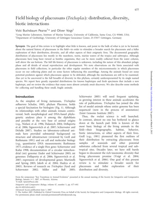
Fig. 4 Phylogenetic relationships (A) and distribution (B) of
placozoan 16S rDNA haplotypes. (A) Maximum likelihood tree,
support values 450 are shown at the corresponding nodes;
bootstrap values (from 500 replicates) are given above Bayesian
clade credibility values (posterior probabilities  100). Length of
the bar indicates 0.05 substitutions per site in the tree.
Sequences generated for this study are labeled with ‘‘Ã’’.
Accession numbers from GenBank sequences are H1: AY652522;
H2: AY652523; H3: AY652524; H4: AY652525; H9 (¼H4-2):
DQ389828 þ DQ389763; H10 (¼H4-3): DQ389825 þ
DQ389760; H5: AY652526; H6: AY652527; H7: AY652528;
H8: AY652529. (B) Geographic distribution of known occur-
rences of haplotypes. Specific locations are listed under
Biogeography. H7 comprises, besides the points shown, a
single specimen of uncertain origin, probably Indo-Pacific,
isolated from an aquarium. See text for notes on the origin
of H11. Unpublished data for Hawaii courtesy of J.M. Ward
and E. Gaidos.
Field biology of placozoans (Trichoplax) 685
byguestonMarch5,2013http://icb.oxfordjournals.org/Downloadedfrom
10. All tubes were then treated as described by
Voigt et al. (2004). They were stored at À208C and
1–3 ml of supernatant was used in PCR to amplify
a fragment of 16S rDNA [primers: fw:
50
-GCCTGCCCARTGRTTGTA-30
; rv:-50
-GGTCGCA
AACATCGTCA-30
; program: 958/5 min, 37 Â (958
C/30 s; 508C/30 s; 728C/1 min)]. The PCR products
were sequenced in both directions with our PCR
primers, using chemistry and equipment as described
by Dohrmann et al. (2006). For the samples from
Lizard Island, cycle sequencing reactions were
modified to sequence over a G–C rich partition
with stable secondary structure by adding DMSO
(5%) and 0.1 ml BIOTAQTM-DNA-polymerase
(5 u/ml; Bioline, http://www.bioline.com) and by an
initial denaturation step (968C/10 min). New
sequences have been submitted to GenBank (http://
www.ncbi.nlm.nih.gov; accession numbers LIZ:
EF421454, H11: EF421455).
Molecular analysis and haplotype distribution
We have chosen a fragment of 16S rDNA as
a molecular marker for our phylogenetic analyses,
including additional sequences from GenBank
(for accession numbers, see caption of Fig. 4A).
This marker is known to yield good phylogenetic
resolution, but also shows considerable polymor-
phism in length among haplotypes (Voigt et al.
2004), hampering alignment of all sequences in three
regions within our analysis. Because some differences
between haplotypes (e.g., H1–H2; H7–H8) appear in
these regions, we locally blocked the alignment to
include the maximum of information by introducing
gaps for nonalignable haplotypes. With this method,
otherwise unsupported or poorly supported relation-
ships of similar, yet not identical haplotypes could
be resolved (H6, H7, H8), while the tree topology
was otherwise unaffected. In all phylogenetic
analyses, we applied a GTRþG model of nucleotide
evolution suggested by the Akaike information
criterion with the software modeltest 3.7 (Posada
and Crandall 1998); likelihood values were calculated
in PAUPÃ 4.0b10 (Swofford 1998). A maximum
likelihood (ML) analysis including bootstrap resam-
pling was carried out with PHYML (Guindon and
Gascuel 2003). In addition, we conducted a Bayesian
analysis with MrBayes 3.1.2 (Huelsenbeck and
Ronquist 2001; Ronquist and Huelsenbeck 2003),
performing a Markov chain Monte Carlo analysis
with two runs (eight chains each) for 10,000,000
generations, with sample frequency set to 100 and
temperature for the heated chains set to 0.2.
From the sampled trees, we discarded the first 25%
(25,000 trees) as burn-in. In the absence of a suitable
outgroup that would result in a well-supported
rooting with our marker (Voigt et al. 2004),
midpoint rooting was applied to display the tree
in Fig. 4A.
Sequences from the two Californian samples
were identical to each other, and sequences from
the 17 samples from Lizard Island were also
identical to each other. In the latter, a stretch in
the middle of the fragment, flanked by several Cs
on the one side and several Gs on the other side,
caused problems in sequencing similar to those
reported by Signorovitch et al. (2006). Even after
applying the modified cycle sequencing protocol,
sequence reads from both sides overlapped only
slightly in the critical region. The new Australian
haplotype is identical to the H9 haplotype, reported
(as H4-2) from Bermuda by Signorovitch et al.
(2006), in parts where information exists for H9.
Our sequence, however, covers a larger part of the
gene including the critical region not available for
the H9 haplotype; thus, it is not clear if our new
sequence is identical or shows base exchanges in
these regions. Therefore, we do not use the label H9
for this haplotype, but refer to it as LIZ (Fig. 4) to
avoid the introduction of another, possibly redun-
dant haplotype number. In contrast, from the two
Californian samples, we can report a new, relatively
divergent haplotype, designated H11.
Our phylogenetic analyses yielded trees that differ
only in the occurrence of a polytomy in the Bayesian
analysis (clade H9/H10/LIZ). All other clades find
high support by bootstrap or clade credibility values
(Fig. 4A). According to our resulting trees and the
applied rooting, the newly reported haplotype H11 is
the sister group to the clade (H5, H4, H9, H10, LIZ),
with high support.
Until distinguishing morphological characters are
discovered, placozoan lineages can be recognized
only by DNA analysis with various sequence
markers. The 16S rDNA fragment we used identifies
at least 11 different haplotypes, including the new
one reported here. Our scant knowledge about the
geographic distribution of placozoan haplotypes is
summarized in Fig. 4B. The known richness of
haplotypes in a region is undoubtedly correlated with
sampling effort: the Caribbean and Sargasso Sea,
where sampling for molecular work has been most
intensive (especially by Signorovitch et al. 2006),
together currently have the highest number of
haplotypes (nine of the 11 known). Thus, we predict
that more sampling will yield not only
new haplotypes but also an increase in the docu-
mented distributions of haplotypes. Clearly, genetic
686 V. B. Pearse and O. Voigt
byguestonMarch5,2013http://icb.oxfordjournals.org/Downloadedfrom
11. distances in Placozoa are not at all correlated
with geographic distances. Several (in the case of
LIZ, presumably) identical haplotypes exhibit a large
distributional range: H1: Caribbean, Central Pacific
(Hawaii), Red Sea; H2: Sargasso Sea (Bermuda),
Caribbean, Central Pacific (Hawaii), Mediterranean;
H4: Sargasso Sea, Caribbean, Eastern Pacific
(Panama), Central Pacific (Hawaii) and H6:
Caribbean, Eastern Pacific (Panama), Central
Pacific (Hawaii); H8: Caribbean, Eastern Pacific
(Panama), Central Pacific (Guam); H9/LIZ:
Sargasso Sea, Western Pacific (Australia). At the
same time, distantly related haplotypes can occur
within the same region, as in the Caribbean (seven
haplotypes), on the Pacific coast of Panama (four
haplotypes), in Bermuda (four haplotypes), in
Hawaii (four haplotypes), and in the Mediterranean
(two haplotypes). Even the hints of geographical
pattern mentioned by Voigt et al. (2004) are,
with additional data, now almost entirely moot.
Although Signorovitch et al. (2006) in their extensive
sampling did not find haplotypes H5 and H11 in the
Caribbean/Sargasso, it appears increasingly likely that
all placozoan lineages may be distributed worldwide.
These are minute animals with a high capacity
for dispersal, easily intermingled through natural
agencies—being carried in currents, on the surfaces
of other animals, or on floating objects. So we may
be observing species whose diversification depends
on isolation in microhabitats: as mentioned,
sympatry of placozoans in the plankton does not
necessarily apply to the sexually reproducing forms
that we assume are on the benthos. The swarmers
observed by Thiemann and Ruthmann (1991) lived
only about 1 week in culture before settling;
however, benthic-phase placozoans probably survive
for longer periods on floating debris.
An alternative, perhaps more likely hypothesis for
the wide distribution of lineages is transfer by human
activities, which have probably at least accelerated the
mixing of lineages and contributed to the global
patchwork of distributions we see today.
For example, in Panama, where identical haplotypes
occur on either side of the isthmus, gene flow is most
parsimoniously explained by transport through the
Panama Canal in ballast water, as placozoans’ sensi-
tivity to reduced salinities would otherwise probably
prevent their surviving passage through the Canal’s
lakes. The ability of placozoans to proliferate rapidly
through fission, fragmentation, and budding (e.g.,
Schulze 1891; Grell and Ruthmann 1991) would
further facilitate invasive events. Thus, even if pla-
cozoan lineages were once geographically isolated, it
may now be impossible to uncover their history.
Taxonomic diversity
The taxonomic status of clades discovered by DNA
analysis has not yet been addressed. The extremely
simple organization of placozoans offers few specific
traits to systematists, and to date, we lack morpho-
logical or other characters that can be used to
distinguish placozoan lineages. Potentially useful
characters include the birefringent granules, which
may or may not be present in placozoans (Pearse et
al. 1994), and the conditions for culture, which differ
for specimens from various sources. For example,
Grell and Lo´pez-Ochoterena (1988) cultured pla-
cozoans from Quintana Roo, Mexico on a green alga
instead of the cryptomonad used for the Red Sea
strain, and Grell (personal communication) was
unable to culture a strain from the Mediterranean.
Electron microscopy may provide ultrastructural
characters; to date, only Grell’s strain from the Red
Sea has been examined in any detail, although hints
of differences have been suggested by Ivanov et al.
(1980) and by Klauser and Ruppert (1981).
The question remains, how such cryptic, yet
distinct, taxa are to be handled by systematists,
because all evidence indicates that at least the more
distant clades in Fig. 4A are indeed different
biological species, if not genera or higher-level taxa;
the level of divergence between placozoan lineages in
the nuclear 18S rRNA gene is similar to or higher
than the levels reported between species of other
diploblast phyla, or even between genera or families
(Voigt et al. 2004). Moreover, Signorovitch et al.
(2007) have published three additional mt genomes
from placozoans having the 16S rDNA haplotypes
H3, H4, and H8 (Signorovitch et al. 2006).
Compared to the already published mt genome of
a H1 haplotype (Dellaporta et al. 2006), several gene
rearrangements were observed (e.g., large inversions),
accompanied by remarkable divergence in length—
the mt genome size varies from 32.7 to 43.1 kb
(Signorovitch et al. 2007). Such rearrangements of
genes and extraordinary differences in length in the
mt genome have not been reported within any
metazoan species. All these findings of unexpected
genetic diversity support the view that Placozoa
comprises at least several deep clades that are
reproductively isolated and highly divergent, com-
parable to species, if not genera or higher taxa of
other phyla (for evidence of sexual reproduction, see
Grell 1972; Signorovitch et al. 2005).
Given the lack of both biogeography and other
distinguishing characters, the eventual description
and naming of placozoan species will challenge the
usual taxonomic conventions. Type locality will often
Field biology of placozoans (Trichoplax) 687
byguestonMarch5,2013http://icb.oxfordjournals.org/Downloadedfrom
12. be meaningless, and place-based names equally so.
The name Trichoplax adhaerens Schulze, 1883 might
best attach, not to a specimen from the Adriatic Sea,
but to the durable laboratory strain from the Red
Sea, which K.G. Grell long ago established in culture
and which survives today. This strain continues as
the basis of most of our knowledge of the phylum
Placozoa Grell (1971), including all of what we know
about fine structure.
Life history
Regarded by their discoverer, F.E. Schulze, as solely
benthic organisms, placozoans inspired O. Bu¨tschli
(1884) to propose the placula theory of metazoan
origins from a holobenthic ancestor. Always a minor-
ity view, in the shadow of Haeckel’s powerful
tradition, the idea of a benthic ancestor has most
recently been reproposed and championed by
Degnan and Degnan (2006), now from the perspec-
tive of sponge development and the well-described
pelagobenthic life histories of sponges. Framing the
processes of gametogenesis, embryogenesis, and
metamorphosis as the essence of a pelagobenthic
cycle, these authors argue persuasively that a pelagic
phase would arise simply and inevitably from
a benthic adult ancestor, practicing sex, whereas the
reverse requires multiple reinvention of a sexual
benthic phase. If only Bu¨tschli had known what we
now know, his placula might not have been so easily
dismissed: placozoans have sex (Grell 1972;
Signorovitch et al. 2005) and they also have
a pelagic phase that is abundant in the water
column in warm seas around the world.
The incompletely known life history of placozoans
thus presents a significant gap in our understanding,
not only of Trichoplax, but of the framework of
metazoan history. For placozoans, not only do
gametogenesis, embryogenesis, and metamorphosis
remain undescribed, but also meiosis, sperm, and
fertilization. Although field studies have revealed
tropical waters teeming with pelagic placozoans, the
nature of what is settling on glass slides remains
a puzzle: small fragments of the benthic phase, or
budded swarmers (Thiemann and Ruthmann 1991),
or sexually produced larvae? Like most invertebrate
larvae, they require the development of a biofilm on
glass slides in order to settle (Pearse 1989), but this
fact alone cannot begin to distinguish between
possible larvae and various asexual products.
Free-swimming placozoans of a variety of forms
have been observed (Fig. 5); although the size of
these seems most consistent with fragments, the
shape has varied from gastrula-like to flat, very much
as Thiemann and Ruthmann (1991) described and
illustrated hollow swarmers preparing to settle.
If V.B.P. was observing swarmers, then this stage is
not an artifact of laboratory culture, an alternative
judiciously discussed by Grell and Ruthmann (1991).
The diameter of the smallest individuals seen on
slides is $120 mm, similar to that reported for both
eggs (Grell 1972) and for swarmers (Thiemann and
Ruthmann 1991). Such small specimens are typically
circular in outline initially, grow rapidly, and can
begin to fission within a few days (Fig. 6). Eggs were
never observed by V.B.P. or by A.Y. Signorovitch
(personal communication, 2006) in placozoans
collected on slides from the field; the field conditions
required for sexual reproduction are completely
unknown.
We may be able to induce sex reliably in the
laboratory, however, by discovering and reproducing
the conditions under which production of eggs
occurs (Grell 1972), e.g., by reducing food or
increasing cadmium (or other heavy metals), as has
been found effective in hydrozoans such as Laomedea
(as Campanularia) flexuosa (Stebbing 1980) and
Eleutheria dichotoma (Schierwater and Hadrys
1998). We might then uncover suitable conditions
for development, which so far has invariably ceased
after a few cleavages (Grell 1972; Signorovitch et al.
2005). Given that no typical animal sperm have ever
been documented in placozoans, the eggs so far
observed may have been unfertilized or failed to
undergo maturation; perhaps the Red Sea strain in
Fig. 5 Drawing of swimming placozoans, estimated at no more
than 200 mm diameter, seen in dishes in the laboratory;
placozoans are too small to be observed directly in the sea.
It may possibly be forms such as these that settle on glass slides,
but their developmental history is unknown. They could be small
fragments of benthic-phase placozoans; a stage of the swarmers
described by Thiemann and Ruthmann (1991); larvae developed
from sexually produced eggs; or some other phase of the
incompletely known life history. (Drawing by J. Keller and
C. Patton, based on observations by V.B.P.)
688 V. B. Pearse and O. Voigt
byguestonMarch5,2013http://icb.oxfordjournals.org/Downloadedfrom
13. culture is female, as Grell once suggested (personal
communication, 1992). Other missing requirements
might be straightforward modifications of culture
conditions, or might conceivably be as complex as
a host organism in which a parasitic placozoan phase
normally completes its development. This speculative
possibility has been suggested by the regularity with
which placozoans turn up in fish mariculture
facilities (observations by V.B.P.; Tomassetti et al.
2005). As parasitism often leads to secondary
reduction, it could explain the extreme simplicity
of placozoans. The coincidence of placozoans with
fishes, however, is far more likely a simple result of
enrichment in a protected environment. Another
remote possibility is that the evolution of placozoans
involved mutations in their equivalent of stem cells
(see Fig. 5 in Schierwater 2005), comparable to
interstitial cells or archeocytes, simplifying their
histology and modifying other aspects of normal
development. While these ideas are purely specula-
tive, they are put forward here to challenge the
assumption that the normal cellular processes of
sexual reproduction known for almost all other
animals must necessarily occur in the familiar
benthic phase of placozoans and have somehow
merely been overlooked.
Finally, even the benthic development of placo-
zoans is still incompletely documented. The smallest
individuals are roughly circular, and they take on
increasingly irregular shapes as they grow, from
somewhat ameba-like (Fig. 1) to the long stringy
forms (see, e.g., Fig. 1 of Schulze 1891) commonly
seen in older laboratory cultures or after placozoans
have been growing and multiplying on aquarium
glass for some time. The occasional development of
a ring-shaped form with a hole in the center has so
far been recorded, to our knowledge, only in
Western Samoa. As documented in film by
K.J. Marschall 1970, the ring subsequently breaks
through, producing a long, stringy shape, and
fragments generated from both free ends crawl
away as small individuals. A ring-shaped placozoan
observed by V.B.P. in 1989 in W. Samoa, however,
reverted to a normal form by closing up the central
hole. Yet to be understood is whether the plastic
spectrum of planar shapes, and of asexual prolifera-
tion (Thiemann and Ruthmann 1991), reflects
developmental stages or environmental conditions
or taxonomic diversity.
Concluding remarks
Placozoans, like sponges, lack a digestive cavity
and nervous system, and like most sponges (except
homoscleromorphs, see e.g., Boury-Esnault et al.
2003; Nichols et al. 2006), also lack a basal lamina.
Yet the body plans and ways of life of the adults of
these two groups could hardly be more different: the
sessile sponges grow to large sizes and filter-feed,
whereas the nearly microscopic placozoans wander
freely over the benthos as active grazers or
scavengers. In this respect, placozoans are more
similar to acoels, which likewise lack a basal lamina
Fig. 6 Growth dynamics of a cohort of placozoans at the
University of Guam Marine Laboratory. Sizes measured as
diameter, plotted in 20 mm increments. On Day 1, 1 day after
preconditioned slides were placed into the outflow of the lab’s
seawater system, a cohort of uniformly small placozoans had
settled; mean diameter Æ SD was 183 Æ 45 mm. On Day 3, the
same number of animals were present, but they had grown
significantly (t-test, P ( 0.001), more than tripled in mean size:
337 Æ 58 mm. By Day 5, mean size had leveled (t-test, P ¼ 0.76,
345 Æ 96 mm), while an increase in total number, and the
reappearance of individuals in small size classes, showed that
some animals had already begun to fission while others continued
to grow.
Field biology of placozoans (Trichoplax) 689
byguestonMarch5,2013http://icb.oxfordjournals.org/Downloadedfrom
14. and digestive cavity (Rieger et al. 1991), and to larval
sponges. Because placozoans are such appealing
models as ancestral metazoans, we are naturally
avid to know where they fit into animal phylogeny.
At the same time, in order to understand them as
functional, living organisms, to be compared with
others and understood in an ecological context, we
need to learn more of the facts of their basic biology.
We hope that the observations collected here might
stimulate others to seek further understanding of
placozoans in their natural haunts.
Acknowledgments
V.B.P. owes special thanks to John S. Pearse and
Devon E. Pearse for their loyal field assistance and
other support throughout her studies. A grant
from the Christensen Foundation to V.B.P. and
NSF INT-8817807 to J.S.P. made much of this field
work possible. The molecular work reported here
was supported by grants of the German Research
Foundation (DFG) to Gert Wo¨rheide. The
Gesellschaft fu¨r wissenschaftliche Datenverarbeitung
Go¨ttingen (GWDG) is acknowledged for providing
computational resources for our Bayesian analysis.
Especially warm thanks are due to all our many hosts
and host institutions.
V.B.P. carried out field work at: Seto Marine
Laboratory, University of Kyoto, Shirahama, Japan.
Sesoko Marine Laboratory, University of the
Ryukyus, Okinawa, Japan. Iriomote Marine
Research Station, Tokai University, Southwest
Ryukyu Islands, Japan. Coastal Marine Laboratory,
Hong Kong University of Science & Technology.
University of Guam Marine Laboratory, Mangilao,
Guam. Palau Mariculture Demonstration Center
(Micronesian Mariculture Development Center),
Palau. Christensen Research Institute, Madang,
Papua New Guinea. Orpheus Island Research
Station, James Cook University, Great Barrier Reef,
Northeastern Australia. Kewalo Marine Laboratory,
Univ. of Hawaii, Oahu, Hawaii. Laboratory of the
late Karl J. Marschall, Apia, Western Samoa. Gump
Field Station, Univ. of California, Berkeley, Moorea,
French Polynesia. Tuna Research and Conservation
Center, Hopkins Marine Station, Stanford University,
Pacific Grove, California. Monterey Bay Aquarium,
Monterey, and Monterey Bay Aquarium Research
Institute, Moss Landing, California. Long Marine
Laboratory, University of California, Santa Cruz,
California. Achotines Laboratory, Azuero Peninsula,
Pacific coast of Panama. Smithsonian Tropical
Research Institute, Panama: Naos Island Laboratories
on the Pacific coast; Bocas del Toro Research Station
and Galeta Marine Laboratory on the Atlantic coast.
Rosenstiel School of Marine and Atmospheric
Science, University of Miami. Roatan Institute of
Marine Biology, Roatan, Honduras. Museo Marino
de Margarita, Boca del Rio, Nueva Esparta,
Venezuela. McMurdo Station, Antarctica.
O.V. carried out field work at: Lizard Island Research
Station, Australian Museum, Great Barrier Reef,
Northeastern Australia.
Finally, we both offer warm thanks to Bernd
Schierwater for his enthusiastic encouragement of
our continuing work on placozoans, and to him and
the other organizers for inviting V.B.P. to participate
in the symposium on ‘‘Key Transitions in Animal
Evolution,’’ January 2007, Phoenix, Arizona, annual
meeting of the Society for Integrative and
Comparative Biology and the American
Microscopical Society.
References
Boury-Esnault N, Ereskovsky A, Be´zac C, Tokina D. 2003.
Larval development in the Homoscleromorpha (Porifera,
Demospongiae). Invertebr Biol 122:187–202.
Bu¨tschli O. 1884. Bemerkungen zur Gastraea-Theorie. Morph
Jahrb 9:415–27.
Degnan SM, Degnan BM. 2006. The origin of the pelago-
benthic metazoan life cycle: what’s sex got to do with it?
Integr Comp Biol 46:683–90.
Dellaporta S, Xu A, Sagasser S, Jakob W, Moreno MA, Buss L,
Schierwater B. 2006. Mitochondrial genome of Trichoplax
adhaerens supports Placozoa as the basal lower metazoan
phylum. Proc Natl Acad Sci 103:8751–6.
Dohrmann M, Voigt O, Erpenbeck D, Wo¨rheide G. 2006.
Non-monophyly of most supraspecific taxa of calcareous
sponges (Porifera, Calcarea) revealed by increased taxon
sampling and partitioned Bayesian analysis of ribosomal
DNA. Mol Phylogenet Evol 40:830–43.
Ender A, Schierwater B. 2003. Placozoa are not derived
cnidarians: evidence from molecular morphology. Mol Biol
Evol 20:130–4.
Grell KG. 1971. Trichoplax adhaerens, F. E. Schulze und die
Entstehung der Metazoen. Naturw Rundschau 24:160–1.
Grell KG. 1972. Eibildung und Furchung von Trichoplax
adhaerens F.E. Schulze (Placozoa). Z Morph Tiere
73:297–314.
Grell KG. 1980. Stamm Placozoa. In: Grell KG, Gruner H-E,
Kilian EF, editors. Lehrbuch der Speziellen Zoologie,
Band I: Wirbellose Tiere, Teil 1: Einfu¨hrung, Protozoa,
Placozoa, Porifera. Jena: VEB Gustav Fisher Verlag.
p 247–50.
Grell KG. 1981. Trichoplax adhaerens and the origin of
Metazoa. In: Origine dei Grandi Phyla dei Metazoi. Acc.
Naz. Lincei, Convegno Intern, p 107–21.
Grell KG. 1983. Ein neues Kulturfahren fu¨r Trichoplax
adhaerens F.E. Schulze. Z Naturforsch 38c:1072.
690 V. B. Pearse and O. Voigt
byguestonMarch5,2013http://icb.oxfordjournals.org/Downloadedfrom
15. Grell KG, Lo´pez-Ochoterena. 1988. A new record of
Trichoplax adhaerens F.E. Schulze (phylum Placozoa) in
the Mexican Caribbean Sea. Anales del instituto de ciencias
del mar y limnologı´a, Universidad Nacional Automa de
Mexico 14: 255–6.
Grell KG, Ruthmann A. 1991. Placozoa. In: Harrison FW,
Westfall JA, editors. Microscopic anatomy of invertebrates,
Vol 2, Placozoa, Porifera, Cnidaria, and Ctenophora.
New York: Wiley-Liss. p 13–27.
Guindon S, Gascuel O. 2003. A simple, fast, and accurate
algorithm to estimate large phylogenies by maximum
likelihood. Syst Biol 52:696–704.
Hadrys T, DeSalle R, Sagasser S, Fischer N, Schierwater B.
2005. The Trichoplax PaxB gene: a putative Proto-PaxA/B/C
gene predating the origin of nerve and sensory cells. Mol
Biol Evol 22:1569–78.
Halanych KM. 2004. The new view of animal phylogeny.
Ann Rev Ecol Evol Syst 35:229–56.
Huelsenbeck JP, Ronquist F. 2001. MRBAYES: Bayesian
inference of phylogenetic trees. Bioinformatics 17:754–5.
Iseto T. 2003. Four new solitary entoprocts (Entoprocta:
Loxosomatidae) from Okinawa Island, the Ryukyu
Archipelago, Japan. Proc Biol Soc Wash 116:1007–20.
Ivanov AV. 1973. Trichoplax adhaerens, a phagocytal animal.
Zool Zh 52:1117–31.
Ivanov DL, Malakhov VV, Tsetlin AB. 1980. A new finding of
primitive multicellular organism Trichoplax sp. Zool Zh
59:1765–7.
Jakob W, Sagasser S, Dellaporta S, Holland P, Kuhn K,
Schierwater B. 2004. The Trox-2 Hox/ParaHox gene of
Trichoplax (Placozoa) marks an epithelial boundary.
Dev Genes Evol 214:170–5.
Joint Genome Institute. 2007. http://www.jgi.doe.gov/
Klauser MD. 1982. An ultrastructural and experimental study
of locomotion in Trichoplax adhaerens (Placozoa).
Unpublished thesis, Clemson, SC: Clemson University.
Klauser MD, Ruppert EE. 1981. Non-flagellar motility in
the phylum Placozoa: ultrastructural analysis of the terminal
web of Trichoplax adhaerens. Am Zool 21:1002(Abstract).
Loya Y, Sakai K, Yamazato K, Nakano Y, Sambali H,
van Woesik R. 2001. Coral bleaching: the winners and
the losers. Ecol Lett 4:122–31.
Martinelli C, Spring J. 2003. Distinct expression patterns of the
two T-box homologues Brachyury and Tbx2/3 in the
placozoan Trichoplax adhaerens. Dev Genes Evol 213:492–9.
Maruyama YK. 2004. Occurrence in the field of a long-term,
year-round, stable population of placozoans. Biol Bull
206:55–60.
Miller D, Ball E. 2005. Animal evolution: the enigmatic
phylum Placozoa revisited. Curr Biol 15:R26–8.
Monticelli FS. 1893. Treptoplax reptans n.g., n.sp. Atti dell’
Academia dei Lincei, Rendiconti II:39–40.
Monticelli FS. 1896. Adelotacta zoologica. 2. Treptoplax
reptans Montic. Mitt Zool Stat Neapel 12:444–62.
Morandini AC, Stampar SN, da Silveira FL. 2006. Trichoplax
from marine cultures in Brazil – first record of the
phylum Placozoa in the South Atlantic Ocean. Zool Anz
254:127–9.
Nichols SA, Dirks W, Pearse JS, King N. 2006. Early evolution
of animal cell signaling and adhesion genes. Proc Natl Acad
Sci 103:12451–6.
Nielsen C, Scharff N, Eibye-Jacobsen D. 1996. Cladistic analyses
of the animal kingdom. Biol J Linn Soc 57:385–410.
Pearse VB. 1988. Field biology of placozoans, August–October
1988. Unpublished report to the Christensen Research
Institute, Madang, Papua New Guinea. (Available as pdf
from V.B.P.).
Pearse VB. 1989. Growth and behavior of Trichoplax
adhaerens: first record of the phylum Placozoa in Hawaii.
Pac Sci 43:117–21.
Pearse VB, Pearse JS. 1991. Year-long settling plate study
yields no antarctic placozoans, and surprisingly little else.
Antarctic J US 26:149–150.
Pearse VB, Uehara T, Miller RL. 1994. Birefringent granules
in placozoans (Trichoplax adhaerens). Trans Am Micr Soc
113:385–9.
Posada D, Crandall KA. 1998. Modeltest: testing the model of
DNA substitution. Bioinformatics 14:817–8.
Rassat J, Ruthmann A. 1979. Trichoplax adhaerens
F.E. Schulze (Placozoa) in the scanning electron micro-
scope. Zoomorphology 93:59–72.
Riedl R. 1959. Beitra¨ge zur Kenntnis der Rhodope veranii,
Teil I. Geschichte und Biologie. Zool Anz 163:107–22.
Rieger RM, Tyler S, Smith JPSIII, Rieger GE. 1991.
Platyhelminthes: Turbellaria. In: Harrison FW, Bogitsh BJ,
editors. Microscopic anatomy of invertebrates, Vol 3,
New York: Wiley-Liss. p 7–140.
Ronquist F, Huelsenbeck JP. 2003. MRBAYES 3.
Bioinformatics 19:475–81.
Ruthmann A. 1977. Cell differentiation, DNA content, and
chromosomes of Trichoplax adhaerens F.E. Schulze.
Cytobiologie 15:58–64.
Schierwater B. 2005. My favorite animal, Trichoplax adhae-
rens. BioEssays 27:1294–1302.
Schierwater B, DeSalle R. 2007. Can we ever identify the
Urmetazoan? Integr Comp Biol 47.
Schierwater B, Hadrys H. 1998. Environmental factors and
metagenesis in the hydroid Eleutheria dichotoma. Invertebr
Reprod Dev 34:139–48.
Schierwater B, Kuhn K. 1998. Homology of Hox genes and
the zootype concept in early metazoan evolution. Mol Phyl
Evol 9:375–81.
Schulze FE. 1883. Trichoplax adhaerens, nov. gen., nov. spec.
Zool Anz 6:92–7.
Schulze FE. 1891. U¨ ber Trichoplax adhaerens. Abhandlungen
der ko¨nigl. preuss. Akad. der Wissenschaften 1–23.
Signorovitch AY, Dellaporta SL, Buss LW. 2005. Molecular
signatures for sex in the Placozoa. Proc Natl Acad Sci
102:15518–22.
Signorovitch AY, Dellaporta SL, Buss LW. 2006. Caribbean
placozoan phylogeography. Biol Bull 211:149–56.
Field biology of placozoans (Trichoplax) 691
byguestonMarch5,2013http://icb.oxfordjournals.org/Downloadedfrom
16. Signorovitch AY, Buss LW, Dellaporta SL. 2007. Comparative
genomics of large mitochondria in placozoans. PloS 3:e13.
Stebbing ARD. 1980. Increase in gonozooid frequency as
a response to stress in Campanularia flexuosa. In:
Tardent P, Tardent R, editors. Development and cellular
biology of coelenterates. New York: Elsevier. p 27–32.
Stiasny G. 1903. Einige histologische Details u¨ber Trichoplax
adhaerens. Zeitschr wiss Zool 75:430–36.
Sudzuki M. 1977. Microscopical marine animals scarcely
known from Japan. II. Occurrence of Trichoplax (Placozoa)
in Shimoda. Proc Jap Soc Syst Zool 13:1–3.
Swofford, DL. 1998. PAUPÃ
. Phylogenetic analysis using
parsimony (Ã
and other methods) Version 4. Sinauer
Associates, Sunderland, MA.
Syed T, Schierwater B. 2002. Trichoplax adhaerens:
discovered as a missing link, forgotten as a hydrozoan,
re-discovered as a key to metazoan evolution. Vie et Milieu
52:177–88.
Thiemann M, Ruthmann A. 1991. Alternative modes of
asexual reproduction in Trichoplax adhaerens (Placozoa).
Zoomorphology 110:165–74.
Tomassetti P, Voigt O, Collins AG, Porrello S, Pearse VB,
Schierwater B. 2005. Placozoans (Trichoplax adhaerens
Schulze, 1883) in the Mediterranean Sea. Meiofauna
Marina 14:5–7.
Voigt O, Collins AG, Pearse VB, Pearse JS, Ender A,
Hadrys H, Schierwater B. 2004. Placozoa—no longer
a phylum of one. Current Biol. 14:R944–5.
692 V. B. Pearse and O. Voigt
byguestonMarch5,2013http://icb.oxfordjournals.org/Downloadedfrom