2. source, they were successful with the green alga Chlorella.
This dietary preference led them to hypothesize that the
animals sampled in Mexico belonged to a different group of
placozoans from the established Red Sea strain.
In 1989, V. B. Pearse reported on the first record of
placozoans in Honolulu, Hawaii. These animals were dis-
covered on glass microscope slides that had been submersed
in running seawater tanks for 2 to 3 weeks. A thin film of
bacteria, algae, or both, and an assemblage of microorgan-
isms such as foraminiferans, sponges, hydroids, ascidians,
copepods, and nematodes characterized these slides. Pearse
suggested that these placozoans most likely belonged to the
only described species, T. adhaerens, because the morpho-
logical features and the growth rate of the placozoans found
in Hawaii were consistent with previous observations by
Grell (1971a) and measurements reported by Ruthmann
(1977).
Placozoans have also been documented at widely distrib-
uted sites in the Caribbean and throughout the tropical
Pacific. The most thorough field study of placozoans to date
is a survey on the north coast of Papua New Guinea (V. B.
Pearse, UC Santa Cruz, pers. comm., 2005). The survey
aimed specifically at understanding the distribution and
abundance of placozoans in a variety of environments rang-
ing from a laboratory seawater system, to rubble, man-
groves, and reefs. Glass microscope slides used as settling
plates were either attached to solid substrata or suspended in
the water column for 2 to 3 weeks before retrieval and
screening. Placozoans were found to be widely distributed
among these sampling locations, with the exception of
sandy or rubble substrates, which were devoid of placozo-
ans. As in the Hawaiian study, placozoans were found
associated with the now familiar thin film of bacteria, algal
crusts and filaments, ciliates, polychaete worms, calcareous
sponges, and hydroids. Like Grell and Lopez-Ochoterena
(1987), Pearse also found survival and growth variability
among the New Guinea isolates.
The longest field study of placozoans was based in Shira-
hama, Japan, from 1989 through 2000 (Maruyama, 2004).
Placozoans were sampled monthly, using glass microscope
slides, from sites about 1 m below the mean tidal level.
Natural substrata were also sampled, such as stones, shells,
and hard corals. A running seawater tank system containing
hard corals was also set up in the laboratory to sample for
placozoans. The 11 years of continuous sampling show that
placozoans are present year-round on both the slides and
substrate material at the field site and on the laboratory
slides in the running seawater system. Variation in the
abundance of placozoans sampled within each year was
reflected in a sharp increase during the late summer and
early winter.
In a seminal paper by Voigt et al. (2004), DNA sequences
from placozoans collected around the world were compared
at four loci (SSU, LSU, ITS, and 16S rDNA) to construct a
phylogeny of this group. From these studies, the investiga-
tors concluded that the Placozoa is composed of at least five
highly divergent clades. Thus, although the past records on
the occurrence and distribution of placozoans do contribute
significantly to our knowledge of the ecology of placozoans,
the genetic information necessary to understand the patterns
in biogeography of these clades has only just begun to
surface from molecular diversity analyses.
The work described here aimed at understanding placo-
zoan phylogeography with a focus on the Caribbean Sea.
Specifically, we report on a molecular survey of partial 16S
ribosomal DNA sequences from 64 samples encompassing
five Caribbean nations and on a detailed analysis of placo-
zoans found in the small mangrove island of Twin Cays,
Belize, where we provide the first high-resolution phylo-
geographic study of the Placozoa.
Materials and Methods
We undertook both a coarse-resolution study, designed to
sample for the presence and diversity of placozoans in
Belize, Bermuda, Grenada, Jamaica, and Panama, and a
fine-resolution study, designed to sample for the presence
and diversity as well as the distribution and abundance of
placozoans in Twin Cays, Belize. All animals were col-
lected using glass slides encased in modified plastic micro-
scope slide boxes, as designed by Pearse (UC Santa Cruz,
pers. comm., 2005). We cut the top and bottom panels from
slide boxes (9.5 cm tall by 8.2 cm wide), leaving a narrow
1-cm border. Five glass slides (7.5 cm by 2.5 cm) were
evenly spaced inside, and the box tops and bottoms were
secured together by cable ties. Either cable ties or rope were
used to attach the slide boxes to mangrove roots or dock
piles at a depth of 40 cm or greater, and the boxes were
exposed to the marine environment for 2 to 7 weeks. Each
slide box was recovered, placed separately in its own dis-
posable plastic bin while still submerged, and returned to
the laboratory for immediate processing. Each slide was
then placed into a large petri dish (14 cm diameter by 2 cm
high) fitted with two parallel runners to prevent slides from
touching the bottom of the dish. Both sides of each slide
were screened for placozoans by using a Zeiss Stemi SR or
Wild dissecting microscope. After all slides from a single
box were screened, the petri dish was thoroughly rinsed
with distilled water before the next box of slides was
screened.
In our Caribbean-wide coarse sampling, we deployed
groups of slide boxes largely in mangrove habitats and boat
docks. The deployment sites were chosen for their proxim-
ity to marine field stations, as easy access to laboratory
facilities was necessary for conducting the animal screens.
Animals were collected near or at the Smithsonian Institu-
tion Marine Station at Carrie Bow Cay in Belize, the Ber-
muda Biological Station for Research (BBSR), St. George’s
University Marine Laboratory in Grenada, Discovery Bay
150 A. Y. SIGNOROVITCH ET AL.
3. Marine Laboratory in Jamaica, and the Smithsonian Tropi-
cal Research Institute (STRI) in Bocas del Toro in Panama.
For details of the deployment sites, habitats, and number of
boxes deployed, see Table 1.
Slides positive for placozoans were individually placed in
50-ml Falcon tubes completely filled with seawater and
transported within 2 days to the laboratory at Yale Univer-
sity. Animals collected from Twin Cays, Belize, during the
summer of 2003 were first cultured at the marine station in
Carrie Bow Cay for 3 weeks, then transported as above to
Yale. In the laboratory, single isolates from each slide were
cultured in glass petri dishes filled with about 250 ml of
filter-sterilized artificial seawater (Reef Crystals, Marine-
land Labs, Moorpark, CA), salinity 37 psu, supplemented
with 250 l of Micro Algae Grow (Florida Aqua Farms,
Dade City, FL) and 2 ml of a stationary phase culture of
Pyrenomonas salina. Genomic DNA was extracted from
successfully cultured isolates following published protocols
(Signorovitch et al., 2005).
Since our second goal was to obtain a fine-resolution
phylogeographic map of Twin Cays, Belize, we systemati-
cally sampled the margins of this island over the course of
two summer field seasons (August 2003 and June 2004). We
chose Twin Cays because of its small size; proximity to
Carrie Bow Cay, the Smithsonian Marine Station in Belize;
and prior knowledge that placozoans occur in this region.
We sampled at intervals of at least 10 m for a total of 150
slide boxes. Although no particular habitat in Twin Cays
was purposely selected for sampling, mangrove trees line
most of the island, and therefore, most of our sampling sites
reflected the fauna and flora associated with a mangrove
ecosystem. One exception was a sandy beach habitat in the
northwest part of the island.
For each single animal isolate, a region of the mitochon-
drial 16S ribosomal DNA (16S rDNA) was amplified by
polymerase chain reaction, using the forward 5Ј-CGAGAA-
GACCCCATTGAGCTTTACTA-3Ј and reverse 5Ј-TACG-
CTGTTATCCCCATGGTAACTTT-3Ј primer pair under
the following PCR conditions: 95 °C denaturation for 2 min;
5 cycles: 95 °C for 30 s, 63 °C for 30 s, and 72 °C for 1 min;
5 cycles: 95 °C for 30 s, 62 °C for 30 s, and 72 °C for 1 min;
20 cycles: 95 °C for 30 s, 61 °C for 30 s, and 72 °C for 1
min; 72 °C final extension for 10 min. The amplification
products were purified using QIAquick (Qiagen) and se-
quenced in both directions with PCR primers by either
dGTP BigDye or TaqFS dye terminator cycle sequencing
reactions using ABI PRISM 3730 DNA sequencers (Ap-
plied Biosystems, Inc.) at the W. M. Keck Biotechnology
Center at Yale University. ABI chromatogram files and
DNA sequences were analyzed by the LaserGene software
package, Ver. 6 (DNASTAR, Inc.) and aligned by CLUST-
ALW (Chenna et al., 2003) with adjustments performed
manually. DNA sequences were deposited in the GenBank
database, searchable by strain name or accession numbers:
DQ389756–DQ389767 and DQ389769–DQ389885. Placo-
zoan samples were also deposited in the Yale University
Peabody Museum and are identified by strain name under
accession number 10653. Maximum parsimony (MP) phy-
logenetic reconstruction was performed only on unique
haplotypes in addition to those available from GenBank
(accession numbers: AY652522–AY652529 and
AY603696) using the heuristic search option in PAUP*
4.0b10 (Swofford, 1998) with default values. Bootstrap
values for the MP tree were obtained by running 10,000
bootstrap replicates under the full heuristic search method.
Bayesian likelihood inference was carried out using the
K80ϩG model of evolution obtained from Modeltest, ver.
3.7 (Posada and Crandall, 1998). Bayesian posterior prob-
abilities were obtained from the software MrBayes, ver.
3.1.2 (Huelsenbeck and Ronquist, 2001; Ronquist and
Huelsenbeck, 2003) using Nchains ϭ 4, temp ϭ 0.5, and
running 1,000,000 Markov Chain Monte Carlo generations,
sampling at every 100 generations with a burn in of 25%
(potential scale reduction factor Ϸ 1.0 for all parameters).
Heretofore, the nomenclature used to describe known
placozoan haplotypes was that introduced by Voigt et al.
(2004). We here build upon this naming convention by
introducing two additional terms. First, we classified the
haplotypes identified by Voigt et al. (2004) into five mono-
phyletic clades: Clade I contains haplotypes H1 and H2;
Clade II contains haplotype H3; Clade III is composed of
haplotypes H6, H7, and H8; Clade IV contains haplotype
H5; and finally, Clade V is represented by haplotype H4.
For any new haplotype discovered in this study, we identi-
fied its closest known haplotype by sequence homology and
named it in a fashion consistent with the already established
haplotype nomenclature. For example, a new haplotype
highly similar to H4 would be named H4-2 and classified as
a member of Clade V. The second term we introduce here
is a nomenclature for the individual strains used in this
study. All individuals were given a name that begins with
their two-letter country code of origin, followed by a labo-
ratory strain identifier. For instance, individuals BZ10101,
JM511, and GD711e originate from Belize, Jamaica, and
Grenada, respectively.
Results
The results of our sampling efforts are summarized in
Table 1. Placozoans were found in all five countries and in
the majority of habitat types sampled, but not at every
deployment site. Mangrove habitats consistently yielded
placozoans regardless of whether the roots possessed a rich
epifauna. For example, placozoans were collected from
mangrove roots in Mangrove Bay and Walsingham Bay,
Bermuda, where the roots were covered with epiphytes but
had few epifauna. Placozoans were even found in the least
pristine habitats sampled, including the muddy Mangrove
151CARIBBEAN PLACOZOAN PHYLOGEOGRAPHY
4. Table 1
Summary of sampling locations and haplotypes discovered
Country Deployment site Habitat type
Deployment
date
(month, year)
Exposure
time
(weeks)
No. boxes
Clade: haplotype (individual strain names)
deployed
positive
No.clones
genotyped
Belize
Carrie Bow Cay boat dock 08/2002 2 12 8 0
Carrie Bow Cay seawater system
tank
08/2002 2 1 0 0
Culew Cay reef 08/2002 2 2 2 0
Twin Cays mangrove roots 08/2002 2 9 4 7 I: H2 (BZ514, BZ516, BZ534, BZ611, BZ613)
III: H8 (BZ10101)
V: H4 (BZ931)
Twin Cays mangrove roots 08/2003 3 71 29 12 II: H3 (BZ12)
III: H6 (BZ227)
III: H7 (BZ45, BZ312, BZ651, BZ672)
III: H8 (BZ264, BZ384, BZ46, BZ413)
V: H4 (BZ42, BZ49)
Twin Cays sandy beach 06/2004 3 5 0 0
Twin Cays mangrove roots 06/2004 3 74 35 20 II: H3 (BZ2423, BZC12, BZD10, BZD12)
III: H6 (BZD5, BZD11)
III: H7 (BZB8, BZE12, BZF2, BZF4)
III: H8 (BZC2, BZC6, BZE8, BZF1)
V: H4 (BZB11, BZD3, BZD8, BZE9, BZF3, BZF5)
Bermuda 08/2005 3
Coot Pond shallow open pond
lined with
Zostera
5 5 4 V: H4-2 (BMCP24, BMCP41, BMCP51)
V: H4-3 (BMCP34)
Private dock in St. David’s boat dock 2 1 1 V: H4-3 (BMJP11)
Mangrove Bay mangrove roots 5 4 3 V: H4 (BMMB11)
V: H4-2 (BMMB42)
V: H4-3 (BMMB33)
BBSR Concrete Beach concrete boat ramp 2 1 0
Mangrove Lake murky enclosed
tidal pond
5 1 0
Walsingham Pond enclosed tidal
pond
5 4 4 V: H4-2 (BMWP53)
V: H4-3 (BMWP12, BMWP22, BMWP45)
Walsingham Bay mangrove roots 5 4 3 I: H2 (BMWB11)
V: H4-2 (BMWB33)
V: H4-3 (BMWB22)
boat dock 2 0 0
Grenada 03/2003 4
Grand Anse buoys in channel
of sandy beach
4 2 0
Grand Mal fish and boat dock 4 4 1 III: H6 (GD711g)
Westerhall Point mangrove roots 3 1 0
Ft. Jeudy mangrove roots 3 0 0
Hog Island mangrove roots 3 3 1 III: H6 (GD1721e)
Jamaica 02/2003 3
Discovery Bay Mar. Lab. mangrove roots
buoy over sandy
bottom
2
2
2
1
0
0
fish dock 2 2 3 I: H1 (JM511)
III: H8 (JM532, JM545)
back reef 2 0 0
Thallasia bed 2 1 0
Panama 06/2002 7
STRI, Bocas del Toro mangrove roots 2 1 5 I: H2 (PNa1, PNb4, PNb5, PNb6)
III: H8 (PNa2)
Thallassia bed 2 0 0
152 A. Y. SIGNOROVITCH ET AL.
5. Lake, also in Bermuda, which emitted foul odors reminis-
cent of sewage, and a “fish dock” at Grand Mal, Grenada,
where the water was polluted with a layer of oil. Placozoans
were not found near the sandy beach habitat in Twin Cays,
but one sample was obtained from a buoy near the extensive
sandy beach of Grand Anse, Grenada, and another from a
buoy over sandy bottom in Jamaica.
Slides at the time of sampling typically displayed an early
successional marine community comprising a biofilm of
bacteria and algae, as well as a rich assembly of epifauna,
including juvenile sponges, foraminiferans, tubeworms, hy-
drozoans, copepods, snails, bryozoans, and ascidians. Pla-
cozoans were often observed crawling over the biofilm and
on hydrozoan stolons. Their colors varied from almost col-
orless to bright pink, depending on the substrate composi-
tion and color. Placozoans varied in size from less than 0.5
mm to as large as 3 mm in diameter, and most were found
in an actively feeding state, as indicated by their flattened,
stationary configuration. Slides on which placozoans were
found typically harbored several individuals.
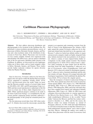
The results of our fine-scale sampling in Twin Cays,
Belize, are mapped in Figure 1: colors indicate whether
placozoans were absent or present at each site. Placozoans
were distributed throughout this mangrove island, with the
exception of the sandy beach habitat in the northwest re-
gion. Of the 159 slide boxes deployed around the perimeter
of Twin Cays during the years 2002 through 2004, 68 were
positive for placozoans. Most mangrove roots to which slide
boxes were attached contained a variety of sponges, colonial
ascidians, anemones, and filamentous algae. Turbidity in the
water was low, and light intensity varied from high to low
throughout the day. Slides displayed an assemblage of or-
ganisms similar to that seen in the coarse-resolution study,
with the addition of the benthic ctenophore Vallicula mul-
tiformis, which was abundant on many of the recovered
slides.
Using the combined coarse- and fine-resolution
16S rDNA data from this study and the 16S rDNA
sequences, available from GenBank, of Voigt et al.
(2004) (AY652522–AY652529, corresponding to haplo-
types H1–H8, respectively) and Aleoshin et al. (2004)
(AY603696), we constructed a partial 16S rDNA phy-
logeny of the Placozoa (Fig. 2). We were unable to
sequence one region within this locus in part of our
samples because of a guanine/cytoseine-rich hairpin sec-
ondary structure; therefore, the data have been parti-
tioned into separate 5Ј and 3Ј regions. We used 73 DNA
samples with an average concatenated length of 358 nt
and a range from 292 to 406 nt. Due to large insertions or
deletions (indels), the aligned data set spanned 480 nt.
Removing indel sites reduced the data to 241 nt. Both
parsimony (MP) and likelihood (Bayesian inference)
methods returned the same tree topology for the well-
supported nodes. On the basis of sequence divergence, the
bootstrap percentages and Bayesian posterior probabilities,
five clades emerged from the combined 73 samples, here-
after referred to as Clade I for haplotypes H1 and H2; Clade
II for haplotype H3; Clade III for haplotypes H6, H7, and
H8; Clade IV for haplotype H5; and Clade V for haplotypes
H4, H4-2, and H4-3 (Fig. 2). Our coarse-and fine-resolution
studies revealed seven of the eight haplotypes already de-
scribed by Voigt et al. (2004), and in addition, two new
haplotypes, H4-2 and H4-3, were discovered in Bermuda.
Haplotype H4-2 differed from the H4 haplotype discovered
by Voigt et al. (2004) by only two nucleotide substitutions,
while haplotype H4-3 differed from H4 by four nucleotide
substitutions. The last column of Table 1 lists the haplotypes
detected during this study. In Bermuda (n ϭ 15), 14 samples
were found to belong to Clade V, while only one belonged
to Clade I. Jamaican samples (n ϭ 3) separated into Clades
I and III. Grenadian samples (n ϭ 2) belonged to Clade III,
Figure 1. Map of Twin Cays, Belize. Dots on the map represent the
sampling sites. Grey dots indicate sites from which placozoans were not
detected. Pink dots represent sites from which placozoans were detected
but not genotyped. The remaining colored dots represent sites positive for
placozoans from which individual strains were genotyped. The habitat type
at the five sampling sites in the northwestern-most part of the map is sandy
beach; all other sampling sites are mangrove roots. (Map modified from a
figure prepared by Molly K. Ryan for the Caribbean Coral Reef Ecosys-
tems Program; used with permission.)
153CARIBBEAN PLACOZOAN PHYLOGEOGRAPHY
6. Figure 2. 16S rDNA phylogeny of combined placozoan samples from this study and sequences available
in GenBank (represented by H1, H2, H3, H4, H5, H6, H7, H8, and AY603896). Haplotypes are represented in
boldface type. Haplotypes H4-2 and H4-3 were newly discovered in this study. Individual strains sequenced in
this study are italicized and labeled by their two-letter country code of origin (BM ϭ Bermuda, BZ ϭ Belize,
GD ϭ Grenada, JM ϭ Jamaica, PN ϭ Panama), followed by laboratory strain identifiers. Localities (countries
and seas) sampled by Voigt et al. (2004) and Aleoshin et al. (2004) are explicitly listed next to haplotypes in
bold. (C) ϭ Caribbean Ocean, (P) ϭ Pacific Ocean. Numbers at each internal node represent % bootstrap under
maximum parsimony and Bayesian posterior probabilities, respectively (only values above 70% and 0.70 are
shown).
154 A. Y. SIGNOROVITCH ET AL.
7. and Panamanian samples (n ϭ 5) fell into Clades I and III.
In Twin Cays (n ϭ 39) we uncovered six haplotypes that
fall into Clades I–III and V. Figure 1 shows this haplotype
distribution among the sampled sites in Twin Cays. The
haplotype groupings (H2), (H3), (H6, H7, H8), (H4) corre-
spond to Clades I–II and V, respectively. The three most
abundant haplotypes were H7, H8, and H4, and the most
diversity was observed in the main channel region that
separates the two land masses composing the island.
Because only a subset of sampled animals were success-
fully established in laboratory culture during the 2003 Twin
Cays field season (41%, see Table 1) and because sequence
data were generated only for those cultured isolates, it is
possible that the molecular data might be biased by differ-
ential viability of different clades. To address this possible
survival bias in our 2003 data, in 2004 we used a Clone-
Saver FTA card (Whatman) to sample 19 placozoan indi-
viduals immediately upon their retrieval from the field, thus
avoiding an intervening step of laboratory culture (for pro-
tocol, see Signorovitch et al., 2005). Sequencing of these 19
DNA samples yielded only representatives of the known
clades.
Discussion
The finding by Voigt et al. (2004) that the Placozoa is not
a monotypic phylum raises a number of immediate ques-
tions that our study was designed to address. For instance,
will extensive sampling result in the discovery of additional
placozoan clades? Do the known clades display obvious
geographic limits or habitat preferences? And finally, are
these clades sympatric on an ecologically relevant spatial
scale?
Our coarse-scale Caribbean-wide sampling revealed four
of the five clades of placozoans previously identified (Voigt
et al., 2004). Clades I, III, and V all were widely distributed
within the Caribbean basin. Voigt et al. had found Clade I
to contain animals from the Red Sea, Italy (Mediterranean),
and Panama (Caribbean side), and Aleoshin et al. (2004)
identified a sample from the Sea of Japan (Fig. 2). Our study
confirmed the presence of Panamanian animals in this clade
and further added Belizean, Bermudian, and Jamaican rep-
resentatives to it. We have also added Belizean animals to
Clade II, which previously contained only a single Carib-
bean sample from Panama. Members of Clade III were
previously identified only from the Pacific in Panama, the
Indo-Pacific, and Guam (Voigt et al., 2004), but we found
that this clade displayed the most geographic diversity
among the four clades. Indeed, Clade III contained animals
from all but one of our sampling locations in the Caribbean
and was widely distributed throughout Twin Cays, Belize.
Finally, Clade V was represented by Panamanian (both
Caribbean and Pacific sides) and Venezuelan samples
(Voigt et al., 2004), and our study has now added Belizean
and Bermudian placozoans to this group. Because of diffi-
culties in establishing animals in laboratory culture, our
sampling efforts in different locales were highly uneven,
preventing us from assessing geographic population struc-
ture across islands. Nevertheless, the fact that our Caribbean-
focused study found identical haplotypes (H2, H3, H4, H6,
H7, and H8) to those discovered in the worldwide sampling
of Voigt et al. (2004) adds further support to the idea that
placozoans, like other microbial eukaryotes (Finlay, 2002),
are geographically unrestricted because of their small body
size, pelagic stage in their life cycle, and large population
size.
In the course of these studies, samplings were concen-
trated around docks, mangrove roots, and to a lesser extent,
patch reefs. These habitat types were similar to those pre-
viously sampled (Voigt et al., 2004). Inspection of Table 1
reveals no clear restriction of placozoans to any one of these
habitats, with the exception of the sandy beach. The absence
of placozoans in the sandy beach habitat of Twin Cays
agrees with Pearse’s observations in Papua New Guinea.
The glass slides recovered from the sandy beach habitat in
Twin Cays were very clean—almost devoid of any epifauna
or flora. Because placozoans are often found on glass slides
covered with a noticeable film of bacteria and algae as well
a rich assemblage of other microorganisms, it is possible
that such a diversity of organisms is necessary for their
recruitment. The fact that we found several known haplo-
types throughout the Caribbean and discovered only two
new haplotypes that fall within an already reported clade
suggests that most of the placozoan diversity in these hab-
itats is adequately characterized in this region. It bears
emphasis, however, that the epifauna of pilings and man-
groves is only a tiny fraction of the possible habitats for
placozoans, and further discoveries of placozoan diversity
might take place in other habitats or in other parts of the
world.
The lack of any obvious clade-specific habitat preference
in our coarse-scale analysis is consistent with the results of
our fine-scale study in the mangrove community of Twin
Cays (Fig. 1). All four clades were isolated from this 92-
hectare island. Members of Clades III and V were wide-
spread throughout the island, whereas members of Clades I
and II were found only within the Main Channel region
dividing the two landmasses composing the island (Fig. 1).
Clades III and V were the most abundant and widely dis-
tributed clades, while Clade I was especially rare. These
results clearly indicate that the four clades occur in sympa-
try in Twin Cays on an ecologically relevant spatial scale.
Furthermore, sympatry is likely to be a widespread phenom-
enon, as indicated by observations of multiple clades in
Panama and Italy (Voigt et al., 2004) and the fact that even
a single slide from our Panamanian collection yielded rep-
resentatives of two different clades (Table 1). The sympatry
155CARIBBEAN PLACOZOAN PHYLOGEOGRAPHY
8. of clades raises questions about the ecological dynamics
among these grossly similar placozoans.
Acknowledgments
We are grateful to Klaus Ru¨tzler, Mike Carpenter, Rafael
Rosengarten, Matthew Nicotra, Casey Dunn, Erika Ed-
wards, Clare Morrall, Oliver Balmer, John Gilbert, Craig
Layne, Christina Glastris, Matt Daren, and Joanna Pitt for
their help during deployment or collection of placozoan
boxes and animal screening. Zack Snable, Aaron Thier, and
Chad Kritzberger helped with animal care. We thank Maria
Moreno, Vicki Pearse, and James Signorovitch for helpful
discussions and critical reading of this manuscript. We also
thank two anonymous reviewers whose suggestion greatly
improved this manuscript. This work was supported in part
by a Caribbean Coral Reef Ecosystem Award (contribution
no. 748) and National Institutes of Health Genetics Training
Grant 5 T32 GM07499-28 to A.Y.S. and a National Science
Foundation Grant EF-0319076 and National Institutes of
Health Grant R21-AI066242-01A1 to L.W.B.
Literature Cited
Aleoshin, V. V., A. V. Konstantinova, M. A. Nikitin, and I. L. Okshtein,
2004. On the genetic uniformity of the genus Trichoplax (Placozoa).
Russ. J. Genet. 40: 1423–1425.
Chenna, R., H. Sugawara, T. Koike, R. Lopez, T. J. Gibson, D. G.
Higgins, and J. D. Thompson. 2003. Multiple sequence alignment
with the CLUSTAL series of programs. Nucleic Acids Res. 31: 3497–
3500.
Finlay, B. J. 2002. Global dispersal of free-living microbial eukaryote
species. Science 296: 1061–1063.
Grell, K. G. 1971a. Trichoplax adhaerens F. E. Schulze und die
Entstehung der Metazoen. Naturwiss. Rundsch. 24: 160–161.
Grell, K. G. 1971b. Embryonic development of Trichoplax adhaerens
F. E. Schulze. Naturwissenschaften 58: 570.
Grell, K. G. 1972. Formation of eggs and cleavage in Trichoplax
adhaerens, F. E. Schulze, Z. Morphol. Tiere 73: 297–314.
Grell, K. G., and G. Benwitz. 1971. Die Ultrastruktur von Trichoplax
adhaerens F. E. Schulze. Cytobiologie 4: 216–240.
Grell, K. G., and E. Lopez-Ochoterena. 1987. A new record of
Trichoplax adhaerens F. E. Schulze (phylum Placozoa) in the Mexican
Caribbean Sea. An. Inst. Cienc. Mar Limnol. 14: 255–256.
Huelsenbeck, J. P., and F. Ronquist. 2001. MRBAYES: Bayesian
inference of phylogeny. Bioinformatics 17: 754–755.
Ivanov, D. L., V. V. Malakhov, and A. B. Tzetlin, 1980. Discovery of
a primitive multicellular organism Trichoplax sp. Zool. Zhurnal 59:
1735–1738.
Martinelli, C., and J. Spring. 2003. Distinct expression patterns of the
two T-box homologues Brachyury and Tbx2/3 in the placozoan
Trichoplax adhaerens. Dev. Genes Evol. 213: 492–499.
Maruyama, Y. K. 2004. Occurrence in the field of a long-term, year-
round, stable population of placozoans. Biol. Bull. 206: 55–60.
Miller, R. L. 1971. Trichoplax adhaerens Schulze, 1883: return of an
enigma Biol. Bull. 141: 374. (Abstract).
Pearse, V. B. 1989. Growth and behavior of Trichoplax adhaerens: first
record of the phylum Placozoa in Hawaii. Pac. Sci. 43: 117–121.
Posada, D., and K. A. Crandall. 1998. Modeltest: testing the model of
DNA substitution. Bioinformatics 14: 817–818.
Ronquist, F., and J. P. Huelsenbeck. 2003. MRBAYES 3. Bioinfor-
matics 19: 475–481.
Ruthmann, A. 1977. Cell-differentiation, DNA content and chromo-
somes of Trichoplax adhaerens F. E. Schulze. Cytobiologie 15: 58–64.
Schulze, F. E. 1883. Trichoplax adhaerens, nov. gen., nov. spec. Zool.
Anz. 6: 92–97.
Signorovitch, A. Y., S. L. Dellaporta, and L. W. Buss. 2005. Molec-
ular signatures for sex in the Placozoa. Proc. Natl. Acad. Sci. USA 102:
15518–15522.
Swofford, D. L. 1998. PAUP*, Phylogenetic Analysis Using Parsimony
(* and other methods). Sinauer, Sunderland, MA.
Voigt, O., A. G. Collins, V. B. Pearse, J. S. Pearse, A. Ender, H.
Hadrys, and B. Schierwater. 2004. Placozoa—no longer a phylum
of one. Curr. Biol. 14: R944–R945.
156 A. Y. SIGNOROVITCH ET AL.