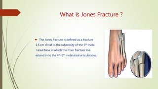
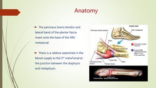
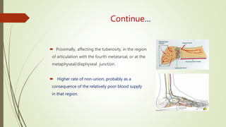
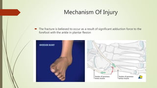
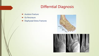
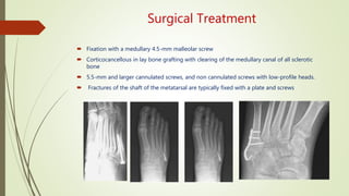
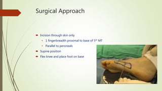
WELCOME
Short Notes Presentation
JONES FRACTURE
Dr. Sheikh Golam Mahbub
D(Ortho)Student
Orthopaedics Surgery
BSMMU
What is Jones Fracture ?
 The Jones fracture is defined as a fracture
1.5 cm distal to the tuberosity of the 5th meta
tarsal base in which the main fracture line
extend in to the 4th-5th metatarsal articulations.
Why it is called jones fracture ?
 First described in 1902 by orthopedic surgeon
Sir Robert Jones.
 He sustained the injury himself(while dancing) as a
fracture of the 5th metatarsal about three-fourths of
an inch from its base.
Anatomy
 The peroneus brevis tendon and
lateral band of the plantar fascia
insert onto the base of the fifth
metatarsal.
 There is a relative watershed in the
blood supply to the 5th metaTarsal at
the junction between the diaphysis
and metaphysis.
Continue…
 Proximally, affecting the tuberosity, in the region
of articulation with the fourth metatarsal, or at the
metaphyseal/diaphyseal junction.
 Higher rate of non-union, probably as a
consequence of the relatively poor blood supply
in that region.
Mechanism Of Injury
 The fracture is believed to occur as a result of significant adduction force to the
forefoot with the ankle in plantar flexion
Clinical Presentation
 Pain over this middle/outside area of
foot
 Swelling
 Difficulty Walking
Radiological Findings
 Diagnostic x-rays include anteroposterior, oblique, and lateral views and should be
made with the foot in full flexion.
 X-Ray shows a transverse fracture near the metatarsal base , usually small
fragment & Minimally displaced.
 Should not be confused with normal apophysis of the proximal 5th metatarsal.
Classification of fracture
(According to site)
Classification of Fracture of 5th Metatarsal
 Type Description
 I Acute fractures at the metaphyseal-diaphyseal junction.
 IA Nondisplaced
 IB Displaced or comminuted or both.
 II Fractures at the metaphseal-diaphyseal junction with clinical or
radiographic evidence of previous injury (i.e., pain, sclerosis).
 III Fractures of the styloid process of the fifth metatarsal.
 IIIA Without involvement of the fifth metatarsocuboid joint.
 IIIB With involvement of the fifth metatarsocuboid joint.
Differntial Diagnosis
 Avulsion Fracture
 Os Peroneum
 Diaphyseal Stress Fractures
Treatment
 Non Surgical
 Until you are able to see a foot & ankle surgeon. the “R.I.C.E”
method of care should be performed
 REST : Stay off the injured foot
 ICE: Apply an icepack to the injured area
 COMPRESSION: An elastic wrap should be used to control swelling
 ELEVATION: The foot should be raised above the level of
Heart to reduce swelling
 If a jones fractures is not significantly displaced, it can be treated with a cast,splint or
walking boot for 4 to 8 weeks.
When is Surgery Needed
 Zone 1 treated without surgery cast, boot or hard-soled shoe
heal within six to eight weeks.
 Zone 2 higher chance of nonunion risk of refracture even after healing.
Surgical treatment is common.
 Zone 3 typically stress fractures in athletes risk of refracture may be
reasons for surgical repair in these fractures.
Type l Fractures
 Type IA fracture (acute)
 Non–weight bearing,
 Short leg cast is worn for 6 to 8 weeks followed by a weight bearing
cast until union has been achieved
 Type IB fractures with displacement and comminuted
 In competitive athletes, consideration should be given to early open reduction
and internal fixation to decrease disability time.
 use of electrical and pulsed ultrasound and bone stimulation for may improve
healing of the fracture.
 Surgery should be considered for type I fractures that are not healing clinically at 8
to 12 weeks
Type ll Fractures
 Type II fractures (partial or complete canal obliteration and sclerosis)
 Non–weight bearing casting may yield satisfactory results.
 Immobilization and non–weight bearing is approximately 8 weeks.
 Refracture is common in this category
 Surgery should be considered for type II fractures in competitive athletes and
others whose occupational demands do not allow prolonged non–weight bearing
immobilization
Type lll Fractures
 Type III fractures
 Short leg cast for 3 weeks followed by a well-molded arch support.
 Nonunions of type III fractures may occur, they rarely are painful and can be
treated with excision of the fragment
Surgical Treatment
 Fixation with a medullary 4.5-mm malleolar screw
 Corticocancellous in lay bone grafting with clearing of the medullary canal of all sclerotic
bone
 5.5-mm and larger cannulated screws, and non cannulated screws with low-profile heads.
 Fractures of the shaft of the metatarsal are typically fixed with a plate and screws
Surgical Approach
 Incision through skin only
• 1 fingerbreadth proximal to base of 5th MT
• Parallel to peroneals
 Supine position
 Flex knee and place foot on base
Post operative care
 A well-padded, short-leg, non-walking cast, extending to the toes, is applied.
 Non weight bearing for 2 weeks.
 Weight bearing in a cast may be started 2 weeks postoperatively.
 Ankle ROM against gravity abduction/adduction, planter and dorsiflexion.
 Return to competitive sports is usually takes 10 to 12 weeks.
Complications
 Refracture
 Screw Failure
 Non union
 Infection
 Sural nerve injury
 Hardware discomfort
Conclusions
 The Jones fracture presents a dilemma in treatment of the active patient.
 Jones fracture has a high rate of nonunion due to low vascular integrity.
 Though cast treatment has been shown to be effective, early screw fixation of the
Jones fracture will results in shorter times to union & return to activity.
 Operatively treated Jones fracture have a high success rate.
 Athlete should not be allowed to return to full activity until full radiographic union
is evidenced.
 Even with non–weight bearing immobilization for 6 to 8 weeks, type I fractures
have a reported nonunion rate of 7% to 28%.
Jone's fracture by Dr.Mahbub

Jone's fracture by Dr.Mahbub

  • 1.
  • 2.
    JONES FRACTURE Dr. SheikhGolam Mahbub D(Ortho)Student Orthopaedics Surgery BSMMU
  • 3.
    What is JonesFracture ?  The Jones fracture is defined as a fracture 1.5 cm distal to the tuberosity of the 5th meta tarsal base in which the main fracture line extend in to the 4th-5th metatarsal articulations.
  • 4.
    Why it iscalled jones fracture ?  First described in 1902 by orthopedic surgeon Sir Robert Jones.  He sustained the injury himself(while dancing) as a fracture of the 5th metatarsal about three-fourths of an inch from its base.
  • 5.
    Anatomy  The peroneusbrevis tendon and lateral band of the plantar fascia insert onto the base of the fifth metatarsal.  There is a relative watershed in the blood supply to the 5th metaTarsal at the junction between the diaphysis and metaphysis.
  • 6.
    Continue…  Proximally, affectingthe tuberosity, in the region of articulation with the fourth metatarsal, or at the metaphyseal/diaphyseal junction.  Higher rate of non-union, probably as a consequence of the relatively poor blood supply in that region.
  • 7.
    Mechanism Of Injury The fracture is believed to occur as a result of significant adduction force to the forefoot with the ankle in plantar flexion
  • 8.
    Clinical Presentation  Painover this middle/outside area of foot  Swelling  Difficulty Walking
  • 9.
    Radiological Findings  Diagnosticx-rays include anteroposterior, oblique, and lateral views and should be made with the foot in full flexion.  X-Ray shows a transverse fracture near the metatarsal base , usually small fragment & Minimally displaced.  Should not be confused with normal apophysis of the proximal 5th metatarsal.
  • 10.
  • 11.
    Classification of Fractureof 5th Metatarsal  Type Description  I Acute fractures at the metaphyseal-diaphyseal junction.  IA Nondisplaced  IB Displaced or comminuted or both.  II Fractures at the metaphseal-diaphyseal junction with clinical or radiographic evidence of previous injury (i.e., pain, sclerosis).  III Fractures of the styloid process of the fifth metatarsal.  IIIA Without involvement of the fifth metatarsocuboid joint.  IIIB With involvement of the fifth metatarsocuboid joint.
  • 12.
    Differntial Diagnosis  AvulsionFracture  Os Peroneum  Diaphyseal Stress Fractures
  • 13.
    Treatment  Non Surgical Until you are able to see a foot & ankle surgeon. the “R.I.C.E” method of care should be performed  REST : Stay off the injured foot  ICE: Apply an icepack to the injured area  COMPRESSION: An elastic wrap should be used to control swelling  ELEVATION: The foot should be raised above the level of Heart to reduce swelling  If a jones fractures is not significantly displaced, it can be treated with a cast,splint or walking boot for 4 to 8 weeks.
  • 14.
    When is SurgeryNeeded  Zone 1 treated without surgery cast, boot or hard-soled shoe heal within six to eight weeks.  Zone 2 higher chance of nonunion risk of refracture even after healing. Surgical treatment is common.  Zone 3 typically stress fractures in athletes risk of refracture may be reasons for surgical repair in these fractures.
  • 15.
    Type l Fractures Type IA fracture (acute)  Non–weight bearing,  Short leg cast is worn for 6 to 8 weeks followed by a weight bearing cast until union has been achieved  Type IB fractures with displacement and comminuted  In competitive athletes, consideration should be given to early open reduction and internal fixation to decrease disability time.  use of electrical and pulsed ultrasound and bone stimulation for may improve healing of the fracture.  Surgery should be considered for type I fractures that are not healing clinically at 8 to 12 weeks
  • 16.
    Type ll Fractures Type II fractures (partial or complete canal obliteration and sclerosis)  Non–weight bearing casting may yield satisfactory results.  Immobilization and non–weight bearing is approximately 8 weeks.  Refracture is common in this category  Surgery should be considered for type II fractures in competitive athletes and others whose occupational demands do not allow prolonged non–weight bearing immobilization
  • 17.
    Type lll Fractures Type III fractures  Short leg cast for 3 weeks followed by a well-molded arch support.  Nonunions of type III fractures may occur, they rarely are painful and can be treated with excision of the fragment
  • 18.
    Surgical Treatment  Fixationwith a medullary 4.5-mm malleolar screw  Corticocancellous in lay bone grafting with clearing of the medullary canal of all sclerotic bone  5.5-mm and larger cannulated screws, and non cannulated screws with low-profile heads.  Fractures of the shaft of the metatarsal are typically fixed with a plate and screws
  • 19.
    Surgical Approach  Incisionthrough skin only • 1 fingerbreadth proximal to base of 5th MT • Parallel to peroneals  Supine position  Flex knee and place foot on base
  • 20.
    Post operative care A well-padded, short-leg, non-walking cast, extending to the toes, is applied.  Non weight bearing for 2 weeks.  Weight bearing in a cast may be started 2 weeks postoperatively.  Ankle ROM against gravity abduction/adduction, planter and dorsiflexion.  Return to competitive sports is usually takes 10 to 12 weeks.
  • 21.
    Complications  Refracture  ScrewFailure  Non union  Infection  Sural nerve injury  Hardware discomfort
  • 22.
    Conclusions  The Jonesfracture presents a dilemma in treatment of the active patient.  Jones fracture has a high rate of nonunion due to low vascular integrity.  Though cast treatment has been shown to be effective, early screw fixation of the Jones fracture will results in shorter times to union & return to activity.  Operatively treated Jones fracture have a high success rate.  Athlete should not be allowed to return to full activity until full radiographic union is evidenced.  Even with non–weight bearing immobilization for 6 to 8 weeks, type I fractures have a reported nonunion rate of 7% to 28%.