Aortic Root SUrgery
•
0 likes•579 views

1) The document discusses various surgical procedures for treating aortic root pathologies. It describes the anatomy of the aortic root and various conditions that can affect it like aneurysms and dissections. 2) Surgical techniques discussed include different types of composite graft replacements, valve sparing procedures, and re-do operations. Specific procedures mentioned are the Bentall procedure and the Ross procedure. 3) Factors that determine whether the aortic valve should be replaced or repaired are discussed. Guidelines for intervention based on aortic root size are also provided.

Recommended
More Related Content
What's hot
What's hot (20)
Similar to Aortic Root SUrgery
Similar to Aortic Root SUrgery (20)
More from Dicky A Wartono
More from Dicky A Wartono (20)
Recently uploaded
Recently uploaded (20)
Aortic Root SUrgery
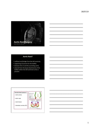
- 1. 28/07/19 1 Aortic Root Surgery Dicky A.Wartono ,MD 2019 Aortic Root? • outflow tract/bridge from the left ventricle, • supporting structures for the leaflets • between the LV and the ascending aorta. • extends from the basal attachments of the leaflets within the left ventricle to the st junction The Aortic Root Consists of : Aortic annulus Aortic cusps Aortic Sinuses Sinotubular Junction (STJ)
- 2. 28/07/19 2
- 3. 28/07/19 3 Too small ( small annulus ) Too large ( Root Aneurysm ) Enlargement - Nicks ( Post. ) procedure - Manouguian ( Post.) Procedure - Konno ( Ant.) Procedure Replacement - Bentall Procedure - Ross Procedure Replacement + Enlargement - Ross – Konno - Modified Ross – Konno Replacement Bentall - Mechanical - Stented tissue - Stentless tissue Valve Sparing - Remodelling - Reimplantation
- 4. 28/07/19 4 Root Abscess ( Endocarditis ) Type A Dissection Replacement - Bentall - Mechanical - Stented tissue - Stentless tissue +/- Mitral / LA Reconstruction Bentall - Mechanical - Stented tissue - Stentless tissue Valve Sparing - Remodelling - Reimplantation Root Aneurysm • Degenerativež • Connective Tissue Disorders ž • Bicuspid Aortopathy • Post Stenotic • Chronic Dissections • 1- Aortic root dilatation secondary to ascending aortic aneurysm • 2-Annulo-aortic ectasia and connective tissue syndromesCTD such as Marfan’s and Ehlers- Danlos • 3-Aortic root and ascending aortic dissection — acute or chronic
- 5. 28/07/19 5 , few data exist on the natural history of iso- rysms, since they are often associated with the ascending or descending aorta. onsidered in patients who have an aortic arch mal diameter ≥55 mm or who present symp- compression. Decision-making should weigh , since aortic arch replacement is associated mortality and stroke than in surgery of the ding aorta. Indications for partial or total nt are more frequently seen in patients who urgeryon anadjacentaneurysmofthe ascend- a. osition (debranching) and TEVAR might be native to conventional surgery in certain clin- ally when there is reluctance to expose mic circulatory arrest; however, especially transposition, as well as in patients with the f acute Type B AD, the risk of retrograde t consequence of the procedure is elevated d against the remaining risk of conventional ic aneurysms descending aortic aneurysms has been e development of TEVAR using stent grafts. exist to guide the choice between open From non-randomized comparisons and mortality is lower after TEVAR than open mortality depends on the extent of repair istics, in particular age and comorbidities. vival does not differ between TEVAR and follow-up, there is a contrast between low rtic complications and relatively high overall om cardiopulmonary causes.331,332 onsidered in patients who have a descending iameter ≥55 mm. When surgery is the only nsidered in patients with a maximal diameter sholds can be considered in patients with dications for treatment and the choice pen surgery should be made by a multidiscip- rtise in both methods, taking into consider- comorbidities, and life expectancy, and h analysis of the arterial tree to assess the ed risks of each technique: extent and size d atheroma, collaterals, and size and length for endovascular grafting and vascular of information on long-term results of pt in mind, in particular in young patients. ay be combined in hybrid approaches. disease, surgery should be preferred over dencesupportinganyuseofTEVARinpatients disease, except in emergency situations in abilization as a bridge to definitive surgical Recommendations on interventions on ascending aortic aneurysms Recommendations Classa Levelb Surgery is indicated in patients who have aortic root aneurysm, with maximal aortic diameterc 50 mm for patients with Marfan syndrome. I C Surgery should be considered in patients who have aortic root aneurysm, with maximal ascending aortic diameters: • 45 mm for patients with Marfan syndrome with risk factors.d • 50 mm for patients with bicuspid valve with risk factors.e,f • 55 mm for other patients with no elastopathy.g,h Lower thresholds for intervention may be considered according to body surface area in patients of small stature or in the case of rapid progression, aortic valve regurgitation, planned pregnancy, and patient’s preference. Interventions on aortic arch aneurysms Surgery should be considered in patients who have isolated aortic arch aneurysm with maximal diameter 55 mm. Aortic arch repair may be considered in patients with aortic arch aneurysm who already have an indication for surgery of an adjacent aneurysm located in the ascending or descending aorta. IIb C Interventions on descending aortic aneurysms TEVAR should be considered, rather than surgery, when anatomy is suitable. IIa C TEVAR should be considered in patients who have descending aortic aneurysm with maximal diameter 55 mm. IIa C When TEVAR is not technically possible, surgery should be considered in patients who have descending aortic aneurysm with maximal diameter 60 mm. IIa C When intervention is indicated, in cases of Marfan syndrome or other elastopathies, surgery should be indicated rather than TEVAR. IIa C IIa C IIb C IIa C a Class of recommendation. b Level of evidence. c Decision should also take into account the shape of the different parts of the aorta. Lower thresholds can be used for combining surgery on the ascending aorta for patients who have an indication for surgery on the aortic valve. d Family history of AD and/or aortic size increase .3 mm/year (on repeated measurements using the same imaging technique, at the same aorta level, with side-by-side comparison and confirmed by another technique), severe aortic or mitral regurgitation, or desire for pregnancy. e Coarctation of the aorta, systemic hypertension, family history of dissection, or increase in aortic diameter .3 mm/year (on repeated measurements using the same imaging technique, measured at the same aorta level, with side-by-side comparison and confirmed by another technique). f Pending comorbidities in the elderly. g See text in section 8. h For patients with LDS or vascular type IV Ehlers-Danlos syndrome (EDS), lower thresholds should be considered, possibly even lower than in Marfan syndrome. Thereare no datato provide figures and a sensible case-by-case approachisthe only option. ESC Guidelines byguestonJuly15,2016http://eurheartj.oxfordjournals.org/Downloadedfrom 2014 ESC Guidelines on the diagnosis and treatment of aortic diseases 30mm = Aneurysm >55mm = Surgery >50mm = Bicuspid >45mm = Marfan • Gold Standard for young patients ( < 65y ) • Permanent Anti coagulation -Contraindications -Life style -Patient preference • Higher risk for TE •Most Durable •Higher risk for infection ( or Re infection) In 1968, Bentall and De Bono reported (in a two page case report), a single patient treated with a composite graft and mechanical valve replacement of the aortic root and ascending aorta with coronary reimplantation Sizes 21 mm - 27mm
- 6. 28/07/19 6 • 0.2% Preserved in glutaraldehyde •Polyester sewing cuff •Alfa amino oleic acid ( anticalcification ) •Zero net pressure fixation of the leaflets Improved Hemodynamics Ideal for Root Abscess Reduced infection (?) Low Thromboembolic Complications ---------------------------------- Availability Risk of Calcification ( >50% SVD in 20 y ) ( immune mediated? ) Homovital ( Fresh) Cryopreserved Reimplantation ( David I) Remodelling ( Yacoub)
- 8. 28/07/19 8 3 essing Video 12 . Testing the seating of the prosthesis. Photo 3 . Commercially available, preclotted composite graft. Video 13 . Suturing the composite graft into position. tons ortic e left Stay sutures are placed in the commissures to facili- tate exposure (Video 11). Valve seating is tested (Vid- eo 12). A standard composite graft is selected (MMCTSLink 4, MMCTSLink 5 and MMCTSLink 6) and the valve implantation is performed using interrupted Ticron Aortotomy & Periaortic dissection (Excise aortic valve leaflets) 2 Composite grafts replacement of the aortic roots Video 2 . Preparing the arch for arterial cannulation. Video 3 . Arterial cannulation. Video 4 . Venous cannulation and placing the LV vent. Video 5 . Crossclamping of the aorta and opening of the aneurysm. Photo 1 . Aortic root after removal of the aorta of the aortic valve, with both coronaries detached and mobilised. Video 7 . Resection of the distal part of aortic aneurysm. Video 8 . Resection of the proximal portion of the aneurysm and resection of the valve leaflets. Video 6 . Antegrade cardioplegia. Cannulation is performed either in the aortic arch or – if the disease extends into the arch – the subclavian artery is cannulated instead (Videos 2, 3). Standard two-stage arterial cannula (MMCTSLink 3) and LV vent are used (Video 4). Surgical procedure At a moderate hypothermia of 268C the ascending aorta is cross-clamped (Video 5), heart is arrested with cardioplegia antegrade cardioplegia (Video 6), and the cardioplegia is continued in retrograde mode, and coronaries are continuously perfused with cold (168C) blood. Aneurysm is totally removed (Photo 1), (Videos 7, 8 ). Coronary button Preparation Composite grafts replacement of the aortic roots Video 14 . Placement of the sutures in the non-coronary sinus. Video 15 . Tying the knots. Video 16 . Cutting the graft to size. Video 17 . Implantation of the left main coronary artery. Video 19 . Implantation of the right coronary into the graft. Video 20 . Implantation of the right coronary into the graft (continued). Video 21. Distal anastomosis with the aortic arch. sutures (MMCTSLink 7) and this surgery can be sup- ported with Teflon felt or pericardium in friable aortic roots (Photo 3) (Videos 13, 14, 15). Prefabricated graft is cut to size to facilitate the implantation of coronary arteries (Video 16). The coronary arteries are implanted into the graft using 4–5 Prolene (MMCTSLink 8) with small needle, starting with left main coronary artery, which must be sufficiently mobilised (Videos 17, 18). Right coronary artery is implanted next (Videos 19, 20); a short clamping of the venous line allows a dil- atation of the right ventricle to determine the exact position of the right coronary artery in relation to the graft. Distal anastomosis is performed either to the arch or to the remnants of the ascending aorta (Video 21) also using a small needle. Graft inclusion technique (Schematic 2) is avoided as Prostetic suture Composite grafts replacement of the aortic roots Schematic 2. Details of the graft inclusion technique from Crawford (Diseases of the Aorta). Video 22. Finished procedure after decannulation. Schematic 3 . Running suture line between the cut edge of the aor- tic wall and the distal portion of the valve sewing ring. The inset shows a cutaway view from the inside. Schematic 4 . Separate grafts to both coronary arteries. Schematic 5 . Detail of the ‘button’-buttress technique used for anastomosis of the coronary ostia to the graft. With proper attention to suturing and buttressing the suture lines with pericardium or Teflon felt, a safe hemostasis is possible without resorting to the graft inclusion (Video 22). Technical innovations an additional suture after placement of the valve fix- ation sutures in the annulus. In case of redo operation, where considerable diffi- culties can be experienced when mobilizing coronary arteries, short segments of graft material can be used to attach coronary arteries to the graft (Schematic 4) w4x. The improvement in coronary anastomosis can be achieved by performing a doubling of tissue at their coronary orifices as described by Hilgenberg et al. w5x and Prateli et al. w6x (Schematic 5). Finally, in difficult redo operations Cabrol technique has to be utilised, employing a graft which connects both coronary ostia and is anastomosed to the com- posite graft itself (Schematic 6). Results ● The mortality on a procedure, when performed electively is considered to be low (less than 5%). Prostetic suture
- 9. 28/07/19 9 Surgical Management of the Aortic Root 135 6.10. Reoperation of the aortic root Structural failure of the root, pseudoaneurysms, or infection may necessitate redo aortic root replacement. This is an operation that typically carries a high risk of mortality and morbidity. Some special considerations when this very difficult operation is undertaken include: calcified homografts or stentless valves, coronary artery length, and infection. In patients with a very calcified neo-aortic wall it is often extremely difficult to dissect out the wall and redo the root as it becomes very adherent to the adjacent structures and pulmonary artery and coronaries can be injured. Replacing just the aortic valve within the calcified root is an option. With the advent of trans aortic valve implantation (TAVI), this may be an excellent option in high risk patients. El-Hamamsy et al compared the Freestyle graft with homograft aortic root replacement in a prospective, randomized trial.[139] One- hundred sixty-six patients with an average age of 65 years had a mean follow-up of 7.6 years. Significant conclusions were made from this data including an improved age of survival (80 vs. 77 years), lower rate of reoperation (100% vs. 90%), and echocardiographically patients had less signs of valvular deterioration (86% vs. 30%) in the FreeStyle group. Figure 14. Aortic root replacement via the Cabrol technique. Coronary buttons are sutured side-to-side to a Dacron interposition graft during root replacement.[140] 5 c roots arate grafts to both coronary arteries. ail of the ‘button’-buttress technique used for e coronary ostia to the graft. uture after placement of the valve fix- n the annulus. o operation, where considerable diffi- experienced when mobilizing coronary segments of graft material can be used nary arteries to the graft (Schematic 4) ent in coronary anastomosis can be erforming a doubling of tissue at their es as described by Hilgenberg et al. w5x al. w6x (Schematic 5). cult redo operations Cabrol technique ed, employing a graft which connects ostia and is anastomosed to the com- elf (Schematic 6). y on a procedure, when performed considered to be low (less than 5%). ncreased in all the patients with con- ue disorders (Marfan) and in patients der emergency conditions. ure also carries considerable risk when acute Type A dissection. 5 Composite grafts replacement of the aortic roots Schematic 2. Details of the graft inclusion technique from Crawford (Diseases of the Aorta). Video 22. Finished procedure after decannulation. Schematic 3 . Running suture line between the cut edge of the aor- tic wall and the distal portion of the valve sewing ring. The inset shows a cutaway view from the inside. Schematic 4 . Separate grafts to both coronary arteries. Schematic 5 . Detail of the ‘button’-buttress technique used for anastomosis of the coronary ostia to the graft. With proper attention to suturing and buttressing the suture lines with pericardium or Teflon felt, a safe hemostasis is possible without resorting to the graft inclusion (Video 22). Technical innovations Several modifications of the technique have been pro- posed in order to eliminate the risk of bleeding (Sche- matic 3). This particular modification by Copeland et al. w3x improves the hemostasis at the aortic root by running an additional suture after placement of the valve fix- ation sutures in the annulus. In case of redo operation, where considerable diffi- culties can be experienced when mobilizing coronary arteries, short segments of graft material can be used to attach coronary arteries to the graft (Schematic 4) w4x. The improvement in coronary anastomosis can be achieved by performing a doubling of tissue at their coronary orifices as described by Hilgenberg et al. w5x and Prateli et al. w6x (Schematic 5). Finally, in difficult redo operations Cabrol technique has to be utilised, employing a graft which connects both coronary ostia and is anastomosed to the com- posite graft itself (Schematic 6). Results ● The mortality on a procedure, when performed electively is considered to be low (less than 5%). ● The risk is increased in all the patients with con- nective tissue disorders (Marfan) and in patients operated under emergency conditions. ● This procedure also carries considerable risk when performed in acute Type A dissection. Implantation of the Left coronary into the graft
- 10. 28/07/19 10 • Conclusions • Aortic Root surgery using Composute Valve graft, is a safe with reasonable outcome • Timing of Surgery, & placement of coronary button is one of the most contributing factor • There are few option for managing fragile tissue • Distal…………