Downloaded 82 times


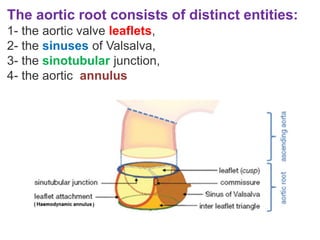


The aortic root connects the left ventricle to the systemic circulation and consists of four distinct components: 1) the aortic valve leaflets, which provide the main sealing mechanism; 2) the sinuses of Valsalva, which host the coronary arteries; 3) the sinotubular junction, which separates the aortic root from the ascending aorta; and 4) the aortic annulus, which defines the separation of ventricular and arterial hemodynamics. Each component contributes to the optimal structure and function of the aortic root, including unidirectional blood flow and maintaining laminar flow under varying cardiac demands.


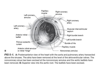
![Mitral_Valve_Anatomy_Surgical_Perspective[1].pptx](https://cdn.slidesharecdn.com/ss_thumbnails/mitralvalveanatomysurgicalperspective1-250701160336-9898ca79-thumbnail.jpg?width=640&height=640&fit=bounds)


