
Recommended
More Related Content
What's hot
What's hot (20)
Similar to Aortic repair ppt
Similar to Aortic repair ppt (20)
More from DR NIKUNJ SHEKHADA
More from DR NIKUNJ SHEKHADA (20)
Recently uploaded
Recently uploaded (20)
Aortic repair ppt
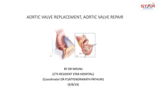
- 1. AORTIC VALVE REPLACEMENT, AORTIC VALVE REPAIR BY DR NIKUNJ (CTS RESIDENT STAR HOSPITAL) (Coordinator:DR P.SATYENDRANATH PATHURI) (9/8/19)
- 5. AORTIC VALVE REPLACEMENT Sharp excision of the aortic valve is performed using a #11 blade, starting with the right coronary cusp.
- 6. Aortic valve exposure through a transverse aortotomy aided by three commissural sutures (arrows) and a suture retracting the distal portion of the ascending aorta cephalad.
- 7. Sizing of the appropriate aortic valve prosthesis using commercial sizers can be accomplished before (as depicted in this picture) or after valve excision.
- 8. • Alternating green and white valve sutures are systematically passed through the aortic valve annulus and then through the sewing ring of the prosthetic valve traveling from the commissure between the left and right coronary cusps to the commissure between the left coronary and noncoronary cusps.
- 9. • All valve sutures have been placed around the annulus and through the sewing ring of the valve.
- 10. • The prosthetic valve is parachuted down into position by the surgeon who holds one set of valve sutures and the prosthetic valve along with the valve holder. The assistant holds the other two sets of valve sutures. The surgeon and assistant pull on the valve sutures while the surgeon parachutes the valve down into position.
- 11. • Picture of prosthetic aortic valve in position after tying all valve sutures systematically beginning with the left coronary cusp, followed by the right coronary cusp, and then the noncoronary cusp.
- 12. • Closure of the aortotomy can be performed using a one- or two-layer technique.
- 13. AORTIC ROOT ENLARGEMENT • In patients with a small aortic annulus, implanting a small stented bioprosthetic valve or a mechanical valve may leave residual gradient across the aortic valve. • Forcing a small prosthetic valve may result in perivalvular leak or disruption of the aorta or left ventricle. • POSTERIOR ENLARGEMENT of the aortic root is performed by either the Nicks-Nunez or the Rittenhouse- Manouguian technique, • ANTERIOR ENLARGEMENT is performed using the Konno- Rastan aortoventriculoplasty technique
- 14. NICKS-NUNEZ TECHNIQUE • a vertical incision through the commissure between the left coronary and noncoronary cusp and extending down in the interleaflet triangle, followed by patch reconstruction of the aortic root. This method can enlarge the aortic root by 2-3 mm.
- 15. • A diamond-shaped patch of autologous pericardium or prosthetic material is fashioned. One end of the patch is inserted into the distal end of the enlargement using interrupted sutures with pledgets. At the level of the aortic annulus the sutures are passed through the sewing ring of the prosthetic valve. The remainder of the valve sutures are placed through the aortic annulus in a standard fashion.
- 16. • The diamond patch is then tailored for closure of the aortotomy. If a transverse aortotomy is used, then the patch is transected at the level of the transverse aortotomy and incorporated as part of the anastomosis of the aortic root to the ascending aorta.
- 17. • Rittenhouse-Manouguian Technique • vertical incision through the middle of the noncoronary sinus extending through the annulus and into the anterior leaflet of the mitral valve.
- 18. KONNO-RASTAN METHOD OF AORTIC ROOT ENLARGEMENT • In the Konno-Rastan method of aortic root enlargement, an incision is made through the right coronary region of the aortic annulus, near the commissure between the left and right coronary cusps. The incision is carried further into the interventricular septum, and a similar incision is made on the right ventricular free wall.
- 19. • One end of a diamond patch is sewed to the deep end of the incision in a continuous fashion to repair the inter- ventricular septum up to the level of the aortic annulus.
- 20. • The base of a triangular patch is attached using interrupted pledgeted mattress sutures to the diamond patch at the level of the annulus. The same sutures are also passed through the sewing ring of the prosthetic valve.
- 21. • The triangular patch is then folded to cover the right ventricular outflow tract defect and sewed using continuous running suture. The left ventricular outflow tract patch is finally used to close the vertical aortotomy using continuous suture technique.
- 23. EXPOSURE AND ASSESSMENT • Full-thickness 4-0 polypropylene traction sutures are placed at the three commissures and retracted using clamps, but not tied, to permit a dynamic assessment of valve anatomy. • Leaflets are inspected to assess mobility, restriction, calcification, and prolapse.
- 24. RECONSTRUCTIVE TECHNIQUES • TYPE I—AORTIC VALVE REGURGITATION • Aortic valve regurgitation with normal leaflet motion is rare and often amenable to valve reconstruction. The techniques vary depending upon the lesions.
- 25. • Isolated Aortic Annular Dilatation : • rare , However, it may occur in dilated cardiomyopathy and in rheumatic valvular diseases, causing mild to moderate regurgitation in association with concomitant mitral valve dysfunction. • If the leaflets display minimal thickening and/or retraction, a circular annuloplasty is carried out to ensure adequate leaflet coaptation.
- 26. LEAFLET PERFORATION • The defect is usually limited and displays a healed fibrotic edge (a). This condition is favorable for valve reconstruction by patching using glutaraldehyde-treated autologous pericardium (b) • The patch is tailored according to the shape and size of the defect, adding a 2-mm margin for suturing. • It is then sutured to the edge of the defect using 5-0 monofilament interrupted sutures tied on the aortic side of the leaflet. • In the presence of a large defect, continuous suture can be used provided that narrow or locked bites are employed to avoid a purse-string effect.
- 27. VEGETATIONS • Small and pedicled vegetations can usually be sharply excised with scissors without producing a perforation . • Larger vegetations must be resected and the valve repaired by patching. • In all instances, the ventricular aspect of the mitral valve should be carefully inspected to detect potential kissing lesions
- 28. LEAFLET TEAR OR AVULSION • The most favorable lesion is a tear of the belly of the leaflet that leaves the commissural areas intact (a). • This type of lesion treated by direct suture. • If a significant lack of tissue is present, the patch is secured to the edges of the tear (c). • Leaflet avulsion is best treated by pericardial patching.
- 29. TYPE II—AORTIC VALVE REGURGITATION • Type II aortic valve regurgitation may be corrected by reconstructive techniques, particularly when the prolapse is limited and involves only one leaflet as in some congenital malformations or rheumatic valve diseases. • Since the mechanism of prolapse is usually a distension of the free edge of the leaflet, appropriate tension can be restored by a triangular resection • The amount of triangular resection is calculated by measuring the length of the free margin of the corresponding halves of the adjacent leaflets and adding 2 mm on each side of the triangle for suturing (b).
- 30. TYPE IIIa—AORTIC REGURGITATION • Type IIIa aortic regurgitation may result from rheumatic valvular disease, valve sclerosis, or bicuspid valve malformation with commissural fusion. • Whenever the amount of leaflet tissue is adequate with preserved pliability, leaflet mobilization by commissurotomy can be attempted. • The commissurotomy should ideally remove a wedge of fibrous tissue.
- 31. • TYPE IIIb—AORTIC VALVE REGURGITATION: VALVE SPARING • In patients with isolated sinotubular junction dilatation and well-preserved leaflets, a supracoronary tube graft interposition is often sufficient to correct aortic valve regurgitation. • The goal of the procedure is to restore the normal ratio between the circumference of the sinotubular junction and the aortic annulus. In those patients with extended aortic root dilatation, aortic valve sparing procedures such as those described by Yacoub and David are an excellent indication.
- 32. SUBCOMMISSURAL ANNULOPLASTY A subcommissural annuloplasty(Cabrol stitch) reduces the width of the interleaflet triangle, improves cusp coaptation, and can help to stabilize the ventriculoaortic junction
- 33. VALVE-SPARING ROOT REPLACEMENT: REIMPLANTATION TECHNIQUE • A valve-sparing root replacement using the reimplantation technique provides the most stable form of functional aortic annuloplasty. • Originally pioneered by David and colleagues, multiple modifications of this procedure have been reported.
- 34. • T. David-I Reimplantation with cylindrical tube graft (1988) • T. David-II Yacoub remodeling • T. David-III Yacoub remodeling + NCC external annuloplasty • T. David-IV Reimplantation with 2-4mm larger graft + narrowed distal end (“STJ”) • T. David-V Reimplantation with ~4-6mm larger graft + necking down both annular end & STJ (May 2001) • T. David-V Stanford modification Reimplantation with 6-8mm larger proximal graft + necking down annular end + small distal graft (December 2002)
- 35. AORTIC ROOT PREPARATION • dissect the aortic root externally as low as possible, given the natural anatomic limitations (i.e., where the root inserts into ventricular muscle). • The root dissection is started along the NC sinus and continued towards the LC/NC commissure • Moving toward the RC/NC commissure and along the right sinus and the RC/LC commissure, • The sinuses of Valsalva are then resected leaving approximately 5 mm of aortic wall attached and the coronary buttons are harvested.
- 36. PROSTHESIS SIZING • a Hagar dilator is used to size the circle that includes the three commissures, and a graft 4 mm larger is chosen as this graft will sit outside the commissural posts. • height of the commissure (measured from the base of the interleaflet triangle to the top of the commissure) is equal to external diameter of the sinotubular junction. • The height of the commissure is most easily measured at the NC/LC commissure by first drawing a connecting line between the nadirs of the two adjacent cusps (base of interleaflet triangle) and measuring the distance between this line and the top of the commissure. • This height corresponds to the diameter of the graft chosen.
- 37. PROXIMAL SUTURE LINE • 2-0 Tycron sutures with pledgets are passed from inside to outside the aorta with the pledgets on the inside, starting from the NC/LC commissure and moving clockwise. • Along the fibrous portion of the aortic annulus, these sutures are inserted along the horizontal plane formed by the base of the inter-leaflet triangles.
- 38. PROSTHESIS PREPARATION AND FIXATION • A Dacron prosthesis with or without builtin neoaortic sinuses can be used. • The three commissures are attached to the prosthesis along the same plane the new sinotubular junction. • First, the distance from the base of the interleaflet triangle to the top of the commissure is measured at the LC/NC commissure and marked on the graft. • Next, at the RC/NC and RC/LC commissures, the distance from the proximal suture to the top of the commissure is measured and used to determine the amount of graft material that needs to be trimmed . • The pledgeted sutures are then passed through the base of the prosthesis, respecting the spaces between sutures and the curvilinear contour of the suture line.
- 39. VALVE REIMPLANTATION. • The commissures are reimplanted first using 4-0 polypropylene sutures while pulling up on the prosthesis and the native commissure and then tied into place.
- 40. LEAFLET ASSESSMENT AND REPAIR • reexamine the leaflets for any unmasked prolapse, symmetry, and the height and depth of coaptation. • Cardioplegia is administered through the distal end of the graft with partial clamping to distend the new aortic root and to assess root pressure and signs of LV dilation. • The cardioplegia solution is then slowly aspirated out of the prosthesis without distorting the leaflets. • This gives another visual assessment of the aortic valve in its physiologic closed state as well as the area and height of coaptation. • Coronary ostia are then reimplanted on the graft, and the distal anastomosis is performed at the level of normal aorta.
- 41. CUSP REPAIR TECHNIQUES • Cusp prolapse is the most frequently encountered lesion and is associated with excess length of the free margin, which can be corrected using either central free margin plication or free margin resuspension. • When a single cusp is prolapsing, the two nonprolapsing cusps serve as the reference and are used to estimate the required reduction in the free margin length. • When two cusps are prolapsing, the third nonprolapsing cusp is used as a reference to indicate the desired height of coaptation. • In the rare instance that all the cusps are prolapsing, the goal is to achieve a cusp coaptation height at the midlevel of the sinuses of Valsalva.
- 44. EARLY COMPLICATIONS • Neurologic Complications • transient ischemic attacks (TIAs), which are fully reversible neurologic events lasting less than 24 hours; • reversible ischemic neurologic deficit, which are fully reversible neurologic events lasting between 24 hours and 3 weeks; • strokes, which are nonreversible neurologic deficits lasting longer than 3 weeks • The rate of stroke for isolated AVR, according to the STS database, is 1.5%. • In high-risk (STS predicted risk of mortality > 10%) and older patients (>80 years old), the risk of stroke rises to between 2% and 4.4%. • The addition of CABG to the AVR also raises the risk to between 4.9% and 5.8%. Risk factors for early postoperative stroke include low LVEF (<40%), ascending aortic calcification, older than 70 years, female sex, diabetes mellitus, bypass time greater than 120 min • a history of stroke, carotid artery disease, and walking less than 300 meters during a 6-minute walk test.
- 45. HEART BLOCK AND PERMANENT PACEMAKER IMPLANTATION • Atrioventricular node block occurs more frequently after heart valve surgery compared with other types of cardiac surgery, with 3% to 8% of patients requiring permanent pacemaker insertion.
- 46. LATE COMPLICATIONS • Thromboembolism, Anticoagulation, and Bleeding Complications • Fluctuations in the international normalized ratio (INR) seem to be an important risk factor for both thromboembolic and bleeding events • close monitoring of the INR to minimize fluctuations may be important to prevent these complication • Anticoagulation with a minimum target INR of 2.0 to 3.0 for low-risk patients and valves with low thrombogenicity is recommended for mechanical AVR • On-X Anticoagulation Clinical Trial (PROACT) suggest that INR may be safely maintained between 1.5 and 2.0 with lower risk of bleeding and without a significant increase in thromboembolism • The target INR should be 2.5 to 3.5 for patients at high risk for thromboembolism or who receive mechanical valves with high thrombogenicity (disc valves).
- 47. PROSTHETIC VALVE ENDOCARDITIS • early PVE if it occurs less than 60 days post implantation and • late PVE if it occurs more than 60 days post implantation. • The rate of PVE is approximately 0.5% to 1% per year, with a slightly higher rate in the first 6 to 12 months compared to less than 12 months. • Early PVE is usually a result of perioperative seeding of the prosthetic valve either intraoperatively or from wound infections or indwelling intravascular catheters postoperatively. • Late PVE usually results from noncardiac sources of bacteremia or some- times from insidious infections with less virulent organsms introduced perioperatively • Early PVE is associated with 30% to 80% mortality, whereas late PVE is associated with 20% to 40% mortality
- 48. PARAVALVULAR LEAK AND HEMOLYSIS • The continuous suture technique of aortic valve implantation was traditionally thought to be a risk factor for paravalvular leak compared with an interrupted suture technique.
- 49. PROSTHESIS-PATIENT MISMATCH • The persistence of left ventricular outflow obstruction postoperatively is termed prosthesis-patient mismatch (PPM) • effective orifice area (EOA) indexed to the patient’s body surface area indexed effective orifice area (IEOA) • The EOA is measured by echocardiography using the continuity equation or by cardiac catheterization using the Gorlin formula. • An IEOA of 0.65 to 0.85 cm2/m2 is generally defined as moderate PPM, and less than 0.65 cm2/m2 is defined as severe PPM. • The prevalence of moderate PPM ranges from 20% to 70% and severe PPM ranges from 2% to 11%.
- 51. BICUSPID AORTIC VALVE REPAIR • Type 0 bicuspid aortic valve do not contain a median raphe, have two symmetric aortic sinuses, two commissures, and a symmetric base of leaflet implantation of the two cusps. • The mechanism of AI in this setting is usually cusp prolapse of one or both cusps because of the presence of excess cusp tissue. • Type 1 bicuspid aortic valve, have a median raphe on the conjoint cusp and an asymmetric distribution of the aortic sinuses, with a large aortic sinus accompanying a large nonconjoint cusp and two smaller cusps fused together with a median raphe. • The raphe often attaches to the cusp base in the form of a “pseudocommissure,” • The raphe may be restrictive, fibrotic, calcified, or prolapsing. • The mechanisms of AI in type 1 valves can be due to a rigid and restrictive raphe associated with small fused cusps resulting in a triangular coaptation defect. • Alternatively, the raphe may be short and nonrestrictive with well-developed cusps and associated prolapse of the conjoint cusp.
- 52. • In type 0 valves, the degree of prolapse is assessed by comparing the prolapsing cusp to the nonprolapsing cusp, similar to trileaflet valves. • If both cusps are prolapsing, the goal is to restore the height of coaptation to the midpoint of the sinuses of Valsalva. • This can be performed using free margin plication, free margin resuspension with 7-0 PTFE suture, or both as previously described for trileaflet valves. • Thickened, fibrotic areas of the leaflet (typically central aspect of the free margin) are shaved, and localized decalcification is performed if calcium is present.
- 53. • In type 1 valves, the median raphe is addressed first. • If the raphe is relatively mobile and only mildly thickened and fibrosed, it is preserved and shaved using a combination of a scalpel and scissors When a severely restrictive or calcified raphe is present, a parsimonious triangular resection of this tissue is performed • Next, the quantity of remaining cusp tissue is assessed by putting the two arms of a 6-0 polypropylene suture on the free margin of the conjoint cusp, on either side of the resected raphe.
- 57. UNICUSPID AND QUADRICUSPID AORTIC VALVES • unicuspid and quadricuspid are rare variants of aortic valve anatomy, and both can manifest with either valve stenosis or insufficiency. • The most common approach to the repair of the regurgitant unicuspid valve involves bicuspidization by creating a neocommissure using a pericardial patch. • Techniques using a single or double patch have been described • These repairs are typically performed in the pediatric or young adult population, and long-term data on valve durability are lacking.
- 58. THANK YOU