SlideShare a Scribd company logo
1 of 50
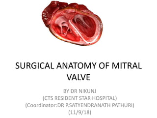
SURGICAL ANATOMY OF MITRAL
VALVE
BY DR NIKUNJ
(CTS RESIDENT STAR HOSPITAL)
(Coordinator:DR P.SATYENDRANATH PATHURI)
(11/9/18)
THE ATRIO-VALVULAR JUNCTION
• The junction between the atrium and the valvular tissue is usually well delineated
by the different colors of these structures: the atrium is slightly pink, and the
leaflets are yellowish.
• This junction defines the hinge where the motion of the leaflets is initiated.
• The hinge allows demar- cation of the annulus fibrosus, which is not visible from
an atrial view.
ANNULUS FIBROSUS
• actually a discontinuous band of connective tissue that exists only in some parts of the
attachment of the posterior leaflet.
• The annulus in actuality does not exist at the attachment of the anterior leaflet because the
leaflet tissue is continuous with the aorto- mitral curtain that extends from the aortic valve
annulus to the base of the anterior leaflet
• At each extremity of the base of the anterior leaflet, the atrio-valvular junction is
reinforced by two dense triangular fibrous structures: the anterolateral and
posteromedial fibrous trigones.
• The shape of the annulus varies throughout the cardiac cycle
• During diastole, the shape is grossly circular . During systole, the annulus has a kidney
shape
• Instead of having a planar configuration, the annulus has a three-dimensional saddle-
shape configuration. The two lowest points are located at the fibrous trigones and the
• two highest points are located at the midpoints of the anterior and posterior annuli.
• The plane of the mitral valve annulus makes a 120° angle with the plane of the aortic valve
annulus.
The 26% ± 3% reduction of the mitral valve orifice area during systole results from the
contraction of the base of the heart and the displacement of the aorto-mitral curtain towards
the center of the orifice.
THE LEAFLETS
• The mitral valve comprises two
leaflets—1)anterior
• 2)posterior
• separated by two commissures.
These leaflets are the opening and
closing structures of the valve
• Optimal closure implies a precise
fitting between the surface area
of the leaflets and the orifice area
of the mitral valve.
• Although the anterior and
posterior leaflets have a dif-
ferent size and shape , with the
anterior leaflet more extended
vertically and the posterior leaflet
more extended transversally, they
have a similar surface area.
• The basal insertion of the
anterior leaflet occupies approx-
imately one third of the
circumference of the mitral
valve
• The remaining two thirds of the
circumference attaches to the
posterior leaflet and the
commissural tissue.
• The anterior leaflet is primarily
related to the left ventricular
outflow tract via the aorto-
mitral curtain whereas the
posterior leaflet is related to the
muscular parietal base of the
left ventricle. As a result of this
configuration, the maximum
stress during systole is concen-
trated at the midline of the
posterior leaflet
ANTERIOR LEAFLET
• ANTERIOR LEAFLET, also called the aortic leaflet, has a trapezoidal shape. Its base,
which measures 32 ± 1.3 mm, is inserted on the aorto-mitral curtain and the
adjacent fibrous trigones. The free edge presents with a slightly convex curvature.
At the midline, the height of the anterior leaflet averages 23 mm.
• From the base to the margin, two zones are clearly appa ent.
• 1)The proximal zone, called the atrial zone, is regular, thin, and translucent.
• 2)The distal zone, called the zone of coaptation or rough zone, is irregular and
thicker because of the numerous chordae attached to its ventricular side. The two
zones have a similar surface area.
• The surface of coaptation, which has a height of 7 to 9 mm,
• During diastole, the anterior leaflet divides the left ventricle into two functional
areas, the inflow chamber and the outflow tract.
• The surface of coaptation, which has a height of 7 to 9 mm
POSTERIOR LEAFLET
• The posterior leaflet is inserted approximately
on two thirds of the annulus, to the crest of
the ventricular wall. The free edge is deeply
scalloped by two indentations ,separating
three segments: the anterior, middle, and
posterior scallops are also called P1, P2, and
P3, respectively, to facilitate valve analysis. By
convention, the corresponding areas of the
anterior leaflet are called A1, A2, and A3, and
the commissures AC and PC.
• The size of the scallops of the posterior leaflet
differs. The largest is the middle scallop (P2)
and the smallest is the anterior scallop (P1)
• the posterior leaflet presents two zones from
its base to the free margin: the atrial zone is
smooth and translucent and the coaptation
zone is thicker
• THE COMMISSURES may be described
as a functional entity consisting of two
different structures: the commissural
leaflet, which provides continuity
between the anterior and posterior
leaflets, and the coaptation surfaces
with adjacent anterior and posterior
leaflets.
• The commissural leaflet is a small,
triangular segment of leaflet tissue. Its
base is attached to the annulus and its
free edge is supported by one or two
characteristic fanlike chordae.
• As a result of this configuration, the
junction between the anterior and
posterior leaflets does not reach the
annulus but forms a Y-shaped line of
coaptation.
THE SUSPENSION SYSTEM
• The leaflets are connected to the
ventricular cavity by a suspension system
called the subvalvular apparatus. The
suspension system has two functions:
• one is to facilitate he opening of the
leaflets during diastole (active opening);
• the other is to prevent the upward
displace- ment of the leaflets above the
plane of the annulus during systole.
• To accomplish these two functions, the
suspension system consists of two
structures with different charcteristics:
• THE PAPILLARY MUSCLES with contractile
properties and
• THE CHORDAE TENDINEAE with elastic
properties.
THE PAPILLARY MUSCLES
• insert on the ventricular wall, are usually
organized into two groups, designated
posteromedial and anterolateral, positioned
below the corresponding commissures
I—Large and bulky with a single head generating
numerous chordae.
II—Large and bulky with multiple heads
attaching numerous chordae.
III—Narrow and having few chordae.
IV—Arch shape from which arise several
chordae; the arch may form an arcade with
several trabeculations attached to the
myocardium.
V—Adherent to the ventricular wall and
generating multiple chordae
.
• The anterior papillary muscle most
often has a type I configuration
with occasionally an adjacent type
III papillary muscle attaching the
commissural chordae.
• The posterior papillary muscle
usually has a type II configuration
with one head attaching the
chordae of the anterior leaflet, one
head attaching the commissural
chordae, and one head attaching
chordae of the posterior leaflet.
• The papillary muscles are implanted on the muscular wall of
the left ventricle at a junction situated approximately 1/3
from the apex and 2/3 from the annulus
• The distance between the tip of the papillary muscle and the
plane of the mitral valve orifice also varies, with an average
of 22 ± 5 mm
THE CHORDAE TENDINEAE
THE CHORDAE TENDINEAE extend from the papillary
muscles to the leaflets . Three types can be described
depending upon their attachment on the leaflets
BASAL (OR TERTIARY) CHORDAE extend from the
papillary muscle or directly from the ventricular wall.
The basal chordae are attached to the base of the
posterior and commissural leaflets or to the annulus.
INTERMEDIARY (OR SECONDARY) CHORDAE extend
from the papillary muscles. The intermediary chordae
are attached to the ventricular side of the leaflets.
MARGINAL (OR PRIMARY) CHORDAE are attached to
the margin of the leaflets. The space between two
marginal chordae does not exceed 3 mm and their
attachment to the leaflet is often bifurcated or
trifurcated
FOUR ANATOMICAL STRUCTURES CLOSE TO THE ANNULUS ARE AT RISK
DURING SURGERY
• The circumflex artery runs
between the base of the left
atrial appendage and the
anterior commissure, 3 to 4 mm
from the leaflet attachment,
and then moves away from the
rest of the posterior annulus.
• The coronary sinus skirts the
attachment of the posterior
leaflet. It is initially in a lateral
position, and then crosses the
artery and becomes medial,
closerTo the posterior leaflet
attachment but 5 mm superior
to the annulus.
• The bundle of His is located
near the posteromedial trigone.
FOUR ANATOMICAL STRUCTURES CLOSE TO THE ANNULUS ARE AT RISK
DURING SURGERY
• The noncoronary and left
coronary aortic cusps are in
close relationship with the base
of the anterior leaflet
• .the nadir of these cusps is 6 to
10 mm away from the anterior
mitral valve annulus, a distance
that repre- sents a safety zone
for the placement of sutures in
this area provided that the
needle is properly oriented
towards the ventricle.
MITRAL VALVE REGURGITATION
• Type I: Mitral valve regurgitation with
normal leaflet motion.
• In type I mitral regurgitation, the course
of the leaflets between systole and
diastole has a normal amplitude and the
free edges of the two leaflets are well
positioned 5 to 10 mm below the plane
of the orifice.
• The regurgitation is due either to a lack
of coaptation between leaflets or to an
annular dilatation or a leaflet perforation,
tear, or vegetation
MITRAL VALVE REGURGITATION
• Type II: Mitral valve regurgitation with excess
leaflet motion: “leaflet prolapse.” Typically, a
leaflet prolapse is a valve dysfunction in which
the free edge of a leaflet overrides the plane of
the mitral valve orifice during systole.
• The resulting lack of leaflet apposition produces
a regurgitant jet, which runs obliquely over the
nonprolapsing leaflet.
• A mitral valve prolapse is due either to chordae
rupture or elongation or to papillary muscle
rupture or elon- gation
MITRAL VALVE REGURGITATION
• Type III: Restricted leaflet motion. In type III
mitral regurgitation , the motion of one or two
leaflets is limited primarily during diastole
(type IIIa) or during systole (type IIIb).
• Type IIIa is typically seen in rheumatic valve
disease.
• The valve motion is limited mostly during
diastole but also to some extent during systole
as a result of commissure fusion, chordae
thickening, and chordae fusion.
• Type IIIb mitral regurgitation displays a leaflet
tethering caused by papillary muscle
displacement resulting either from a localized
ventricular wall dyskinesia, as seen in ischemic
cardiomyopathy, or from a global dilatation of
the ventricle, as seen in end-stage
cardiomyopathy
• Another type III systolic dysfunction with a com- pletely different mechanism is
systolic anterior motion of the anterior leaflet (SAM) observed in obstructive
hypertrophic cardiomyopathy or in repaired Barlow’s disease. In these two settings
the motion of the anterior leaflet is first limited by the posterior leaflet and
inverted towards the left outflow tract
APPROACHES TO THE LEFT ATRIUM AND MITRAL VALVE
• TRADITIONAL APPROACH: VERTICAL LEFT ATRIOTOMY.
• The site of the vertical left atriotomy may be at the fatty interatrial junction or may be located
closer to the mitral valve by , separating the left atrium from the right, and allowing the
surgeon to perform left atriotomy 2 to 4 cm mediall.This latter incision requires closure where
the left atrium is thinner and may carry a greater risk of suture line bleeding.Accidental entry
in to the right atrium can allow air lock and obstruction of venous drainage. This is easily
controlled by oversewing the entry point.
TRANSECTION OF THE SVC also allows extension of the cephalad limb of the
atriotomy on to the superior roof and permits further rotation of the right atrium and atrial
septum to the left and away from the surgeon.The transection should leave at least a 1-to 2-cm
cuff on the right atrium and may require moving the SVC cannulation site from the right atrium to
the SVC or innominate vein.Air lock or compromised venous drainage may occur during this
transfer. SVC stenosis or thrombosis and sinoatrial node injury have been described with
this'technique.
TRANSSEPTAL APPROACH
(A) The vertical left atriotomy may also be converted to a transseptal approach if exposure is
inadequate. Begin by performing a vertical right atriotomy parallel to the left atriotmy .then,
open into the left atrium through the septum, with a transverse incision through the fossa ovalis,
perpendicular to the two atriotomies, and transecting the bridge of tissue between them.
(B) Alternatively, open into the left atrium through a vertical septal incision through the fossa
ovalis, parallel to the two atriotomies. With this incision, avoid encroaching on the coronary sinus
inferiorly or the dome of the left atrium superiorly. With either of these transseptal incisions,
avoid excessive medial displacement that may impinge on the mitral annulus
If exposure can be predicted to be difficult, as in the case of a small left atrium or deep chest, a
PLANNED TRANSSEPTAL APPROACH may be used.With a vertical right
atriotomy, parallel to the atrioventricular sulcus, make a secondary, vertical septal incision
through the fossa ovalis, avoiding the coronary sinus.
THE CLASSIC DUBOST APPROACH. Make a transverse right atriotomy and extend
it laterally into the superior pulmonary vein or between the pulmonary veins into the left atrium.
Then, beginning at the edge of the septa1 incision, carry the incision medially through the fossa
ovalis to expose the mitral valve
• SUPERIOR SEPTAL APPROACH-
The superior septal approach gives superb exposure.
Its liability is the possibility of atrial dysrhythmia.
Because this may be caused by disruption of the
sinoatrial node artery, Three variations of this arterial
supply
Pathway A, the most common (48% to 80%) courses
along the inner anterior border of the right atrium.
Pathway B courses anteriorly then behind the SVC to
the sino atrial (SA) node.
Pathway C arises from the circumflex and also
courses posteriorly over the left atrium, behind the
SVC to the SA node.
• THE SUPERIOR SEPTAL APPROACH. ’Begin by placing the SVC cannula lateral to the right
atrial appendage or in the SVC directly. Make a longitudinal (vertical) right atriotomy anterior to the
sulcus terminalis. Carry the incision cephalad around the superior base of the atrial appendage, or
directly through the appendage, to reach the atrial septum. Avoid the sino-atrial node, but keep the
incision 1to 2 cm from the right ventricle to allow safe closure. Incise the septum vertically through the
fossa ovalis, directly under and parallel to the right atriotomy, extending the incision superiorly to the
superior apex of the right atriotomy. Continue the confluence of these two incisions superiorly into the
dome of the left atrium. Retract the atrial septum gently to avoid atrioventricular nodal injury.
• THE SUPERIOR APPROACH TO THE MITRAL VALVE
• This approach allows adequate exposure of the mitral valve, with minimal to moderate
retraction of the aorta and SVC. It also allows a view of the mitral annulus more
perpendicular to its plane than do other approaches.
• Dissect the aorta and the SVC from the right pulmonary artery behind them. Avoid
injuring the left coronary artery behind the aorta. Mobilize the SVC from cephalad to the
right pulmonary artery inferiorly to the groove between the SVC and the right atrium
(sulcus terminalis). Incise the superior dome of the left atrium transversely as shown.
Avoid extending the incision too far to the left under the aortic root and into the thin-
walled left atrial appendage. Instead, provide greater exposure if necessary by extending
the incision to the right and into the right superior pulmonary vein.
TRANSAORTIC APPROACH
• transaortic approach-Perform a standard aortotomy. After excising the aortic leaflets, retract
the aortic root and annulus to expose the anterior leaflet of the mitral valve. Place the valve
sutures from the left atrium into the left ventricle and then through the mitral prosthetic
annulus from the superior to the inferior (ventricular) side, where the sutures are tied. The
prosthetic annulus thus lies below the native mitral annulus. Then proceed with the aortic
valve replacement.
LEFT VENTRICULAR APPROACH
• Concomitant mitral valve and left ventricular operations for ventricular septal defect or
aneurysm may both be performed through the ventriculotomy.
• Open the left ventricle parallel to the septum, through the scarred or infarcted tissue. Extend
the incision towards the apex to improve exposure without endangering the mitral
subvalvular apparatus. Either mitral valve repair or replacement may be performed through
this approach .With, mitral replacement, remove the prosthesis from the valve holder, which,
if used with this approach, may result in the valve being inserted upside down. Place the
valve sutures from the atrial side to the ventricular side and then through the prosthetic
annulus from its superior to inferior (ventricular) side.
RIGHT THORACOTOMY APPROACH.
LEFT THORACOTOMY APPROACH
AUTOTRANSPLANTATION
MINIMALLY INVASIVE APPROACHES
THANK YOU

More Related Content

What's hot

Aortic valve anatomy presentation
Aortic valve anatomy presentationAortic valve anatomy presentation
Aortic valve anatomy presentationIndia CTVS
 
Echocardiography of Aortic stenosis
Echocardiography of Aortic stenosis Echocardiography of Aortic stenosis
Echocardiography of Aortic stenosis Dr. Muhammad AzAm Shah
 
Approch to bifurcation lesion
Approch to bifurcation lesionApproch to bifurcation lesion
Approch to bifurcation lesionRamachandra Barik
 
Congenitally corrected transposition of great arteries
Congenitally corrected transposition of great arteriesCongenitally corrected transposition of great arteries
Congenitally corrected transposition of great arteriesDheeraj Sharma
 
CORONARY ARTERY PERFORATION DURING PCI
CORONARY ARTERY PERFORATION DURING PCICORONARY ARTERY PERFORATION DURING PCI
CORONARY ARTERY PERFORATION DURING PCIVishal Vanani
 
Sinus of valsalva aneurysm
Sinus of valsalva aneurysmSinus of valsalva aneurysm
Sinus of valsalva aneurysmRamachandra Barik
 
Coronary artery dissection and perforation
Coronary artery dissection and perforationCoronary artery dissection and perforation
Coronary artery dissection and perforationFuad Farooq
 
Segmental analysis of heart anatomy
Segmental analysis of heart anatomySegmental analysis of heart anatomy
Segmental analysis of heart anatomyIndia CTVS
 
Coronary Calcium Modification
Coronary Calcium ModificationCoronary Calcium Modification
Coronary Calcium ModificationNAJEEB ULLAH SOFI
 
Shunt Detection And Quantification
Shunt Detection And QuantificationShunt Detection And Quantification
Shunt Detection And QuantificationDang Thanh Tuan
 
PCI complications
PCI complicationsPCI complications
PCI complicationsIqbal Dar
 
Cath hemodynamics vir
Cath hemodynamics virCath hemodynamics vir
Cath hemodynamics virDr Virbhan Balai
 
Mitral valve anatomy - ppt by kunwar sidharth
Mitral valve    anatomy - ppt by kunwar sidharthMitral valve    anatomy - ppt by kunwar sidharth
Mitral valve anatomy - ppt by kunwar sidharthkunwar sidharth
 
Intravascular Ultrasound (IVUS)
Intravascular Ultrasound (IVUS)Intravascular Ultrasound (IVUS)
Intravascular Ultrasound (IVUS)Dr.Sayeedur Rumi
 

What's hot (20)

Aortic valve anatomy presentation
Aortic valve anatomy presentationAortic valve anatomy presentation
Aortic valve anatomy presentation
 
Looping of heart
Looping of heartLooping of heart
Looping of heart
 
Echocardiography of Aortic stenosis
Echocardiography of Aortic stenosis Echocardiography of Aortic stenosis
Echocardiography of Aortic stenosis
 
Intravenous Ultrasound
Intravenous UltrasoundIntravenous Ultrasound
Intravenous Ultrasound
 
Approch to bifurcation lesion
Approch to bifurcation lesionApproch to bifurcation lesion
Approch to bifurcation lesion
 
SINGLE VENTRICLE: MANAGEMENT
SINGLE VENTRICLE: MANAGEMENTSINGLE VENTRICLE: MANAGEMENT
SINGLE VENTRICLE: MANAGEMENT
 
Congenitally corrected transposition of great arteries
Congenitally corrected transposition of great arteriesCongenitally corrected transposition of great arteries
Congenitally corrected transposition of great arteries
 
CORONARY ARTERY PERFORATION DURING PCI
CORONARY ARTERY PERFORATION DURING PCICORONARY ARTERY PERFORATION DURING PCI
CORONARY ARTERY PERFORATION DURING PCI
 
Sinus of valsalva aneurysm
Sinus of valsalva aneurysmSinus of valsalva aneurysm
Sinus of valsalva aneurysm
 
Coronary artery dissection and perforation
Coronary artery dissection and perforationCoronary artery dissection and perforation
Coronary artery dissection and perforation
 
SEGMENTAL ANALYSIS OF CONGENITAL HEART DISEASE
SEGMENTAL ANALYSIS OF CONGENITAL HEART DISEASE SEGMENTAL ANALYSIS OF CONGENITAL HEART DISEASE
SEGMENTAL ANALYSIS OF CONGENITAL HEART DISEASE
 
Segmental analysis of heart anatomy
Segmental analysis of heart anatomySegmental analysis of heart anatomy
Segmental analysis of heart anatomy
 
Coronary Calcium Modification
Coronary Calcium ModificationCoronary Calcium Modification
Coronary Calcium Modification
 
CONGENITAL CORONARY ANOMALIES AND VARIANTS, ALCAPA
CONGENITAL CORONARY ANOMALIES AND VARIANTS, ALCAPACONGENITAL CORONARY ANOMALIES AND VARIANTS, ALCAPA
CONGENITAL CORONARY ANOMALIES AND VARIANTS, ALCAPA
 
Shunt Detection And Quantification
Shunt Detection And QuantificationShunt Detection And Quantification
Shunt Detection And Quantification
 
PCI complications
PCI complicationsPCI complications
PCI complications
 
Cath hemodynamics vir
Cath hemodynamics virCath hemodynamics vir
Cath hemodynamics vir
 
ECHO ASSESSMENT OF ASD FOR DEVICE CLOSURE
ECHO ASSESSMENT OF ASD FOR DEVICE CLOSURE ECHO ASSESSMENT OF ASD FOR DEVICE CLOSURE
ECHO ASSESSMENT OF ASD FOR DEVICE CLOSURE
 
Mitral valve anatomy - ppt by kunwar sidharth
Mitral valve    anatomy - ppt by kunwar sidharthMitral valve    anatomy - ppt by kunwar sidharth
Mitral valve anatomy - ppt by kunwar sidharth
 
Intravascular Ultrasound (IVUS)
Intravascular Ultrasound (IVUS)Intravascular Ultrasound (IVUS)
Intravascular Ultrasound (IVUS)
 

Similar to Mitral valve surgical anatomy DR NIKUNJ R SHEKHADA (MBBS ,MS GRN SURG , DNB CTS SR

Anatomy tricuspid valve DR NIKUNJ R SHEKHADA (MBBS,MS GEN SURG ,DNB CTS SR)
Anatomy tricuspid valve DR NIKUNJ R SHEKHADA (MBBS,MS GEN SURG ,DNB CTS SR)Anatomy tricuspid valve DR NIKUNJ R SHEKHADA (MBBS,MS GEN SURG ,DNB CTS SR)
Anatomy tricuspid valve DR NIKUNJ R SHEKHADA (MBBS,MS GEN SURG ,DNB CTS SR)DR NIKUNJ SHEKHADA
 
Anatomy of heart
Anatomy of heartAnatomy of heart
Anatomy of heartnmonty02
 
Normal anatomy of tricuspid valve
Normal anatomy of tricuspid valve Normal anatomy of tricuspid valve
Normal anatomy of tricuspid valve India CTVS
 
Dr Garva Ameta anatomy Of Left Heart.pptx
Dr Garva Ameta anatomy Of Left Heart.pptxDr Garva Ameta anatomy Of Left Heart.pptx
Dr Garva Ameta anatomy Of Left Heart.pptxPooja Jakhar
 
Aortic root anatomy DR NIKUNJ R SHEKHADA (MBBS,MS GEN SURG,DNB CTS SR)
Aortic root anatomy DR NIKUNJ R SHEKHADA (MBBS,MS GEN SURG,DNB CTS SR)Aortic root anatomy DR NIKUNJ R SHEKHADA (MBBS,MS GEN SURG,DNB CTS SR)
Aortic root anatomy DR NIKUNJ R SHEKHADA (MBBS,MS GEN SURG,DNB CTS SR)DR NIKUNJ SHEKHADA
 
Echocardiographic evaluation of aortic valve and Aortic stenosis
Echocardiographic evaluation of aortic valve  and Aortic stenosisEchocardiographic evaluation of aortic valve  and Aortic stenosis
Echocardiographic evaluation of aortic valve and Aortic stenosissruthiMeenaxshiSR
 
Arterial supply of head and neck
Arterial supply of head and neckArterial supply of head and neck
Arterial supply of head and neckAvinash Rathore
 
Surgery for atrial fibrillation abhijit presentation
Surgery for atrial fibrillation abhijit presentationSurgery for atrial fibrillation abhijit presentation
Surgery for atrial fibrillation abhijit presentationAbhijit Joshi
 
Mitral valve
Mitral valveMitral valve
Mitral valveUphar Gupta
 
The valves of the heart
The valves of the heartThe valves of the heart
The valves of the heartIdris Siddiqui
 
Anatomy of oesophagus.ppt
Anatomy of oesophagus.pptAnatomy of oesophagus.ppt
Anatomy of oesophagus.pptYtchechy
 
Chordal preservation DR NIKUNJ R SHEKHADA (MBBS,MS GEN SURG ,DNB CTS SR)
Chordal preservation DR  NIKUNJ R SHEKHADA (MBBS,MS GEN SURG ,DNB CTS SR)Chordal preservation DR  NIKUNJ R SHEKHADA (MBBS,MS GEN SURG ,DNB CTS SR)
Chordal preservation DR NIKUNJ R SHEKHADA (MBBS,MS GEN SURG ,DNB CTS SR)DR NIKUNJ SHEKHADA
 
2.Anatomy of oesophagus.ppt
2.Anatomy of oesophagus.ppt2.Anatomy of oesophagus.ppt
2.Anatomy of oesophagus.pptDr.arun Naragund
 
Anatomy and embryology of valves
Anatomy and embryology of valvesAnatomy and embryology of valves
Anatomy and embryology of valvesDrvasanthi
 

Similar to Mitral valve surgical anatomy DR NIKUNJ R SHEKHADA (MBBS ,MS GRN SURG , DNB CTS SR (20)

Anatomy tricuspid valve DR NIKUNJ R SHEKHADA (MBBS,MS GEN SURG ,DNB CTS SR)
Anatomy tricuspid valve DR NIKUNJ R SHEKHADA (MBBS,MS GEN SURG ,DNB CTS SR)Anatomy tricuspid valve DR NIKUNJ R SHEKHADA (MBBS,MS GEN SURG ,DNB CTS SR)
Anatomy tricuspid valve DR NIKUNJ R SHEKHADA (MBBS,MS GEN SURG ,DNB CTS SR)
 
Anatomy of heart
Anatomy of heartAnatomy of heart
Anatomy of heart
 
Normal anatomy of tricuspid valve
Normal anatomy of tricuspid valve Normal anatomy of tricuspid valve
Normal anatomy of tricuspid valve
 
Aortic valve disease
Aortic valve diseaseAortic valve disease
Aortic valve disease
 
Aortic valve disease
Aortic valve diseaseAortic valve disease
Aortic valve disease
 
Dr Garva Ameta anatomy Of Left Heart.pptx
Dr Garva Ameta anatomy Of Left Heart.pptxDr Garva Ameta anatomy Of Left Heart.pptx
Dr Garva Ameta anatomy Of Left Heart.pptx
 
Aortic root anatomy DR NIKUNJ R SHEKHADA (MBBS,MS GEN SURG,DNB CTS SR)
Aortic root anatomy DR NIKUNJ R SHEKHADA (MBBS,MS GEN SURG,DNB CTS SR)Aortic root anatomy DR NIKUNJ R SHEKHADA (MBBS,MS GEN SURG,DNB CTS SR)
Aortic root anatomy DR NIKUNJ R SHEKHADA (MBBS,MS GEN SURG,DNB CTS SR)
 
Mitral valve disease
Mitral valve diseaseMitral valve disease
Mitral valve disease
 
Echocardiographic evaluation of aortic valve and Aortic stenosis
Echocardiographic evaluation of aortic valve  and Aortic stenosisEchocardiographic evaluation of aortic valve  and Aortic stenosis
Echocardiographic evaluation of aortic valve and Aortic stenosis
 
Arterial supply of head and neck
Arterial supply of head and neckArterial supply of head and neck
Arterial supply of head and neck
 
Surgery for atrial fibrillation abhijit presentation
Surgery for atrial fibrillation abhijit presentationSurgery for atrial fibrillation abhijit presentation
Surgery for atrial fibrillation abhijit presentation
 
Mitral valve
Mitral valveMitral valve
Mitral valve
 
The valves of the heart
The valves of the heartThe valves of the heart
The valves of the heart
 
Aortic repair ppt
Aortic repair pptAortic repair ppt
Aortic repair ppt
 
Aortic repair ppt
Aortic repair pptAortic repair ppt
Aortic repair ppt
 
Anatomy of oesophagus.ppt
Anatomy of oesophagus.pptAnatomy of oesophagus.ppt
Anatomy of oesophagus.ppt
 
Chordal preservation DR NIKUNJ R SHEKHADA (MBBS,MS GEN SURG ,DNB CTS SR)
Chordal preservation DR  NIKUNJ R SHEKHADA (MBBS,MS GEN SURG ,DNB CTS SR)Chordal preservation DR  NIKUNJ R SHEKHADA (MBBS,MS GEN SURG ,DNB CTS SR)
Chordal preservation DR NIKUNJ R SHEKHADA (MBBS,MS GEN SURG ,DNB CTS SR)
 
Triangle of koch
Triangle of kochTriangle of koch
Triangle of koch
 
2.Anatomy of oesophagus.ppt
2.Anatomy of oesophagus.ppt2.Anatomy of oesophagus.ppt
2.Anatomy of oesophagus.ppt
 
Anatomy and embryology of valves
Anatomy and embryology of valvesAnatomy and embryology of valves
Anatomy and embryology of valves
 

More from DR NIKUNJ SHEKHADA

Mitral valve surgical treatment
Mitral valve surgical treatmentMitral valve surgical treatment
Mitral valve surgical treatmentDR NIKUNJ SHEKHADA
 
Cardiac cycle DR NIKUNJ R SHEKHADA (MBBS,MS GEN SURG ,DNB CTS SR)
Cardiac cycle DR  NIKUNJ R SHEKHADA (MBBS,MS GEN SURG ,DNB CTS SR)Cardiac cycle DR  NIKUNJ R SHEKHADA (MBBS,MS GEN SURG ,DNB CTS SR)
Cardiac cycle DR NIKUNJ R SHEKHADA (MBBS,MS GEN SURG ,DNB CTS SR)DR NIKUNJ SHEKHADA
 
History of cardiac surgery DR NIKUNJ R SHEKHADA (MBBS,MS GEN SURG ,DNB CTS SR)
History of cardiac surgery  DR  NIKUNJ R SHEKHADA (MBBS,MS GEN SURG ,DNB CTS SR)History of cardiac surgery  DR  NIKUNJ R SHEKHADA (MBBS,MS GEN SURG ,DNB CTS SR)
History of cardiac surgery DR NIKUNJ R SHEKHADA (MBBS,MS GEN SURG ,DNB CTS SR)DR NIKUNJ SHEKHADA
 
Cpb oxygenators DR NIKUNJ R SHEKHADA (MBBS,MS GEN SURG ,DNB CTS SR)
Cpb oxygenators  DR  NIKUNJ R SHEKHADA (MBBS,MS GEN SURG ,DNB CTS SR)Cpb oxygenators  DR  NIKUNJ R SHEKHADA (MBBS,MS GEN SURG ,DNB CTS SR)
Cpb oxygenators DR NIKUNJ R SHEKHADA (MBBS,MS GEN SURG ,DNB CTS SR)DR NIKUNJ SHEKHADA
 
Post oprative care 1 DR NIKUNJ R SHEKHADA (MBBS,MS GEN SURG,DNB CTS SR)
Post oprative care 1 DR NIKUNJ R SHEKHADA (MBBS,MS GEN SURG,DNB CTS SR)Post oprative care 1 DR NIKUNJ R SHEKHADA (MBBS,MS GEN SURG,DNB CTS SR)
Post oprative care 1 DR NIKUNJ R SHEKHADA (MBBS,MS GEN SURG,DNB CTS SR)DR NIKUNJ SHEKHADA
 
Ecg1 DR NIKUNJ R SHEKHADA (MBBS,MS GEN SURG,DNB CTS SR)
Ecg1 DR NIKUNJ R SHEKHADA (MBBS,MS GEN SURG,DNB CTS SR)Ecg1 DR NIKUNJ R SHEKHADA (MBBS,MS GEN SURG,DNB CTS SR)
Ecg1 DR NIKUNJ R SHEKHADA (MBBS,MS GEN SURG,DNB CTS SR)DR NIKUNJ SHEKHADA
 
CARDIO PLEGIA DELIVERY DR NIKUNJ R SHEKHADA (MBBS,MS GEN SURG,DNB CTS SR
CARDIO PLEGIA DELIVERY  DR NIKUNJ R SHEKHADA (MBBS,MS GEN SURG,DNB CTS SRCARDIO PLEGIA DELIVERY  DR NIKUNJ R SHEKHADA (MBBS,MS GEN SURG,DNB CTS SR
CARDIO PLEGIA DELIVERY DR NIKUNJ R SHEKHADA (MBBS,MS GEN SURG,DNB CTS SRDR NIKUNJ SHEKHADA
 
Atrial septal defect DR NIKUNJ R SHEKHADA (MBBS,MS GEN SURG,DNB CTS SR
Atrial septal defect DR NIKUNJ R SHEKHADA (MBBS,MS GEN SURG,DNB CTS SRAtrial septal defect DR NIKUNJ R SHEKHADA (MBBS,MS GEN SURG,DNB CTS SR
Atrial septal defect DR NIKUNJ R SHEKHADA (MBBS,MS GEN SURG,DNB CTS SRDR NIKUNJ SHEKHADA
 
Asd managment DR NIKUNJ R SHEKHADA (MBBS,MS GEN SURG,DNB CTS SR)
Asd managment DR NIKUNJ R SHEKHADA (MBBS,MS GEN SURG,DNB CTS SR)Asd managment DR NIKUNJ R SHEKHADA (MBBS,MS GEN SURG,DNB CTS SR)
Asd managment DR NIKUNJ R SHEKHADA (MBBS,MS GEN SURG,DNB CTS SR)DR NIKUNJ SHEKHADA
 
Myocardial protection DR NIKUNJ R SHEKHADA (MBBS ,MS GRN SURG , DNB CTS SR
Myocardial protection DR NIKUNJ R SHEKHADA (MBBS ,MS GRN SURG , DNB CTS SRMyocardial protection DR NIKUNJ R SHEKHADA (MBBS ,MS GRN SURG , DNB CTS SR
Myocardial protection DR NIKUNJ R SHEKHADA (MBBS ,MS GRN SURG , DNB CTS SRDR NIKUNJ SHEKHADA
 
CPB(CARDIO PULMONARY BYPASS)
CPB(CARDIO PULMONARY BYPASS)CPB(CARDIO PULMONARY BYPASS)
CPB(CARDIO PULMONARY BYPASS)DR NIKUNJ SHEKHADA
 
CARDIO PULMONARYBYPASS BLOOD PUMPS DR NIKUNJ R SHEKHADA (MBBS,MSGEN SURG ,DN...
CARDIO PULMONARYBYPASS BLOOD PUMPS  DR NIKUNJ R SHEKHADA (MBBS,MSGEN SURG ,DN...CARDIO PULMONARYBYPASS BLOOD PUMPS  DR NIKUNJ R SHEKHADA (MBBS,MSGEN SURG ,DN...
CARDIO PULMONARYBYPASS BLOOD PUMPS DR NIKUNJ R SHEKHADA (MBBS,MSGEN SURG ,DN...DR NIKUNJ SHEKHADA
 
Ventricular septal defect DR NIKUNJ .R .SHRKHADA (MBBS,MS GEN SURG DNB CTS SR)
Ventricular septal defect  DR NIKUNJ .R .SHRKHADA (MBBS,MS GEN SURG DNB CTS SR)Ventricular septal defect  DR NIKUNJ .R .SHRKHADA (MBBS,MS GEN SURG DNB CTS SR)
Ventricular septal defect DR NIKUNJ .R .SHRKHADA (MBBS,MS GEN SURG DNB CTS SR)DR NIKUNJ SHEKHADA
 
Coronary arteries and veins DR NIKUNJ .R .SHRKHADA (MBBS,MS GEN SURG DNB CTS SR)
Coronary arteries and veins DR NIKUNJ .R .SHRKHADA (MBBS,MS GEN SURG DNB CTS SR)Coronary arteries and veins DR NIKUNJ .R .SHRKHADA (MBBS,MS GEN SURG DNB CTS SR)
Coronary arteries and veins DR NIKUNJ .R .SHRKHADA (MBBS,MS GEN SURG DNB CTS SR)DR NIKUNJ SHEKHADA
 
Anatomy of lungs 3DR NIKUNJ R SHEKHADA (MBBS,MS GEN SURG DNB CTS SR)
Anatomy of lungs 3DR NIKUNJ R SHEKHADA (MBBS,MS GEN SURG DNB CTS SR)Anatomy of lungs 3DR NIKUNJ R SHEKHADA (MBBS,MS GEN SURG DNB CTS SR)
Anatomy of lungs 3DR NIKUNJ R SHEKHADA (MBBS,MS GEN SURG DNB CTS SR)DR NIKUNJ SHEKHADA
 
Arrhythmias 1DR NIKUNJ R SHEKHADA (MBBS,MS GEN SURG DNB CTS SR)
Arrhythmias 1DR NIKUNJ R SHEKHADA (MBBS,MS GEN SURG DNB CTS SR)Arrhythmias 1DR NIKUNJ R SHEKHADA (MBBS,MS GEN SURG DNB CTS SR)
Arrhythmias 1DR NIKUNJ R SHEKHADA (MBBS,MS GEN SURG DNB CTS SR)DR NIKUNJ SHEKHADA
 
Anatomy of heart dr nikunj shekhada (mbbs,ms gen surg ,dnb cts SR) 11 6-18
Anatomy of heart dr nikunj shekhada (mbbs,ms gen surg ,dnb cts SR) 11 6-18Anatomy of heart dr nikunj shekhada (mbbs,ms gen surg ,dnb cts SR) 11 6-18
Anatomy of heart dr nikunj shekhada (mbbs,ms gen surg ,dnb cts SR) 11 6-18DR NIKUNJ SHEKHADA
 

More from DR NIKUNJ SHEKHADA (19)

Mitral valve surgical treatment
Mitral valve surgical treatmentMitral valve surgical treatment
Mitral valve surgical treatment
 
Mitral valve repair
Mitral valve repairMitral valve repair
Mitral valve repair
 
Surgical emphsema
Surgical emphsemaSurgical emphsema
Surgical emphsema
 
Cardiac cycle DR NIKUNJ R SHEKHADA (MBBS,MS GEN SURG ,DNB CTS SR)
Cardiac cycle DR  NIKUNJ R SHEKHADA (MBBS,MS GEN SURG ,DNB CTS SR)Cardiac cycle DR  NIKUNJ R SHEKHADA (MBBS,MS GEN SURG ,DNB CTS SR)
Cardiac cycle DR NIKUNJ R SHEKHADA (MBBS,MS GEN SURG ,DNB CTS SR)
 
History of cardiac surgery DR NIKUNJ R SHEKHADA (MBBS,MS GEN SURG ,DNB CTS SR)
History of cardiac surgery  DR  NIKUNJ R SHEKHADA (MBBS,MS GEN SURG ,DNB CTS SR)History of cardiac surgery  DR  NIKUNJ R SHEKHADA (MBBS,MS GEN SURG ,DNB CTS SR)
History of cardiac surgery DR NIKUNJ R SHEKHADA (MBBS,MS GEN SURG ,DNB CTS SR)
 
Cpb oxygenators DR NIKUNJ R SHEKHADA (MBBS,MS GEN SURG ,DNB CTS SR)
Cpb oxygenators  DR  NIKUNJ R SHEKHADA (MBBS,MS GEN SURG ,DNB CTS SR)Cpb oxygenators  DR  NIKUNJ R SHEKHADA (MBBS,MS GEN SURG ,DNB CTS SR)
Cpb oxygenators DR NIKUNJ R SHEKHADA (MBBS,MS GEN SURG ,DNB CTS SR)
 
Post oprative care 1 DR NIKUNJ R SHEKHADA (MBBS,MS GEN SURG,DNB CTS SR)
Post oprative care 1 DR NIKUNJ R SHEKHADA (MBBS,MS GEN SURG,DNB CTS SR)Post oprative care 1 DR NIKUNJ R SHEKHADA (MBBS,MS GEN SURG,DNB CTS SR)
Post oprative care 1 DR NIKUNJ R SHEKHADA (MBBS,MS GEN SURG,DNB CTS SR)
 
Ecg1 DR NIKUNJ R SHEKHADA (MBBS,MS GEN SURG,DNB CTS SR)
Ecg1 DR NIKUNJ R SHEKHADA (MBBS,MS GEN SURG,DNB CTS SR)Ecg1 DR NIKUNJ R SHEKHADA (MBBS,MS GEN SURG,DNB CTS SR)
Ecg1 DR NIKUNJ R SHEKHADA (MBBS,MS GEN SURG,DNB CTS SR)
 
CARDIO PLEGIA DELIVERY DR NIKUNJ R SHEKHADA (MBBS,MS GEN SURG,DNB CTS SR
CARDIO PLEGIA DELIVERY  DR NIKUNJ R SHEKHADA (MBBS,MS GEN SURG,DNB CTS SRCARDIO PLEGIA DELIVERY  DR NIKUNJ R SHEKHADA (MBBS,MS GEN SURG,DNB CTS SR
CARDIO PLEGIA DELIVERY DR NIKUNJ R SHEKHADA (MBBS,MS GEN SURG,DNB CTS SR
 
Atrial septal defect DR NIKUNJ R SHEKHADA (MBBS,MS GEN SURG,DNB CTS SR
Atrial septal defect DR NIKUNJ R SHEKHADA (MBBS,MS GEN SURG,DNB CTS SRAtrial septal defect DR NIKUNJ R SHEKHADA (MBBS,MS GEN SURG,DNB CTS SR
Atrial septal defect DR NIKUNJ R SHEKHADA (MBBS,MS GEN SURG,DNB CTS SR
 
Asd managment DR NIKUNJ R SHEKHADA (MBBS,MS GEN SURG,DNB CTS SR)
Asd managment DR NIKUNJ R SHEKHADA (MBBS,MS GEN SURG,DNB CTS SR)Asd managment DR NIKUNJ R SHEKHADA (MBBS,MS GEN SURG,DNB CTS SR)
Asd managment DR NIKUNJ R SHEKHADA (MBBS,MS GEN SURG,DNB CTS SR)
 
Myocardial protection DR NIKUNJ R SHEKHADA (MBBS ,MS GRN SURG , DNB CTS SR
Myocardial protection DR NIKUNJ R SHEKHADA (MBBS ,MS GRN SURG , DNB CTS SRMyocardial protection DR NIKUNJ R SHEKHADA (MBBS ,MS GRN SURG , DNB CTS SR
Myocardial protection DR NIKUNJ R SHEKHADA (MBBS ,MS GRN SURG , DNB CTS SR
 
CPB(CARDIO PULMONARY BYPASS)
CPB(CARDIO PULMONARY BYPASS)CPB(CARDIO PULMONARY BYPASS)
CPB(CARDIO PULMONARY BYPASS)
 
CARDIO PULMONARYBYPASS BLOOD PUMPS DR NIKUNJ R SHEKHADA (MBBS,MSGEN SURG ,DN...
CARDIO PULMONARYBYPASS BLOOD PUMPS  DR NIKUNJ R SHEKHADA (MBBS,MSGEN SURG ,DN...CARDIO PULMONARYBYPASS BLOOD PUMPS  DR NIKUNJ R SHEKHADA (MBBS,MSGEN SURG ,DN...
CARDIO PULMONARYBYPASS BLOOD PUMPS DR NIKUNJ R SHEKHADA (MBBS,MSGEN SURG ,DN...
 
Ventricular septal defect DR NIKUNJ .R .SHRKHADA (MBBS,MS GEN SURG DNB CTS SR)
Ventricular septal defect  DR NIKUNJ .R .SHRKHADA (MBBS,MS GEN SURG DNB CTS SR)Ventricular septal defect  DR NIKUNJ .R .SHRKHADA (MBBS,MS GEN SURG DNB CTS SR)
Ventricular septal defect DR NIKUNJ .R .SHRKHADA (MBBS,MS GEN SURG DNB CTS SR)
 
Coronary arteries and veins DR NIKUNJ .R .SHRKHADA (MBBS,MS GEN SURG DNB CTS SR)
Coronary arteries and veins DR NIKUNJ .R .SHRKHADA (MBBS,MS GEN SURG DNB CTS SR)Coronary arteries and veins DR NIKUNJ .R .SHRKHADA (MBBS,MS GEN SURG DNB CTS SR)
Coronary arteries and veins DR NIKUNJ .R .SHRKHADA (MBBS,MS GEN SURG DNB CTS SR)
 
Anatomy of lungs 3DR NIKUNJ R SHEKHADA (MBBS,MS GEN SURG DNB CTS SR)
Anatomy of lungs 3DR NIKUNJ R SHEKHADA (MBBS,MS GEN SURG DNB CTS SR)Anatomy of lungs 3DR NIKUNJ R SHEKHADA (MBBS,MS GEN SURG DNB CTS SR)
Anatomy of lungs 3DR NIKUNJ R SHEKHADA (MBBS,MS GEN SURG DNB CTS SR)
 
Arrhythmias 1DR NIKUNJ R SHEKHADA (MBBS,MS GEN SURG DNB CTS SR)
Arrhythmias 1DR NIKUNJ R SHEKHADA (MBBS,MS GEN SURG DNB CTS SR)Arrhythmias 1DR NIKUNJ R SHEKHADA (MBBS,MS GEN SURG DNB CTS SR)
Arrhythmias 1DR NIKUNJ R SHEKHADA (MBBS,MS GEN SURG DNB CTS SR)
 
Anatomy of heart dr nikunj shekhada (mbbs,ms gen surg ,dnb cts SR) 11 6-18
Anatomy of heart dr nikunj shekhada (mbbs,ms gen surg ,dnb cts SR) 11 6-18Anatomy of heart dr nikunj shekhada (mbbs,ms gen surg ,dnb cts SR) 11 6-18
Anatomy of heart dr nikunj shekhada (mbbs,ms gen surg ,dnb cts SR) 11 6-18
 

Recently uploaded

Housewife Call Girls Hoskote | 7001305949 At Low Cost Cash Payment Booking
Housewife Call Girls Hoskote | 7001305949 At Low Cost Cash Payment BookingHousewife Call Girls Hoskote | 7001305949 At Low Cost Cash Payment Booking
Housewife Call Girls Hoskote | 7001305949 At Low Cost Cash Payment Bookingnarwatsonia7
 
Call Girls ITPL Just Call 7001305949 Top Class Call Girl Service Available
Call Girls ITPL Just Call 7001305949 Top Class Call Girl Service AvailableCall Girls ITPL Just Call 7001305949 Top Class Call Girl Service Available
Call Girls ITPL Just Call 7001305949 Top Class Call Girl Service Availablenarwatsonia7
 
Call Girl Bangalore Nandini 7001305949 Independent Escort Service Bangalore
Call Girl Bangalore Nandini 7001305949 Independent Escort Service BangaloreCall Girl Bangalore Nandini 7001305949 Independent Escort Service Bangalore
Call Girl Bangalore Nandini 7001305949 Independent Escort Service Bangalorenarwatsonia7
 
Mumbai Call Girls Service 9910780858 Real Russian Girls Looking Models
Mumbai Call Girls Service 9910780858 Real Russian Girls Looking ModelsMumbai Call Girls Service 9910780858 Real Russian Girls Looking Models
Mumbai Call Girls Service 9910780858 Real Russian Girls Looking Modelssonalikaur4
 
Sonagachi Call Girls Services 9907093804 @24x7 High Class Babes Here Call Now
Sonagachi Call Girls Services 9907093804 @24x7 High Class Babes Here Call NowSonagachi Call Girls Services 9907093804 @24x7 High Class Babes Here Call Now
Sonagachi Call Girls Services 9907093804 @24x7 High Class Babes Here Call NowRiya Pathan
 
VIP Call Girls Mumbai Arpita 9910780858 Independent Escort Service Mumbai
VIP Call Girls Mumbai Arpita 9910780858 Independent Escort Service MumbaiVIP Call Girls Mumbai Arpita 9910780858 Independent Escort Service Mumbai
VIP Call Girls Mumbai Arpita 9910780858 Independent Escort Service Mumbaisonalikaur4
 
College Call Girls Vyasarpadi Whatsapp 7001305949 Independent Escort Service
College Call Girls Vyasarpadi Whatsapp 7001305949 Independent Escort ServiceCollege Call Girls Vyasarpadi Whatsapp 7001305949 Independent Escort Service
College Call Girls Vyasarpadi Whatsapp 7001305949 Independent Escort ServiceNehru place Escorts
 
Bangalore Call Girls Majestic đź“ž 9907093804 High Profile Service 100% Safe
Bangalore Call Girls Majestic đź“ž 9907093804 High Profile Service 100% SafeBangalore Call Girls Majestic đź“ž 9907093804 High Profile Service 100% Safe
Bangalore Call Girls Majestic đź“ž 9907093804 High Profile Service 100% Safenarwatsonia7
 
Aspirin presentation slides by Dr. Rewas Ali
Aspirin presentation slides by Dr. Rewas AliAspirin presentation slides by Dr. Rewas Ali
Aspirin presentation slides by Dr. Rewas AliRewAs ALI
 
Asthma Review - GINA guidelines summary 2024
Asthma Review - GINA guidelines summary 2024Asthma Review - GINA guidelines summary 2024
Asthma Review - GINA guidelines summary 2024Gabriel Guevara MD
 
Low Rate Call Girls Pune Esha 9907093804 Short 1500 Night 6000 Best call girl...
Low Rate Call Girls Pune Esha 9907093804 Short 1500 Night 6000 Best call girl...Low Rate Call Girls Pune Esha 9907093804 Short 1500 Night 6000 Best call girl...
Low Rate Call Girls Pune Esha 9907093804 Short 1500 Night 6000 Best call girl...Miss joya
 
Call Girls Hosur Just Call 7001305949 Top Class Call Girl Service Available
Call Girls Hosur Just Call 7001305949 Top Class Call Girl Service AvailableCall Girls Hosur Just Call 7001305949 Top Class Call Girl Service Available
Call Girls Hosur Just Call 7001305949 Top Class Call Girl Service Availablenarwatsonia7
 
VIP Call Girls Pune Vrinda 9907093804 Short 1500 Night 6000 Best call girls S...
VIP Call Girls Pune Vrinda 9907093804 Short 1500 Night 6000 Best call girls S...VIP Call Girls Pune Vrinda 9907093804 Short 1500 Night 6000 Best call girls S...
VIP Call Girls Pune Vrinda 9907093804 Short 1500 Night 6000 Best call girls S...Miss joya
 
Call Girls Electronic City Just Call 7001305949 Top Class Call Girl Service A...
Call Girls Electronic City Just Call 7001305949 Top Class Call Girl Service A...Call Girls Electronic City Just Call 7001305949 Top Class Call Girl Service A...
Call Girls Electronic City Just Call 7001305949 Top Class Call Girl Service A...narwatsonia7
 
Call Girls Whitefield Just Call 7001305949 Top Class Call Girl Service Available
Call Girls Whitefield Just Call 7001305949 Top Class Call Girl Service AvailableCall Girls Whitefield Just Call 7001305949 Top Class Call Girl Service Available
Call Girls Whitefield Just Call 7001305949 Top Class Call Girl Service Availablenarwatsonia7
 
Call Girls Frazer Town Just Call 7001305949 Top Class Call Girl Service Avail...
Call Girls Frazer Town Just Call 7001305949 Top Class Call Girl Service Avail...Call Girls Frazer Town Just Call 7001305949 Top Class Call Girl Service Avail...
Call Girls Frazer Town Just Call 7001305949 Top Class Call Girl Service Avail...narwatsonia7
 
Call Girls Thane Just Call 9910780858 Get High Class Call Girls Service
Call Girls Thane Just Call 9910780858 Get High Class Call Girls ServiceCall Girls Thane Just Call 9910780858 Get High Class Call Girls Service
Call Girls Thane Just Call 9910780858 Get High Class Call Girls Servicesonalikaur4
 
Book Call Girls in Kasavanahalli - 7001305949 with real photos and phone numbers
Book Call Girls in Kasavanahalli - 7001305949 with real photos and phone numbersBook Call Girls in Kasavanahalli - 7001305949 with real photos and phone numbers
Book Call Girls in Kasavanahalli - 7001305949 with real photos and phone numbersnarwatsonia7
 
Housewife Call Girls Bangalore - Call 7001305949 Rs-3500 with A/C Room Cash o...
Housewife Call Girls Bangalore - Call 7001305949 Rs-3500 with A/C Room Cash o...Housewife Call Girls Bangalore - Call 7001305949 Rs-3500 with A/C Room Cash o...
Housewife Call Girls Bangalore - Call 7001305949 Rs-3500 with A/C Room Cash o...narwatsonia7
 
College Call Girls Pune Mira 9907093804 Short 1500 Night 6000 Best call girls...
College Call Girls Pune Mira 9907093804 Short 1500 Night 6000 Best call girls...College Call Girls Pune Mira 9907093804 Short 1500 Night 6000 Best call girls...
College Call Girls Pune Mira 9907093804 Short 1500 Night 6000 Best call girls...Miss joya
 

Recently uploaded (20)

Housewife Call Girls Hoskote | 7001305949 At Low Cost Cash Payment Booking
Housewife Call Girls Hoskote | 7001305949 At Low Cost Cash Payment BookingHousewife Call Girls Hoskote | 7001305949 At Low Cost Cash Payment Booking
Housewife Call Girls Hoskote | 7001305949 At Low Cost Cash Payment Booking
 
Call Girls ITPL Just Call 7001305949 Top Class Call Girl Service Available
Call Girls ITPL Just Call 7001305949 Top Class Call Girl Service AvailableCall Girls ITPL Just Call 7001305949 Top Class Call Girl Service Available
Call Girls ITPL Just Call 7001305949 Top Class Call Girl Service Available
 
Call Girl Bangalore Nandini 7001305949 Independent Escort Service Bangalore
Call Girl Bangalore Nandini 7001305949 Independent Escort Service BangaloreCall Girl Bangalore Nandini 7001305949 Independent Escort Service Bangalore
Call Girl Bangalore Nandini 7001305949 Independent Escort Service Bangalore
 
Mumbai Call Girls Service 9910780858 Real Russian Girls Looking Models
Mumbai Call Girls Service 9910780858 Real Russian Girls Looking ModelsMumbai Call Girls Service 9910780858 Real Russian Girls Looking Models
Mumbai Call Girls Service 9910780858 Real Russian Girls Looking Models
 
Sonagachi Call Girls Services 9907093804 @24x7 High Class Babes Here Call Now
Sonagachi Call Girls Services 9907093804 @24x7 High Class Babes Here Call NowSonagachi Call Girls Services 9907093804 @24x7 High Class Babes Here Call Now
Sonagachi Call Girls Services 9907093804 @24x7 High Class Babes Here Call Now
 
VIP Call Girls Mumbai Arpita 9910780858 Independent Escort Service Mumbai
VIP Call Girls Mumbai Arpita 9910780858 Independent Escort Service MumbaiVIP Call Girls Mumbai Arpita 9910780858 Independent Escort Service Mumbai
VIP Call Girls Mumbai Arpita 9910780858 Independent Escort Service Mumbai
 
College Call Girls Vyasarpadi Whatsapp 7001305949 Independent Escort Service
College Call Girls Vyasarpadi Whatsapp 7001305949 Independent Escort ServiceCollege Call Girls Vyasarpadi Whatsapp 7001305949 Independent Escort Service
College Call Girls Vyasarpadi Whatsapp 7001305949 Independent Escort Service
 
Bangalore Call Girls Majestic đź“ž 9907093804 High Profile Service 100% Safe
Bangalore Call Girls Majestic đź“ž 9907093804 High Profile Service 100% SafeBangalore Call Girls Majestic đź“ž 9907093804 High Profile Service 100% Safe
Bangalore Call Girls Majestic đź“ž 9907093804 High Profile Service 100% Safe
 
Aspirin presentation slides by Dr. Rewas Ali
Aspirin presentation slides by Dr. Rewas AliAspirin presentation slides by Dr. Rewas Ali
Aspirin presentation slides by Dr. Rewas Ali
 
Asthma Review - GINA guidelines summary 2024
Asthma Review - GINA guidelines summary 2024Asthma Review - GINA guidelines summary 2024
Asthma Review - GINA guidelines summary 2024
 
Low Rate Call Girls Pune Esha 9907093804 Short 1500 Night 6000 Best call girl...
Low Rate Call Girls Pune Esha 9907093804 Short 1500 Night 6000 Best call girl...Low Rate Call Girls Pune Esha 9907093804 Short 1500 Night 6000 Best call girl...
Low Rate Call Girls Pune Esha 9907093804 Short 1500 Night 6000 Best call girl...
 
Call Girls Hosur Just Call 7001305949 Top Class Call Girl Service Available
Call Girls Hosur Just Call 7001305949 Top Class Call Girl Service AvailableCall Girls Hosur Just Call 7001305949 Top Class Call Girl Service Available
Call Girls Hosur Just Call 7001305949 Top Class Call Girl Service Available
 
VIP Call Girls Pune Vrinda 9907093804 Short 1500 Night 6000 Best call girls S...
VIP Call Girls Pune Vrinda 9907093804 Short 1500 Night 6000 Best call girls S...VIP Call Girls Pune Vrinda 9907093804 Short 1500 Night 6000 Best call girls S...
VIP Call Girls Pune Vrinda 9907093804 Short 1500 Night 6000 Best call girls S...
 
Call Girls Electronic City Just Call 7001305949 Top Class Call Girl Service A...
Call Girls Electronic City Just Call 7001305949 Top Class Call Girl Service A...Call Girls Electronic City Just Call 7001305949 Top Class Call Girl Service A...
Call Girls Electronic City Just Call 7001305949 Top Class Call Girl Service A...
 
Call Girls Whitefield Just Call 7001305949 Top Class Call Girl Service Available
Call Girls Whitefield Just Call 7001305949 Top Class Call Girl Service AvailableCall Girls Whitefield Just Call 7001305949 Top Class Call Girl Service Available
Call Girls Whitefield Just Call 7001305949 Top Class Call Girl Service Available
 
Call Girls Frazer Town Just Call 7001305949 Top Class Call Girl Service Avail...
Call Girls Frazer Town Just Call 7001305949 Top Class Call Girl Service Avail...Call Girls Frazer Town Just Call 7001305949 Top Class Call Girl Service Avail...
Call Girls Frazer Town Just Call 7001305949 Top Class Call Girl Service Avail...
 
Call Girls Thane Just Call 9910780858 Get High Class Call Girls Service
Call Girls Thane Just Call 9910780858 Get High Class Call Girls ServiceCall Girls Thane Just Call 9910780858 Get High Class Call Girls Service
Call Girls Thane Just Call 9910780858 Get High Class Call Girls Service
 
Book Call Girls in Kasavanahalli - 7001305949 with real photos and phone numbers
Book Call Girls in Kasavanahalli - 7001305949 with real photos and phone numbersBook Call Girls in Kasavanahalli - 7001305949 with real photos and phone numbers
Book Call Girls in Kasavanahalli - 7001305949 with real photos and phone numbers
 
Housewife Call Girls Bangalore - Call 7001305949 Rs-3500 with A/C Room Cash o...
Housewife Call Girls Bangalore - Call 7001305949 Rs-3500 with A/C Room Cash o...Housewife Call Girls Bangalore - Call 7001305949 Rs-3500 with A/C Room Cash o...
Housewife Call Girls Bangalore - Call 7001305949 Rs-3500 with A/C Room Cash o...
 
College Call Girls Pune Mira 9907093804 Short 1500 Night 6000 Best call girls...
College Call Girls Pune Mira 9907093804 Short 1500 Night 6000 Best call girls...College Call Girls Pune Mira 9907093804 Short 1500 Night 6000 Best call girls...
College Call Girls Pune Mira 9907093804 Short 1500 Night 6000 Best call girls...
 

Mitral valve surgical anatomy DR NIKUNJ R SHEKHADA (MBBS ,MS GRN SURG , DNB CTS SR

  • 1. SURGICAL ANATOMY OF MITRAL VALVE BY DR NIKUNJ (CTS RESIDENT STAR HOSPITAL) (Coordinator:DR P.SATYENDRANATH PATHURI) (11/9/18)
  • 2.
  • 3.
  • 4. THE ATRIO-VALVULAR JUNCTION • The junction between the atrium and the valvular tissue is usually well delineated by the different colors of these structures: the atrium is slightly pink, and the leaflets are yellowish. • This junction defines the hinge where the motion of the leaflets is initiated. • The hinge allows demar- cation of the annulus fibrosus, which is not visible from an atrial view.
  • 5. ANNULUS FIBROSUS • actually a discontinuous band of connective tissue that exists only in some parts of the attachment of the posterior leaflet. • The annulus in actuality does not exist at the attachment of the anterior leaflet because the leaflet tissue is continuous with the aorto- mitral curtain that extends from the aortic valve annulus to the base of the anterior leaflet
  • 6. • At each extremity of the base of the anterior leaflet, the atrio-valvular junction is reinforced by two dense triangular fibrous structures: the anterolateral and posteromedial fibrous trigones.
  • 7. • The shape of the annulus varies throughout the cardiac cycle • During diastole, the shape is grossly circular . During systole, the annulus has a kidney shape
  • 8. • Instead of having a planar configuration, the annulus has a three-dimensional saddle- shape configuration. The two lowest points are located at the fibrous trigones and the • two highest points are located at the midpoints of the anterior and posterior annuli. • The plane of the mitral valve annulus makes a 120° angle with the plane of the aortic valve annulus.
  • 9. The 26% ± 3% reduction of the mitral valve orifice area during systole results from the contraction of the base of the heart and the displacement of the aorto-mitral curtain towards the center of the orifice.
  • 10. THE LEAFLETS • The mitral valve comprises two leaflets—1)anterior • 2)posterior • separated by two commissures. These leaflets are the opening and closing structures of the valve • Optimal closure implies a precise fitting between the surface area of the leaflets and the orifice area of the mitral valve. • Although the anterior and posterior leaflets have a dif- ferent size and shape , with the anterior leaflet more extended vertically and the posterior leaflet more extended transversally, they have a similar surface area.
  • 11. • The basal insertion of the anterior leaflet occupies approx- imately one third of the circumference of the mitral valve • The remaining two thirds of the circumference attaches to the posterior leaflet and the commissural tissue. • The anterior leaflet is primarily related to the left ventricular outflow tract via the aorto- mitral curtain whereas the posterior leaflet is related to the muscular parietal base of the left ventricle. As a result of this configuration, the maximum stress during systole is concen- trated at the midline of the posterior leaflet
  • 12. ANTERIOR LEAFLET • ANTERIOR LEAFLET, also called the aortic leaflet, has a trapezoidal shape. Its base, which measures 32 ± 1.3 mm, is inserted on the aorto-mitral curtain and the adjacent fibrous trigones. The free edge presents with a slightly convex curvature. At the midline, the height of the anterior leaflet averages 23 mm. • From the base to the margin, two zones are clearly appa ent. • 1)The proximal zone, called the atrial zone, is regular, thin, and translucent. • 2)The distal zone, called the zone of coaptation or rough zone, is irregular and thicker because of the numerous chordae attached to its ventricular side. The two zones have a similar surface area. • The surface of coaptation, which has a height of 7 to 9 mm, • During diastole, the anterior leaflet divides the left ventricle into two functional areas, the inflow chamber and the outflow tract.
  • 13. • The surface of coaptation, which has a height of 7 to 9 mm
  • 14. POSTERIOR LEAFLET • The posterior leaflet is inserted approximately on two thirds of the annulus, to the crest of the ventricular wall. The free edge is deeply scalloped by two indentations ,separating three segments: the anterior, middle, and posterior scallops are also called P1, P2, and P3, respectively, to facilitate valve analysis. By convention, the corresponding areas of the anterior leaflet are called A1, A2, and A3, and the commissures AC and PC. • The size of the scallops of the posterior leaflet differs. The largest is the middle scallop (P2) and the smallest is the anterior scallop (P1) • the posterior leaflet presents two zones from its base to the free margin: the atrial zone is smooth and translucent and the coaptation zone is thicker
  • 15. • THE COMMISSURES may be described as a functional entity consisting of two different structures: the commissural leaflet, which provides continuity between the anterior and posterior leaflets, and the coaptation surfaces with adjacent anterior and posterior leaflets. • The commissural leaflet is a small, triangular segment of leaflet tissue. Its base is attached to the annulus and its free edge is supported by one or two characteristic fanlike chordae. • As a result of this configuration, the junction between the anterior and posterior leaflets does not reach the annulus but forms a Y-shaped line of coaptation.
  • 16.
  • 17. THE SUSPENSION SYSTEM • The leaflets are connected to the ventricular cavity by a suspension system called the subvalvular apparatus. The suspension system has two functions: • one is to facilitate he opening of the leaflets during diastole (active opening); • the other is to prevent the upward displace- ment of the leaflets above the plane of the annulus during systole. • To accomplish these two functions, the suspension system consists of two structures with different charcteristics: • THE PAPILLARY MUSCLES with contractile properties and • THE CHORDAE TENDINEAE with elastic properties.
  • 18. THE PAPILLARY MUSCLES • insert on the ventricular wall, are usually organized into two groups, designated posteromedial and anterolateral, positioned below the corresponding commissures I—Large and bulky with a single head generating numerous chordae. II—Large and bulky with multiple heads attaching numerous chordae. III—Narrow and having few chordae. IV—Arch shape from which arise several chordae; the arch may form an arcade with several trabeculations attached to the myocardium. V—Adherent to the ventricular wall and generating multiple chordae .
  • 19. • The anterior papillary muscle most often has a type I configuration with occasionally an adjacent type III papillary muscle attaching the commissural chordae. • The posterior papillary muscle usually has a type II configuration with one head attaching the chordae of the anterior leaflet, one head attaching the commissural chordae, and one head attaching chordae of the posterior leaflet.
  • 20. • The papillary muscles are implanted on the muscular wall of the left ventricle at a junction situated approximately 1/3 from the apex and 2/3 from the annulus • The distance between the tip of the papillary muscle and the plane of the mitral valve orifice also varies, with an average of 22 ± 5 mm
  • 21. THE CHORDAE TENDINEAE THE CHORDAE TENDINEAE extend from the papillary muscles to the leaflets . Three types can be described depending upon their attachment on the leaflets BASAL (OR TERTIARY) CHORDAE extend from the papillary muscle or directly from the ventricular wall. The basal chordae are attached to the base of the posterior and commissural leaflets or to the annulus. INTERMEDIARY (OR SECONDARY) CHORDAE extend from the papillary muscles. The intermediary chordae are attached to the ventricular side of the leaflets. MARGINAL (OR PRIMARY) CHORDAE are attached to the margin of the leaflets. The space between two marginal chordae does not exceed 3 mm and their attachment to the leaflet is often bifurcated or trifurcated
  • 22.
  • 23.
  • 24. FOUR ANATOMICAL STRUCTURES CLOSE TO THE ANNULUS ARE AT RISK DURING SURGERY • The circumflex artery runs between the base of the left atrial appendage and the anterior commissure, 3 to 4 mm from the leaflet attachment, and then moves away from the rest of the posterior annulus. • The coronary sinus skirts the attachment of the posterior leaflet. It is initially in a lateral position, and then crosses the artery and becomes medial, closerTo the posterior leaflet attachment but 5 mm superior to the annulus. • The bundle of His is located near the posteromedial trigone.
  • 25. FOUR ANATOMICAL STRUCTURES CLOSE TO THE ANNULUS ARE AT RISK DURING SURGERY • The noncoronary and left coronary aortic cusps are in close relationship with the base of the anterior leaflet • .the nadir of these cusps is 6 to 10 mm away from the anterior mitral valve annulus, a distance that repre- sents a safety zone for the placement of sutures in this area provided that the needle is properly oriented towards the ventricle.
  • 26.
  • 27.
  • 28. MITRAL VALVE REGURGITATION • Type I: Mitral valve regurgitation with normal leaflet motion. • In type I mitral regurgitation, the course of the leaflets between systole and diastole has a normal amplitude and the free edges of the two leaflets are well positioned 5 to 10 mm below the plane of the orifice. • The regurgitation is due either to a lack of coaptation between leaflets or to an annular dilatation or a leaflet perforation, tear, or vegetation
  • 29. MITRAL VALVE REGURGITATION • Type II: Mitral valve regurgitation with excess leaflet motion: “leaflet prolapse.” Typically, a leaflet prolapse is a valve dysfunction in which the free edge of a leaflet overrides the plane of the mitral valve orifice during systole. • The resulting lack of leaflet apposition produces a regurgitant jet, which runs obliquely over the nonprolapsing leaflet. • A mitral valve prolapse is due either to chordae rupture or elongation or to papillary muscle rupture or elon- gation
  • 30. MITRAL VALVE REGURGITATION • Type III: Restricted leaflet motion. In type III mitral regurgitation , the motion of one or two leaflets is limited primarily during diastole (type IIIa) or during systole (type IIIb). • Type IIIa is typically seen in rheumatic valve disease. • The valve motion is limited mostly during diastole but also to some extent during systole as a result of commissure fusion, chordae thickening, and chordae fusion. • Type IIIb mitral regurgitation displays a leaflet tethering caused by papillary muscle displacement resulting either from a localized ventricular wall dyskinesia, as seen in ischemic cardiomyopathy, or from a global dilatation of the ventricle, as seen in end-stage cardiomyopathy
  • 31. • Another type III systolic dysfunction with a com- pletely different mechanism is systolic anterior motion of the anterior leaflet (SAM) observed in obstructive hypertrophic cardiomyopathy or in repaired Barlow’s disease. In these two settings the motion of the anterior leaflet is first limited by the posterior leaflet and inverted towards the left outflow tract
  • 32.
  • 33. APPROACHES TO THE LEFT ATRIUM AND MITRAL VALVE • TRADITIONAL APPROACH: VERTICAL LEFT ATRIOTOMY.
  • 34. • The site of the vertical left atriotomy may be at the fatty interatrial junction or may be located closer to the mitral valve by , separating the left atrium from the right, and allowing the surgeon to perform left atriotomy 2 to 4 cm mediall.This latter incision requires closure where the left atrium is thinner and may carry a greater risk of suture line bleeding.Accidental entry in to the right atrium can allow air lock and obstruction of venous drainage. This is easily controlled by oversewing the entry point.
  • 35. TRANSECTION OF THE SVC also allows extension of the cephalad limb of the atriotomy on to the superior roof and permits further rotation of the right atrium and atrial septum to the left and away from the surgeon.The transection should leave at least a 1-to 2-cm cuff on the right atrium and may require moving the SVC cannulation site from the right atrium to the SVC or innominate vein.Air lock or compromised venous drainage may occur during this transfer. SVC stenosis or thrombosis and sinoatrial node injury have been described with this'technique.
  • 36. TRANSSEPTAL APPROACH (A) The vertical left atriotomy may also be converted to a transseptal approach if exposure is inadequate. Begin by performing a vertical right atriotomy parallel to the left atriotmy .then, open into the left atrium through the septum, with a transverse incision through the fossa ovalis, perpendicular to the two atriotomies, and transecting the bridge of tissue between them. (B) Alternatively, open into the left atrium through a vertical septal incision through the fossa ovalis, parallel to the two atriotomies. With this incision, avoid encroaching on the coronary sinus inferiorly or the dome of the left atrium superiorly. With either of these transseptal incisions, avoid excessive medial displacement that may impinge on the mitral annulus
  • 37. If exposure can be predicted to be difficult, as in the case of a small left atrium or deep chest, a PLANNED TRANSSEPTAL APPROACH may be used.With a vertical right atriotomy, parallel to the atrioventricular sulcus, make a secondary, vertical septal incision through the fossa ovalis, avoiding the coronary sinus.
  • 38. THE CLASSIC DUBOST APPROACH. Make a transverse right atriotomy and extend it laterally into the superior pulmonary vein or between the pulmonary veins into the left atrium. Then, beginning at the edge of the septa1 incision, carry the incision medially through the fossa ovalis to expose the mitral valve
  • 39. • SUPERIOR SEPTAL APPROACH- The superior septal approach gives superb exposure. Its liability is the possibility of atrial dysrhythmia. Because this may be caused by disruption of the sinoatrial node artery, Three variations of this arterial supply Pathway A, the most common (48% to 80%) courses along the inner anterior border of the right atrium. Pathway B courses anteriorly then behind the SVC to the sino atrial (SA) node. Pathway C arises from the circumflex and also courses posteriorly over the left atrium, behind the SVC to the SA node.
  • 40.
  • 41. • THE SUPERIOR SEPTAL APPROACH. ’Begin by placing the SVC cannula lateral to the right atrial appendage or in the SVC directly. Make a longitudinal (vertical) right atriotomy anterior to the sulcus terminalis. Carry the incision cephalad around the superior base of the atrial appendage, or directly through the appendage, to reach the atrial septum. Avoid the sino-atrial node, but keep the incision 1to 2 cm from the right ventricle to allow safe closure. Incise the septum vertically through the fossa ovalis, directly under and parallel to the right atriotomy, extending the incision superiorly to the superior apex of the right atriotomy. Continue the confluence of these two incisions superiorly into the dome of the left atrium. Retract the atrial septum gently to avoid atrioventricular nodal injury.
  • 42. • THE SUPERIOR APPROACH TO THE MITRAL VALVE • This approach allows adequate exposure of the mitral valve, with minimal to moderate retraction of the aorta and SVC. It also allows a view of the mitral annulus more perpendicular to its plane than do other approaches. • Dissect the aorta and the SVC from the right pulmonary artery behind them. Avoid injuring the left coronary artery behind the aorta. Mobilize the SVC from cephalad to the right pulmonary artery inferiorly to the groove between the SVC and the right atrium (sulcus terminalis). Incise the superior dome of the left atrium transversely as shown. Avoid extending the incision too far to the left under the aortic root and into the thin- walled left atrial appendage. Instead, provide greater exposure if necessary by extending the incision to the right and into the right superior pulmonary vein.
  • 43. TRANSAORTIC APPROACH • transaortic approach-Perform a standard aortotomy. After excising the aortic leaflets, retract the aortic root and annulus to expose the anterior leaflet of the mitral valve. Place the valve sutures from the left atrium into the left ventricle and then through the mitral prosthetic annulus from the superior to the inferior (ventricular) side, where the sutures are tied. The prosthetic annulus thus lies below the native mitral annulus. Then proceed with the aortic valve replacement.
  • 44. LEFT VENTRICULAR APPROACH • Concomitant mitral valve and left ventricular operations for ventricular septal defect or aneurysm may both be performed through the ventriculotomy. • Open the left ventricle parallel to the septum, through the scarred or infarcted tissue. Extend the incision towards the apex to improve exposure without endangering the mitral subvalvular apparatus. Either mitral valve repair or replacement may be performed through this approach .With, mitral replacement, remove the prosthesis from the valve holder, which, if used with this approach, may result in the valve being inserted upside down. Place the valve sutures from the atrial side to the ventricular side and then through the prosthetic annulus from its superior to inferior (ventricular) side.