SlideShare a Scribd company logo
1 of 15
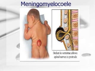
Meningomyelocoele
Humongous Insurance
Introduction
• Meningomyelocoele is the most severe type of SPINA
  BIFIDA.
• Spina Bifida means "split spine”
• 4 types of Spina Bifida are:
   – Spina bifida occulta,
   – Meningocele
   – Spina bifida cystica (Myelomeningocele) and
   – Lipomeningocele
• Meningomyelocoele
    – occurs due to Failure of closure of the neural tube
      during the third week of gestation
    – abnormal differentiation of the embryonic neural tube
• In Meningomyelocoele, a cystic swelling occurs over
  the site of the spinal defect which contains meninges,
  nerve roots, and the spinal cord itself which has left
  the vertebral canal
Spina bifida
Spinal Dysraphism


Spina bifida occulta   Meningocele   Myelomeningocele
Statistics Spina Bifida
Incidence - Meningomyelocoele
  • 1-2/1000 live birth
  • Neural tube defects are
    second most common type
    of birth defect
  • Myelomeningocele is the
    most common form of neural
    tube defect.
  • Slightly higher in females
    than in males (1.2:1)
  • 85% occur in lumbosacral
  • region
Aetiology
•   Poor nutrition - Folic acid deficiency
•   Genetics (People of Northern European and Hungarian
    ancestry have the highest rates of the disease)
•   Chromosome abnormalities:
     –   Trisomies 13 and 18
     –   Triploidy
     –   Single gene mutations
•   Maternal obesity, hyperthermia
•   pregnancies complicated by diabetes at conception
•   Drugs: valproate, carbamazepine and drugs to induce
    ovulation
•   Maternal exposures to fumonisins, electromagnetic fields,
    hazardous waste sites, disinfection by-products found in
    drinking water and pesticides
Meningomyelocele – Clinical
        Features
• Both meninges and spinal cord protrude
  into the skin of the back
• Leak of cerebrospinal fluid (CSF) is common
• Severe neurological deficits are common
• Risk for bacterial meningitis
• Paraplegia
• Diminished control of lower limbs, bladder Hydrocephalus
  and bowel
• Hydrocephalus often accompanies
• Short stature and precocious puberty


                                                Paraplegia
Primary Functional           System Congenital
      deficits                    Anomalies
• Lower limb paralysis
                              • Facial clefts
• Sensory loss
                              • Heart
• Bladder and bowel             malformations, and
  dysfunction
                              • Genitourinary tract
• Cognitive dysfunction         anomalies
• Cranial nerve               • Urinary tract
  dysfunction                   anomalies
   – Ocular muscle palsies,
                                 – solitary kidney or
   – Swallowing andeating
                                 – malformed ureters
     problems and
   – Abnormal phonation
Arnold Chiari II in 95% of
       myelomeningocoele
• Herniation of cerebella vermis through
  foramen Magnum (cerebella tonsils extend
  down into the spinal cord, lower CN run up
  instead of down)
• Dislocation of the 4th ventricle towards
  neural canal
• Associated with hydrocephalus 85%
• Developmental abnormalities in CV, GI,
  GU systems 10%
DIAGNOSIS
• Ultrasound during the second trimester
• The diagnosis of myelomeningocele is certain when 3 classic
  central findings are present in ultrasonography
   – concavity of the frontal bones,
   – ventriculomegaly, and
   – Chiari II malformation.
• Positive screening for maternal serum alpha-fetoprotein (AFP)
• Screening of the amniotic fluid for AFP, as well as for the
  presence of acetylcholinesterase
    Ultra Sound


     Coronal view of spine                              Lemon" shaped skull
                               Sagittal view of spine
Treatment
• Intrauterine surgery
• Post-natal surgery:- Closure of the Myelomeningocele is performed
  immediately after birth if external CSF leakage is present and
  typically within the first 24-48 hours in the absence of CSF leakage
• Multidisciplinary interventions needed to prevent progressive
  deterioration of multiple body systems
• Treatment team consists of pediatric specialists in
    –   Physical medicine and rehabilitation
    –   Neurosurgery
    –   Urology
    –   Orthopedics along with
    –   Pediatric nursing
    –   Physical therapy
    –   occupational and recreational therapy
    –   psychology and
    –   medical social work
Medication
• Anticholinergics: - to suppress detrusor
  overactivity eg. Oxybutynin chloride
• Tricyclic antidepressants, eg.
  Imipramine hydrochloride
• Alpha-adrenergic antagonists: -
  decrease bladder outlet resistance,
  increase urinary flow rate, and improve
  bladder emptying eg. Terazosin
Locomotion improves the
        cognitive profile of children
         with Meningomyelocele?
  • Twenty-nine children with MMC and shunted hydrocephalus
    were studied.
  • All had motor impairment, but after physiotherapy and training,
    walking was possible in 23 of them (5 autonomously and 18
    with an aid), while 6 had recourse to a wheelchair.
  • Even though global I.Q. in these MMC children was within the
    normal range, the characteristic splitting between verbal I.Q.
    (VIQ) and performance I.Q. (PIQ) was observed, but the most
    interesting observation was a significant performance-related
    difference (P=0.044 and P=0.012) between ambulatory patients
    (both with and without aids) and those who were dependent on
    wheelchairs (PIQ: 83-85 vs 63).
Reference: Rendeli C. et.al; Does locomotion improve the cognitive profile of children with
meningomyelocele?; Child’s Nerv Syst (2002) 18:231–234
Meningomyelocoele

More Related Content

What's hot (20)

Club foot
Club footClub foot
Club foot
 
Encephaloceles
EncephalocelesEncephaloceles
Encephaloceles
 
Guillain - Barré syndrome
Guillain -  Barré syndrome  Guillain -  Barré syndrome
Guillain - Barré syndrome
 
Guillain Barre Syndrome
Guillain Barre SyndromeGuillain Barre Syndrome
Guillain Barre Syndrome
 
Chorea
Chorea Chorea
Chorea
 
Guillain barre syndrome
Guillain barre syndromeGuillain barre syndrome
Guillain barre syndrome
 
Imperforate Anus
Imperforate Anus Imperforate Anus
Imperforate Anus
 
Paraplegia
ParaplegiaParaplegia
Paraplegia
 
Erbs palsy
Erbs palsyErbs palsy
Erbs palsy
 
Febrile seizures
Febrile seizuresFebrile seizures
Febrile seizures
 
Congenital diaphragmatic hernia (CDH)
Congenital diaphragmatic hernia (CDH)Congenital diaphragmatic hernia (CDH)
Congenital diaphragmatic hernia (CDH)
 
Epidural haematoma extradural haemorrhage
Epidural haematoma extradural haemorrhageEpidural haematoma extradural haemorrhage
Epidural haematoma extradural haemorrhage
 
Dandy Walker syndrome
Dandy Walker syndromeDandy Walker syndrome
Dandy Walker syndrome
 
Foot Drop
Foot DropFoot Drop
Foot Drop
 
Hemiplegia
HemiplegiaHemiplegia
Hemiplegia
 
Congenital Anomalies of Nervous System
Congenital Anomalies of Nervous SystemCongenital Anomalies of Nervous System
Congenital Anomalies of Nervous System
 
Cerebral palsy
Cerebral palsyCerebral palsy
Cerebral palsy
 
CONGENITAL TALIPES EQUINO VARUS
CONGENITAL TALIPES EQUINO VARUSCONGENITAL TALIPES EQUINO VARUS
CONGENITAL TALIPES EQUINO VARUS
 
Microcephaly
MicrocephalyMicrocephaly
Microcephaly
 
Hydrocephalus
HydrocephalusHydrocephalus
Hydrocephalus
 

Similar to Meningomyelocoele

Dementia-final+++++++++++++++
Dementia-final+++++++++++++++Dementia-final+++++++++++++++
Dementia-final+++++++++++++++dhavalshah4424
 
HYDROCEPHALUS (2)222.ppt
HYDROCEPHALUS (2)222.pptHYDROCEPHALUS (2)222.ppt
HYDROCEPHALUS (2)222.pptItratFatima21
 
microcephaly-150601111803-lva1-app6891 (1).pptx
microcephaly-150601111803-lva1-app6891 (1).pptxmicrocephaly-150601111803-lva1-app6891 (1).pptx
microcephaly-150601111803-lva1-app6891 (1).pptxSahilVerma19852
 
Spinal dysraphism and its management
Spinal dysraphism and its managementSpinal dysraphism and its management
Spinal dysraphism and its managementMukhtar Khan
 
Spina Bifida physiotherapy for pediatrics.pdf
Spina Bifida physiotherapy for pediatrics.pdfSpina Bifida physiotherapy for pediatrics.pdf
Spina Bifida physiotherapy for pediatrics.pdfPTMAAbdelrahman
 
Birth defects
Birth defectsBirth defects
Birth defectsEneutron
 
Myelomeningeocele by Dr. Raheel Gohar.pptx
Myelomeningeocele by Dr. Raheel Gohar.pptxMyelomeningeocele by Dr. Raheel Gohar.pptx
Myelomeningeocele by Dr. Raheel Gohar.pptxRaheel Gohar Qazi
 
Neurologic Causes Of Stransimus
Neurologic Causes Of StransimusNeurologic Causes Of Stransimus
Neurologic Causes Of Stransimusneurophq8
 
HYDROCEPHALUS presentation.docx
HYDROCEPHALUS presentation.docxHYDROCEPHALUS presentation.docx
HYDROCEPHALUS presentation.docxosmanconteh4
 
Clival Pathology.ppt
Clival Pathology.pptClival Pathology.ppt
Clival Pathology.pptmhmodsaad2
 
Neural Tube Defects for Undergraduate Medical Students
Neural Tube Defects for Undergraduate Medical StudentsNeural Tube Defects for Undergraduate Medical Students
Neural Tube Defects for Undergraduate Medical StudentsFarrukh Javeed
 

Similar to Meningomyelocoele (20)

Meningomyelocoele
MeningomyelocoeleMeningomyelocoele
Meningomyelocoele
 
Dementia-final+++++++++++++++
Dementia-final+++++++++++++++Dementia-final+++++++++++++++
Dementia-final+++++++++++++++
 
Meningomyelocoele
MeningomyelocoeleMeningomyelocoele
Meningomyelocoele
 
Spina bifida
Spina bifidaSpina bifida
Spina bifida
 
HYDROCEPHALUS (2)222.ppt
HYDROCEPHALUS (2)222.pptHYDROCEPHALUS (2)222.ppt
HYDROCEPHALUS (2)222.ppt
 
microcephaly-150601111803-lva1-app6891 (1).pptx
microcephaly-150601111803-lva1-app6891 (1).pptxmicrocephaly-150601111803-lva1-app6891 (1).pptx
microcephaly-150601111803-lva1-app6891 (1).pptx
 
Spina bifida
Spina bifidaSpina bifida
Spina bifida
 
Microcephaly
MicrocephalyMicrocephaly
Microcephaly
 
Spinal dysraphism and its management
Spinal dysraphism and its managementSpinal dysraphism and its management
Spinal dysraphism and its management
 
Spina Bifida physiotherapy for pediatrics.pdf
Spina Bifida physiotherapy for pediatrics.pdfSpina Bifida physiotherapy for pediatrics.pdf
Spina Bifida physiotherapy for pediatrics.pdf
 
Birth defects
Birth defectsBirth defects
Birth defects
 
Myelomeningeocele by Dr. Raheel Gohar.pptx
Myelomeningeocele by Dr. Raheel Gohar.pptxMyelomeningeocele by Dr. Raheel Gohar.pptx
Myelomeningeocele by Dr. Raheel Gohar.pptx
 
Neurologic Causes Of Stransimus
Neurologic Causes Of StransimusNeurologic Causes Of Stransimus
Neurologic Causes Of Stransimus
 
Mylomeningocele
MylomeningoceleMylomeningocele
Mylomeningocele
 
HYDROCEPHALUS presentation.docx
HYDROCEPHALUS presentation.docxHYDROCEPHALUS presentation.docx
HYDROCEPHALUS presentation.docx
 
Newborn examination
Newborn examinationNewborn examination
Newborn examination
 
Clival Pathology.ppt
Clival Pathology.pptClival Pathology.ppt
Clival Pathology.ppt
 
Ntd
NtdNtd
Ntd
 
Neural Tube Defects for Undergraduate Medical Students
Neural Tube Defects for Undergraduate Medical StudentsNeural Tube Defects for Undergraduate Medical Students
Neural Tube Defects for Undergraduate Medical Students
 
Ataxia
AtaxiaAtaxia
Ataxia
 

More from dhavalshah4424

More from dhavalshah4424 (20)

CAD+And+ACS
CAD+And+ACSCAD+And+ACS
CAD+And+ACS
 
Cardiac+Electrophysiology
Cardiac+ElectrophysiologyCardiac+Electrophysiology
Cardiac+Electrophysiology
 
Hypertension
HypertensionHypertension
Hypertension
 
Management+of+Diabetic+Neuropathy
Management+of+Diabetic+NeuropathyManagement+of+Diabetic+Neuropathy
Management+of+Diabetic+Neuropathy
 
Diabetes+and+Pregnancy
Diabetes+and+PregnancyDiabetes+and+Pregnancy
Diabetes+and+Pregnancy
 
Legg+Calve+Perthes+Disease
Legg+Calve+Perthes+DiseaseLegg+Calve+Perthes+Disease
Legg+Calve+Perthes+Disease
 
Osteoid+Osteoma
Osteoid+OsteomaOsteoid+Osteoma
Osteoid+Osteoma
 
Osteoid+Osteoma
Osteoid+OsteomaOsteoid+Osteoma
Osteoid+Osteoma
 
Osteoid+Osteoma
Osteoid+OsteomaOsteoid+Osteoma
Osteoid+Osteoma
 
Osteoid+Osteoma
Osteoid+OsteomaOsteoid+Osteoma
Osteoid+Osteoma
 
Legg+Calve+Perthes+Disease
Legg+Calve+Perthes+DiseaseLegg+Calve+Perthes+Disease
Legg+Calve+Perthes+Disease
 
Osteoid+Osteoma
Osteoid+OsteomaOsteoid+Osteoma
Osteoid+Osteoma
 
Complications+of+Diabetes
Complications+of+DiabetesComplications+of+Diabetes
Complications+of+Diabetes
 
Interventional+Procedures
Interventional+ProceduresInterventional+Procedures
Interventional+Procedures
 
Angiogram
AngiogramAngiogram
Angiogram
 
Polypharmacy+in+Schizophrenia
Polypharmacy+in+SchizophreniaPolypharmacy+in+Schizophrenia
Polypharmacy+in+Schizophrenia
 
Depression+and+Diabetes
Depression+and+DiabetesDepression+and+Diabetes
Depression+and+Diabetes
 
Antidepressants+in+Psychiatric+Disorder
Antidepressants+in+Psychiatric+DisorderAntidepressants+in+Psychiatric+Disorder
Antidepressants+in+Psychiatric+Disorder
 
Osteoarthritis
OsteoarthritisOsteoarthritis
Osteoarthritis
 
Juvenile+Rheumatoid+Arthritis+slides
Juvenile+Rheumatoid+Arthritis+slidesJuvenile+Rheumatoid+Arthritis+slides
Juvenile+Rheumatoid+Arthritis+slides
 

Meningomyelocoele

  • 2. Introduction • Meningomyelocoele is the most severe type of SPINA BIFIDA. • Spina Bifida means "split spine” • 4 types of Spina Bifida are: – Spina bifida occulta, – Meningocele – Spina bifida cystica (Myelomeningocele) and – Lipomeningocele • Meningomyelocoele – occurs due to Failure of closure of the neural tube during the third week of gestation – abnormal differentiation of the embryonic neural tube • In Meningomyelocoele, a cystic swelling occurs over the site of the spinal defect which contains meninges, nerve roots, and the spinal cord itself which has left the vertebral canal
  • 4. Spinal Dysraphism Spina bifida occulta Meningocele Myelomeningocele
  • 6. Incidence - Meningomyelocoele • 1-2/1000 live birth • Neural tube defects are second most common type of birth defect • Myelomeningocele is the most common form of neural tube defect. • Slightly higher in females than in males (1.2:1) • 85% occur in lumbosacral • region
  • 7. Aetiology • Poor nutrition - Folic acid deficiency • Genetics (People of Northern European and Hungarian ancestry have the highest rates of the disease) • Chromosome abnormalities: – Trisomies 13 and 18 – Triploidy – Single gene mutations • Maternal obesity, hyperthermia • pregnancies complicated by diabetes at conception • Drugs: valproate, carbamazepine and drugs to induce ovulation • Maternal exposures to fumonisins, electromagnetic fields, hazardous waste sites, disinfection by-products found in drinking water and pesticides
  • 8. Meningomyelocele – Clinical Features • Both meninges and spinal cord protrude into the skin of the back • Leak of cerebrospinal fluid (CSF) is common • Severe neurological deficits are common • Risk for bacterial meningitis • Paraplegia • Diminished control of lower limbs, bladder Hydrocephalus and bowel • Hydrocephalus often accompanies • Short stature and precocious puberty Paraplegia
  • 9. Primary Functional System Congenital deficits Anomalies • Lower limb paralysis • Facial clefts • Sensory loss • Heart • Bladder and bowel malformations, and dysfunction • Genitourinary tract • Cognitive dysfunction anomalies • Cranial nerve • Urinary tract dysfunction anomalies – Ocular muscle palsies, – solitary kidney or – Swallowing andeating – malformed ureters problems and – Abnormal phonation
  • 10. Arnold Chiari II in 95% of myelomeningocoele • Herniation of cerebella vermis through foramen Magnum (cerebella tonsils extend down into the spinal cord, lower CN run up instead of down) • Dislocation of the 4th ventricle towards neural canal • Associated with hydrocephalus 85% • Developmental abnormalities in CV, GI, GU systems 10%
  • 11. DIAGNOSIS • Ultrasound during the second trimester • The diagnosis of myelomeningocele is certain when 3 classic central findings are present in ultrasonography – concavity of the frontal bones, – ventriculomegaly, and – Chiari II malformation. • Positive screening for maternal serum alpha-fetoprotein (AFP) • Screening of the amniotic fluid for AFP, as well as for the presence of acetylcholinesterase Ultra Sound Coronal view of spine Lemon" shaped skull Sagittal view of spine
  • 12. Treatment • Intrauterine surgery • Post-natal surgery:- Closure of the Myelomeningocele is performed immediately after birth if external CSF leakage is present and typically within the first 24-48 hours in the absence of CSF leakage • Multidisciplinary interventions needed to prevent progressive deterioration of multiple body systems • Treatment team consists of pediatric specialists in – Physical medicine and rehabilitation – Neurosurgery – Urology – Orthopedics along with – Pediatric nursing – Physical therapy – occupational and recreational therapy – psychology and – medical social work
  • 13. Medication • Anticholinergics: - to suppress detrusor overactivity eg. Oxybutynin chloride • Tricyclic antidepressants, eg. Imipramine hydrochloride • Alpha-adrenergic antagonists: - decrease bladder outlet resistance, increase urinary flow rate, and improve bladder emptying eg. Terazosin
  • 14. Locomotion improves the cognitive profile of children with Meningomyelocele? • Twenty-nine children with MMC and shunted hydrocephalus were studied. • All had motor impairment, but after physiotherapy and training, walking was possible in 23 of them (5 autonomously and 18 with an aid), while 6 had recourse to a wheelchair. • Even though global I.Q. in these MMC children was within the normal range, the characteristic splitting between verbal I.Q. (VIQ) and performance I.Q. (PIQ) was observed, but the most interesting observation was a significant performance-related difference (P=0.044 and P=0.012) between ambulatory patients (both with and without aids) and those who were dependent on wheelchairs (PIQ: 83-85 vs 63). Reference: Rendeli C. et.al; Does locomotion improve the cognitive profile of children with meningomyelocele?; Child’s Nerv Syst (2002) 18:231–234