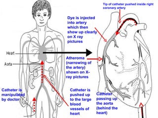
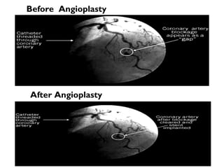
Angiogram
Road Map of Angiogram What is Angiogram? How is the procedure done? For what it is performed? Types of Angiography. Need of Angiography Conclusion
What is Angiogram? It is an imaging test that uses x-rays to poduce the rood map of  body’s blood vessels.  Physicians often use this test to study narrow, blocked, enlarged, or malformed arteries or veins in many parts of your body, including your brain, heart, abdomen, and legs.  When the arteries are studied, the test is also called an arteriogram, if the veins are studied, it is called a venogram. 
HOW IS THE PROCEDURE DONE?
How is the procedure done? During this procedure also known as coronary angiography , the patient is positioned on an x-ray table. Depending on the condition of the arteries, either the under arm or groin area will be disinfected and then numbed with an anesthetic. A long, narrow tube (catheter) is then inserted into a large artery (in either the upper leg or upper arm) and gently threaded up into the coronary arteries around the heart muscle.  Contd…
4.  A special type of dye that is visible on x-rays is then released from the catheter and the x-rays are then taken of the heart and surrounding vessels. 5.  The doctor will review the x-rays taken during the angiogram to pinpoint the location and severity of any narrowing or blockages within the coronary arteries. 6.  If a blockage is found, the doctor can then determine the best way to remove the blockage material or dilate (widen) the narrowed blood vessel(s).
Catheter is manipulated by doctor Catheter is  pushed up to the large blood vessels of heart  Catheter passing up the aorta (behind the heart) Tip of catheter pushed inside right coronary artery Dye is injected into artery which then show up clearly on X ray pictures  Atheroma (narrowing of the artery) shown on X-ray pictures
Before  Angioplasty After Angioplasty
For what it is performed? Angiography is performed for a variety of reasons, including: Detection of occlusions of blood vessels, in patients with chest pain and suspected as having heart disease. For the presence of an aneurysm For a possible tear in a blood vessel and bleeding. For use in vascular surgery or percutaneous vascular intervention Organ tumours can be diagnosed and surgical therapy can be planned In internal medicine patients, used  in evaluating hypertension, gastrointestinal bleeding, and pulmonary embolus.
Types of Angiograms Coronary angiography Cerebral angiography Peripheral angiography (arm or leg)  Visceral angiography(the abdominal organs, or viscera)  Pulmonary angiography (lungs)  Lymphangiography (lymph vessels)  Aortography (looking at the aorta, the major artery  from the heart)  Retinal angiography Magnetic Resonance Angiography (MRI study of the blood vessels)
BENEFITS AND RISKS……
Benefits of Angiogram Shows no of coronary arteries  blocked by fatty plaque accumulations (atherosclerosis)  Pinpoint the area of blockages  located in the blood vessels  Indicate the extent ( %)  of blockages  Also eliminates the need for surgery. If surgery remains necessary, it can be performed more accurately.  It presents a very detailed, clear and accurate picture of the blood vessels. This is especially helpful when a surgical procedure is being considered.
Risks of Angiogram Bleeding, pain, or swelling where the catheter was inserted Rarely, severe allergic reactions can occur, especially among people who have had previous allergic reactions to the contrast dye.  Infrequently, a patient with CHF may experience shortness of breath or fluid overload due to poor pumping action of heart
CONCLUSION
Conclusion It is an imaging test that uses x-rays to pinpoint the location and severity of any narrowing or blockages within the coronary arteries Shows number of blockages  Indicates % of blockages Pinpoint the area of blockages  located in the blood vessels
Thank You

Angiogram

  • 1.
  • 2.
    Road Map ofAngiogram What is Angiogram? How is the procedure done? For what it is performed? Types of Angiography. Need of Angiography Conclusion
  • 3.
    What is Angiogram?It is an imaging test that uses x-rays to poduce the rood map of body’s blood vessels. Physicians often use this test to study narrow, blocked, enlarged, or malformed arteries or veins in many parts of your body, including your brain, heart, abdomen, and legs. When the arteries are studied, the test is also called an arteriogram, if the veins are studied, it is called a venogram. 
  • 4.
    HOW IS THEPROCEDURE DONE?
  • 5.
    How is theprocedure done? During this procedure also known as coronary angiography , the patient is positioned on an x-ray table. Depending on the condition of the arteries, either the under arm or groin area will be disinfected and then numbed with an anesthetic. A long, narrow tube (catheter) is then inserted into a large artery (in either the upper leg or upper arm) and gently threaded up into the coronary arteries around the heart muscle. Contd…
  • 6.
    4. Aspecial type of dye that is visible on x-rays is then released from the catheter and the x-rays are then taken of the heart and surrounding vessels. 5. The doctor will review the x-rays taken during the angiogram to pinpoint the location and severity of any narrowing or blockages within the coronary arteries. 6. If a blockage is found, the doctor can then determine the best way to remove the blockage material or dilate (widen) the narrowed blood vessel(s).
  • 7.
    Catheter is manipulatedby doctor Catheter is pushed up to the large blood vessels of heart Catheter passing up the aorta (behind the heart) Tip of catheter pushed inside right coronary artery Dye is injected into artery which then show up clearly on X ray pictures Atheroma (narrowing of the artery) shown on X-ray pictures
  • 8.
    Before AngioplastyAfter Angioplasty
  • 9.
    For what itis performed? Angiography is performed for a variety of reasons, including: Detection of occlusions of blood vessels, in patients with chest pain and suspected as having heart disease. For the presence of an aneurysm For a possible tear in a blood vessel and bleeding. For use in vascular surgery or percutaneous vascular intervention Organ tumours can be diagnosed and surgical therapy can be planned In internal medicine patients, used in evaluating hypertension, gastrointestinal bleeding, and pulmonary embolus.
  • 10.
    Types of AngiogramsCoronary angiography Cerebral angiography Peripheral angiography (arm or leg) Visceral angiography(the abdominal organs, or viscera) Pulmonary angiography (lungs) Lymphangiography (lymph vessels) Aortography (looking at the aorta, the major artery from the heart) Retinal angiography Magnetic Resonance Angiography (MRI study of the blood vessels)
  • 11.
  • 12.
    Benefits of AngiogramShows no of coronary arteries blocked by fatty plaque accumulations (atherosclerosis) Pinpoint the area of blockages located in the blood vessels Indicate the extent ( %) of blockages Also eliminates the need for surgery. If surgery remains necessary, it can be performed more accurately. It presents a very detailed, clear and accurate picture of the blood vessels. This is especially helpful when a surgical procedure is being considered.
  • 13.
    Risks of AngiogramBleeding, pain, or swelling where the catheter was inserted Rarely, severe allergic reactions can occur, especially among people who have had previous allergic reactions to the contrast dye.  Infrequently, a patient with CHF may experience shortness of breath or fluid overload due to poor pumping action of heart
  • 14.
  • 15.
    Conclusion It isan imaging test that uses x-rays to pinpoint the location and severity of any narrowing or blockages within the coronary arteries Shows number of blockages Indicates % of blockages Pinpoint the area of blockages located in the blood vessels
  • 16.