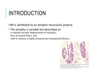
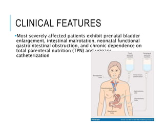
2. INTRODUCTION
Visceral myopathy (VM) is a rare, severe, and often
misdiagnosed pathological condition.
It is a rare inherited form of myopathic pseudo-
obstruction, characterized by impaired function of
enteric smooth muscle cells and sometimes the urinary
tract.
3. INTRODUCTION
VM is attributed to an atrophic muscularis propria.
The atrophy is variable but described as:
a marked vacuolar degeneration of myocytes,
loss of muscle fibers, and
with or without a highly characteristic honeycomb fibrosis.
4. TYPES
The cases are either familial or
sporadic.
It may be:
1. Primary
1. familial and Sporadic
2. secondary to the other diseases:
1. scleroderma
2. systemic lupus
3. stroke
4. encephalitis
5. CLINICAL FEATURES
Patients suffering from VM vary greatly in their clinical
manifestations.
Some patients are completely asymptomatic
•Nausea and vomiting
•postprandial pain
•abdominal distension and diarrhea
Some patients will experience light
abdominal symptoms
Few patients experience repetitive attacks of
bowel obstruction resembling small bowel
or colonic obstruction
Few patients may present with recurrent
urinary tract infections or urinary retention
when visceral myopathy involves the
bladder muscles
6. CLINICAL FEATURES
The usual clinical picture of a patient with VM is
characterized by a chronic course dominated by:
recurrent episodes of abdominal pain,
vomiting
abdominal distension
7. CLINICAL FEATURES
Most severely affected patients exhibit prenatal bladder
enlargement, intestinal malrotation, neonatal functional
gastrointestinal obstruction, and chronic dependence on
total parenteral nutrition (TPN) and urinary
catheterization
8. DIAGNOSIS
The diagnosis may be delayed due to the rarity of VM,
great variation in symptoms, and the many similarities it
has with other more common gastrointestinal diseases.
VM should be considered as differential diagnosis
whenever the patient presents with acute appendicitis,
uncharacteristic abdominal symptoms, recurrent attacks
of abdominal distention, and pain with no radiological
evidence of intestinal obstruction.
9. DIAGNOSIS
1. Radiological studies
There is almost always absence of specific radiological features
that indicates the diagnosis VM.
Radiological studies (X-ray, CT Abdomen) should be done to
exclude bowel obstruction or pseudo-obstruction.
2. Histopathological studies:
To confirm the diagnosis of VM marked vacuolar
degeneration of myocytes
loss of muscle fibers
with or without a highly
characteristic honeycomb
fibrosis
10. PRENATAL DIAGNOSIS
prenatal diagnosis by ultrasound before 20 weeks could
be possible (Vezina et al., 1979).
Farrell (1988) described an affected sib pair with
intrauterine death of 1 of the sibs.
Young et al. (1989) made the diagnosis by
ultrasonography in a pregnancy that was monitored due
to the previous pregnancy resulting in a male infant who
died at 4 hours of age as a consequence of multiple
anomalies; these anomalies were believed to include
urethral atresia and possibly intestinal atresia.
Termination of pregnancy was performed at 18 weeks.
Necropsy of the male fetus showed vacuolation and
degeneration in smooth muscle of bowel and bladder
wall.
12. TREATMENT
Treatment of VM consists of fluid and electrolytes by IV
infusions and gastric and colonic decompression by
nasogastric and rectal tubes.
Pitt et al. showed that total parenteral nutrition (TPN)
and ventral enterostomy greatly reduced the number of
required admissions in a group of patients with chronic
intestinal pseudoobstruction.
Surgical resections and enterostomies is an option in
patients with localized involvement of the
gastrointestinal tract who do not respond to conservative
treatment
13. LEARNING POINTS
The symptoms of visceral myopathy are unspecific and
often mimic other more common gastrointestinal
diseases.
In patients that present with recurrent episodes of
abdominal distension with no evidence of
mechanical obstruction, VM should be considered.
It is important to consider VM no matter the patients
age.
1
2
3
14. REFERENCES
1. Burcharth J, Olsen C, Rosenberg J. Acute abdomen and perforated bowel with a rare
pathology: Nonfamilial visceral myopathy. Case reports in surgery. 2011 Dec
8;2011.
2. Kharbuja P, Thakur R, Suo J. Visceral Myopathy Presenting as Acute Appendicitis
and Ogilvie Syndrome. Case reports in surgery. 2013 Apr 30;2013.
3. Sen R, Gupta V, Hasija S, Ralli M, Garg S. Visceral Myopathy Causing Intestinal
Obstruction: A Case Report. Journal of Gastroenterology and Hepatology Research.
2014 Aug 21;3(8):1213-5.
4. Pitt HA, Mann LL, Berquist WE, Ament ME, Fonkalsrud EW, DenBesten L. Chronic
intestinal pseudo-obstruction: management with total parenteral nutrition and a
venting enterostomy. Archives of Surgery. 1985 May 1;120(5):614-8.
5. Vezina WC, Morin FR, Winsberg F. Megacystis-microcolon-intestinal hypoperistalsis
syndrome: antenatal ultrasound appearance. American Journal of Roentgenology.
1979 Oct 1;133(4):749-50.
6. Farrell SA. Intrauterine death in megacystis-microcolon-intestinal hypoperistalsis
syndrome. Journal of medical genetics. 1988 May 1;25(5):350-1.
7. Young ID, McKeever PA, Brown LA, Lang GD. Prenatal diagnosis of the megacystis-
microcolon-intestinal hypoperistalsis syndrome. Journal of medical genetics. 1989
Jun 1;26(6):403-6.
8. https://www.omim.org/entry/155310#diagnosis
Editor's Notes
Pseudoobstruction ( impaired intestinal function and motility in absence of mechanical obstruction )