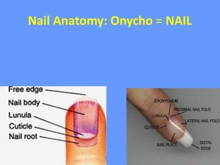
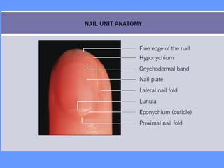
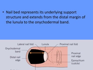
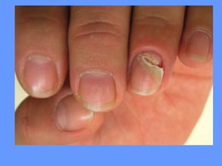
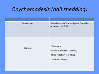
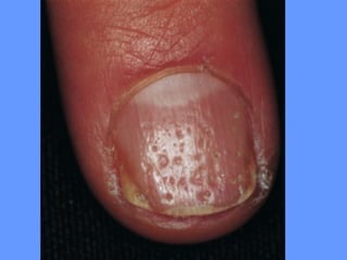
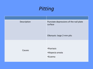
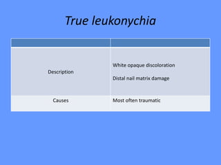
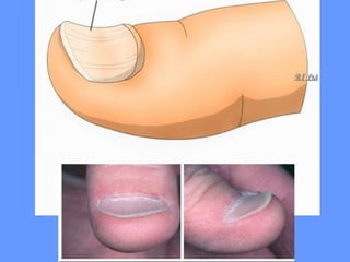
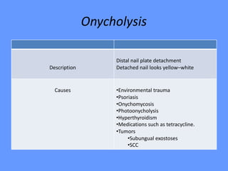
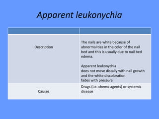
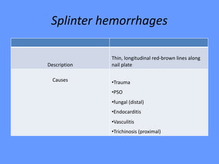
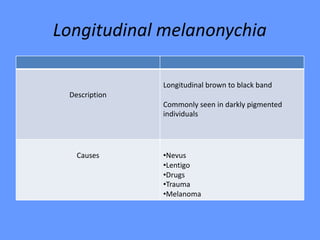
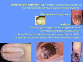
4. Nail plate
The nail plate is a hard, semitransparent, slightly convex sheet
comprised of tightly packed corneocytes termed onychocytes.
• It contains abundant “hard” hair-type keratins, which are embedded
in a matrix of sulfur-rich high-cysteine and high-glycine/tyrosine
proteins.
• Pink color of nail plate due to longitudinally situated subungual
capillaries
5. • The nail plate is surrounded on three edges by
nail folds (one proximal and two lateral).
6. • Nail bed represents its underlying support
structure and extends from the distal margin of
the lunula to the onychodermal band.
7. • The proximal nail fold, a double layer of skin
overlying the proximal part of the nail.
• Protects this area from trauma, solvents and infectious agents
that might otherwise alter nail shape and structure.
8. The cuticle (eponychium)
Is a thin strip of cornified epithelium that adheres to the nail at
the edge of the proximal nail fold and acts as a seal against
ingress.
• Damage to the cuticle results in a predisposition to inflammation of the
nail fold; conversely, inflammation of the nail folds disrupts normal
maturation of the epithelium and cuticle formation
9. The dermis of the nail apparatus
• Is a fibrocollagenous network that is “sandwiched” between
the hard tissues of the nail plate and the underlying bone,
lacking associated subcutaneous soft tissues or pilosebaceous
units.
10. • Bacterial infections in this region have a higher
risk of spread to the adjacent bone (i.e.
osteomyelitis of the distal phalanx) than in
most other sites of the body.
11. Lunula
• The half-moon-shaped lunula, which is most
prominent on the thumb, demarcates the
junction between the matrix and the nail bed.
12. Nail Matrix
• Is the germinative epithelium from which the nail plate is
derived.
• Wedge-shaped area of specialized epithelium, divided into
proximal and distal portion
• Lunula demarcates distal portion of nail matrix
• Melanocytes found in high concentration in nail matrix
(mainly seen in the distal matrix)
13. • Proximal matrix → dorsal nail plate
• Distal matrix (lunula) → ventral nail plate
15. Growth
• Fingernails usually grow faster than toenails
• Fingernails: 2–3 mm/month
• Toenails: 1 mm/month
• Complete replacement of a nail requires 6 (fingernails)
to 18 months (toenails)
• In children the growth is rapid 6 – 8 weeks
• Nail growth is quicker:
· During pregnancy
· In the summer than in the winter
· On the hands than the feet
21. Beau’s lines
Single horizontal ridge OR Transverse depressions
More evident in the central portion of the nail plate
Description
Mechanical trauma (e.g. manicures, onychotillomania)
Dermatologic disease of the proximal nail fold (e.g.
eczema, chronic paronychia).
The presence of Beau’s lines at the same level in all nails
suggests a systemic cause (e.g. severe or febrile illness,
erythroderma, drugs).
Causes
22.
23. Onychomadesis (nail shedding)
Detachment of the nail plate from the
proximal nail fold
Description
•Traumatic
•Medications (i.e. chemo),
•Drug reaction (i.e. TEN),
•Systemic illness
Causes
24.
25. Pitting
Punctate depressions of the nail plate
surface
Elkonyxis: large 2-mm pits
Description
•Psoriasis
•Alopecia areata
•Eczema
Causes
26.
27. Onychorrhexis
Thinning
Longitudinal ridging and fissuring
Description
•Lichen planus
•Impaired vascular supply
•Trauma
•Tumors that compress the nail matrix.
•Repeated wet/dry cycles,
• Normal with aging
Causes
28.
29. Trachyonychia (twenty-nail dystrophy,
sandpapered nails)
Nail roughness
Often associated with thinning
Description
•Alopecia areata (common)
•Lichen planus (rare)
•Psoriasis (rare)
•Eczema (very rare)
Causes
34. Punctate leukonychia
• The nail plate shows small opaque white
spots
• Move distally with nail growth and sometimes
disappear before reaching the distal nail.
• It is caused by trauma and is most commonly
observed in the fingernails of children.
35.
36. Striate leukonychia
The nail plate shows one or more transverse
white opaque parallel lines.
• Observed in the fingernails of women, due to
matrix trauma secondary to manicures.
37.
38. Diffuse leukonychia
• The nail plate is completely or almost
completely opaque and white.
• Chemotherapy
• White superficial onychomycosis
• Congenital disease
39. Needs to be distinguished
• Apparent leukonychia (nail bed discoloration)
( White discoloration (fades with pressure) nail plate
looks white but normal color)
• Pseudoleukonychia (nail plate invasion by fungi), which is
typical of superficial white onychomycosis In this case the nail
plate surface is friable due to the presence of keratin debris
and fungal elements
40.
41. Koilonychia (spoon nails)
Thinned concave nails
The nail plate is thinned, flat and
spoonshaped due to upward eversion
of its lateral edges.
Description
•Physiologic in the toenails of children
•Adults -- occupational
•Severe iron deficiency.
Causes
44. Onycholysis
Distal nail plate detachment
Detached nail looks yellow–whiteDescription
•Environmental trauma
•Psoriasis
•Onychomycosis
•Photoonycholysis
•Hyperthyroidism
•Medications such as tetracycline.
•Tumors
•Subungual exostoses
•SCC
Causes
45.
46. Onychauxis
The nail plate appears thickened due to
the presence of subungual scales.Description
•Psoriasis,
•Onychomycosis
•Eczema
Causes
47.
48. Apparent leukonychia
The nails are white because of
abnormalities in the color of the nail
bed and this is usually due to nail bed
edema.
Apparent leukonychia
does not move distally with nail growth
and the white discoloration
fades with pressure
Description
Drugs (i.e. chemo agents) or systemic
diseaseCauses
49.
50. Splinter hemorrhages
Thin, longitudinal red-brown lines along
nail plateDescription
•Trauma
•PSO
•fungal (distal)
•Endocarditis
•Vasculitis
•Trichinosis (proximal)
Causes
55. Hutchinson’s sign
•Brown–black periungual pigmentation
• Needs to be distinguished from pseudo-
Hutchinson’s sign (dark bands producing
“illusory” pigmentation of the proximal
nail fold due to cuticle transparency)
Description
•Possible sign of nail melanomaCauses
56.
57. Green nail syndrome
nail has a greenishblack or greenishblue
color due to the deposition of pyocyanin,
a blue–green pigment produced
by Pseudomonas aeruginosa,
Predisposing factors
• Prolonged exposure to water,
• Detergents and soaps,
• Nail trauma
in barbers, dishwashers, bakers and
medical personnel.
Description
•Pseudomonas aeruginosaCauses
58.
59. Half and half nails
(Lindsay’s nails)
Proximal ½ with white zone, distal ½ with
red-brown zoneDescription
•Chronic renal diseaseCauses
60.
61. Terry’s nails
Proximal 2/3 white nail color, distal
1/3 brown-pink bandDescription
•Cirrhosis,
•Hypoalbuminemia
•Diabetes
•cardiac disease
Causes
64. Color changes due
to drugs:
• Chloroquine may
produce blue-black
pigmentation of the nail
• Arsenic may produce
longitudinal bands of
pigment or transverse
white stripes (Mees'
stripes) across the nail
69. Absent lunula
• No visible lunula
• Yellow nail syndrome,
• Renal failure,
• Trauma
70. Yellow nail syndrome
• The nails are yellow due to thickening, sometimes with a tinge of green
• The lunula is obscured and there is increased transverse and longitudinal
curvature and loss of cuticle
• It is usually accompanied by lymphoedema and pleural effusions
71.
72. Anonychia
• Absence of nail
• Nail patella syndrome
• COIF (Congenital Onychodysplasia of the Index
Finger) scarring
73.
74. Blue lunula
• Blue discoloration of lunula
• Wilson’s
• Drugs
• PUVA
• Argyria
77. Causes of clubbing
1-Primary (idiopathic) clubbing e.g. familial clubbing
2-Secondary clubbing include the following:
• Pulmonary disease e.g. Lung cancer, cystic fibrosis
• Cardiac disease e.g. Cyanotic congenital heart disease
• GIT disease e.g. inflammatory bowel disease
• Skin disease e.g. Pachydermoperiostosis
• Malignancies e.g. Thyroid cancer, Hodgkin disease, leukemia
• Miscellaneous conditions e.g. Acromegaly, pregnancy, and
hypoxemia possibly related to long-term smoking of cannabis
86. Ingrowing toenail (Onychocryptosis):
• The edge of the nail plate penetrates the
lateral nail fold
• Due to compression of the toe from the side
due to ill-fitting footwear
94. – Keratosis follicularis (Darier’s disease) is
associated with each of the following nail changes
EXCEPT:
A. “V-shaped” notch at the nail plate’s distal free
edge.
B. Splinter hemorrohages.
C. Proximal nail fold keratotic papules.
D. “V-shaped” lunula
E. Longitudinal dyschromic lunular bands.
95. • Nail changes in Pachyonychia congenita are
most consistent with Which of the following
descriptions?
A. Congenital malalignment
B. Leukonychia
C. Koilonychias
D. Subungual hyperkeratosis
E. Onychomadesis
96. • Hutchinson nail sign is periungual extension of
pigment to the proximal and lateral folds of
the nail and is noted in:
A. LP
B. Traumatic purpura
C. Onychomycosis
D. Subungual melanoma
E. Periungual fibroma
97. • Which One of the following definition about nail
changes is not true :
A. Roughened surface of the nail plate is
trachyonychia
B. Longitudinal striations of the nail plate is
onychmadesis
C. Ingrown nail is onychocryptosis
D. A traumatic, familial non- inflammatory nail
loss is onychoptosis
E. Superficial splitting of the nail plate or near the
free edge is onychoschizia
98. • The dorsal nail plate is produced by :
a. Proximal matrix
b. Nail bed
c. Nail fol
d. Hyponychium
e. Distal matrix
99. • Pitting of the nail plate in psoriasis is
associated with involvement of which portion
of the nail apparatus?
• A. Proximal nail matrix.
• B. Distal nail matrix.
• C. Cuticle.
• D. Nail bed.
• E. Nail plate.
100.
101. • One of the following is not true about yellow
nail syndrome:
A. yellow thickened nails
B. Increased longitudinal and transverse
curvature
C. Triangular lunula
D. Decreased growth rate of nails
E. Lymphedema mostly in legs
102. • Transverse grooves on nail following systemic
disease are called:
• A. Mee’s line.
B. Coenen’s line.
C. Trachyonychia.
D. Beau’s line
E. Grove sign.
103. • The nail findings shown here are most
characteristic of what condition?
• A. Lichen planus.
• B. Lichen striatus.
• C. Darier’s disease.
• D. Dyskeratosis congenita.
• E. Pachyonychia congenita.
105. Nail infections
Paronychia: is a soft tissue infection around a fingernail
• is the most common hand infection
Acute paronychia
• nail biting breaks down the physical barrier between the nail bed and the nail
allowing the infiltration of infectious organisms
• S. aureus is the most common infecting organism.
• pain, tenderness, and swelling in the lateral folds of the nail
• erythematous and swollen, pus collects under the skin of the lateral fold
• Oral antibiotics
Chronic paronychia
• After 6 weeks or longer
• The nail folds are swollen, erythematous, and tender with pronounced
transverse ridges
• Cause is a mixture of C. albicans and bacteria
• Can be a complication of eczema
• In housekeepers, dishwashers, and swimmers
106. Pseudomonas infection
• It is always a complication of onycholysis or chronic paronychia
• The nail plate has a characteristic bluish-black or green color due to
accumulation of the pigment pyocyanin below the nail which may remain after
the organism has been removed
• Treatment is as for paronychia
ONYCHOMYCOSIS (TINEA UNGUIUM)
• An infectious fungal disease mainly seen as white spots that can be scraped off
the surface, or long yellowish streaks within the nail substance.
• attacks the free edge and moves its way to the matrix.
• The infected portion is thick and discoloured.
107. 4-Tumors
Warts
Fibrokeratoma: periungual hyperkeratotic tip
Subungual exostosis: bony outgrowth of the distal part of the toe
Glomus tumour:
•is painful, (pain may be spontaneous or evoked by mild trauma or temperature
change)
•Nail-plate changes depend on the location of the tumour:
- Matrix tumours cause splitting and distortion of the nail plate.
- Nail bed lesions appear as bluish or red foci of 1-5mm diameter beneath
the nail
108. Squamous cell carcinoma: hyperkeratotic, warty changes, erosions and
fissuring, macerated cuticle, periungual swelling & erythema
Melanocytic nevi: longitudinal melanonychia
Malignant melanoma:
features suggest the possibility of malignant melanoma:
•75% will have Longitudinal melanonychia
•Brown-black periungual pigmentation in a single digit in adult life
•The pigmentation becomes darker and broader and has blurred edges