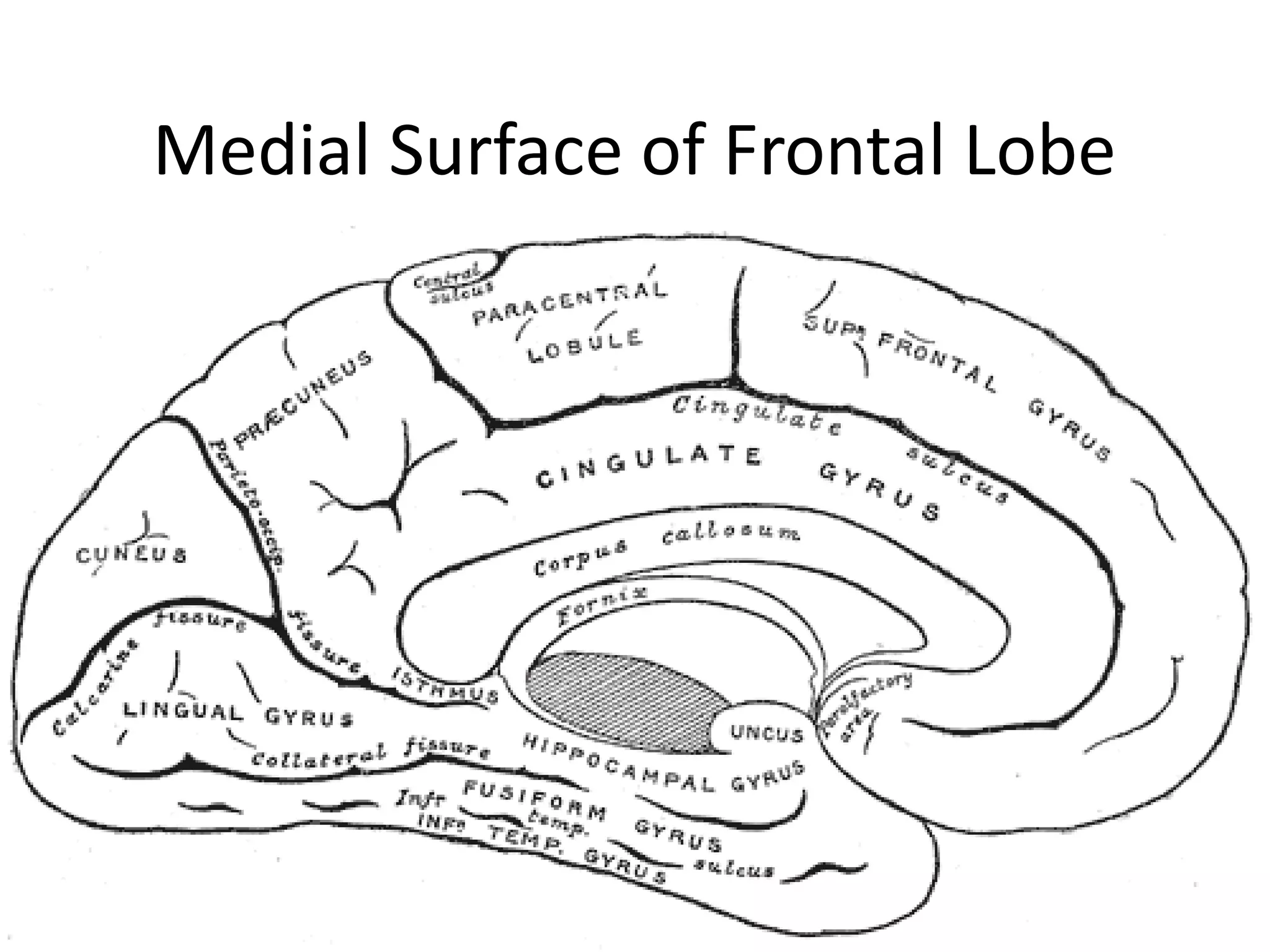
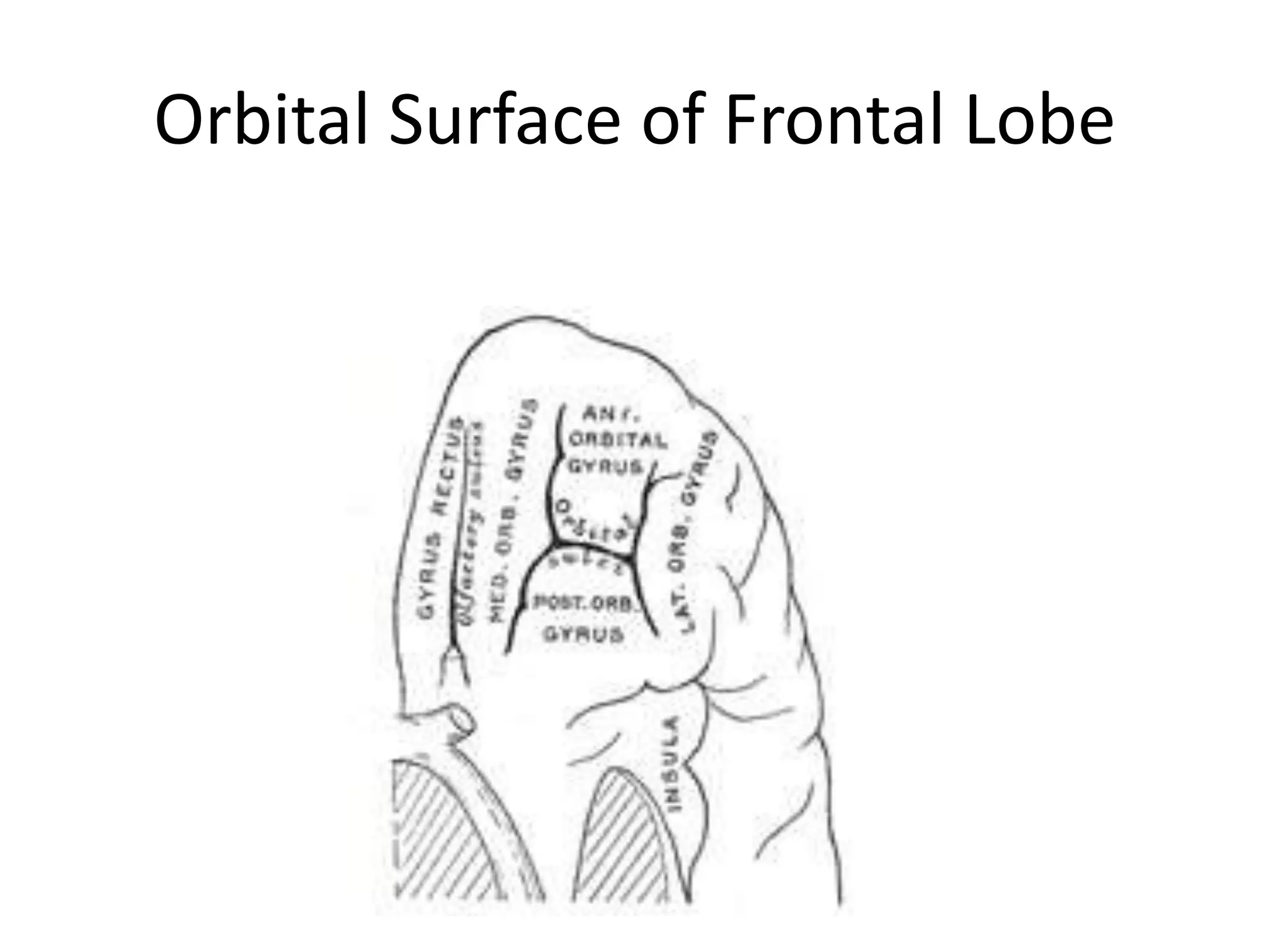
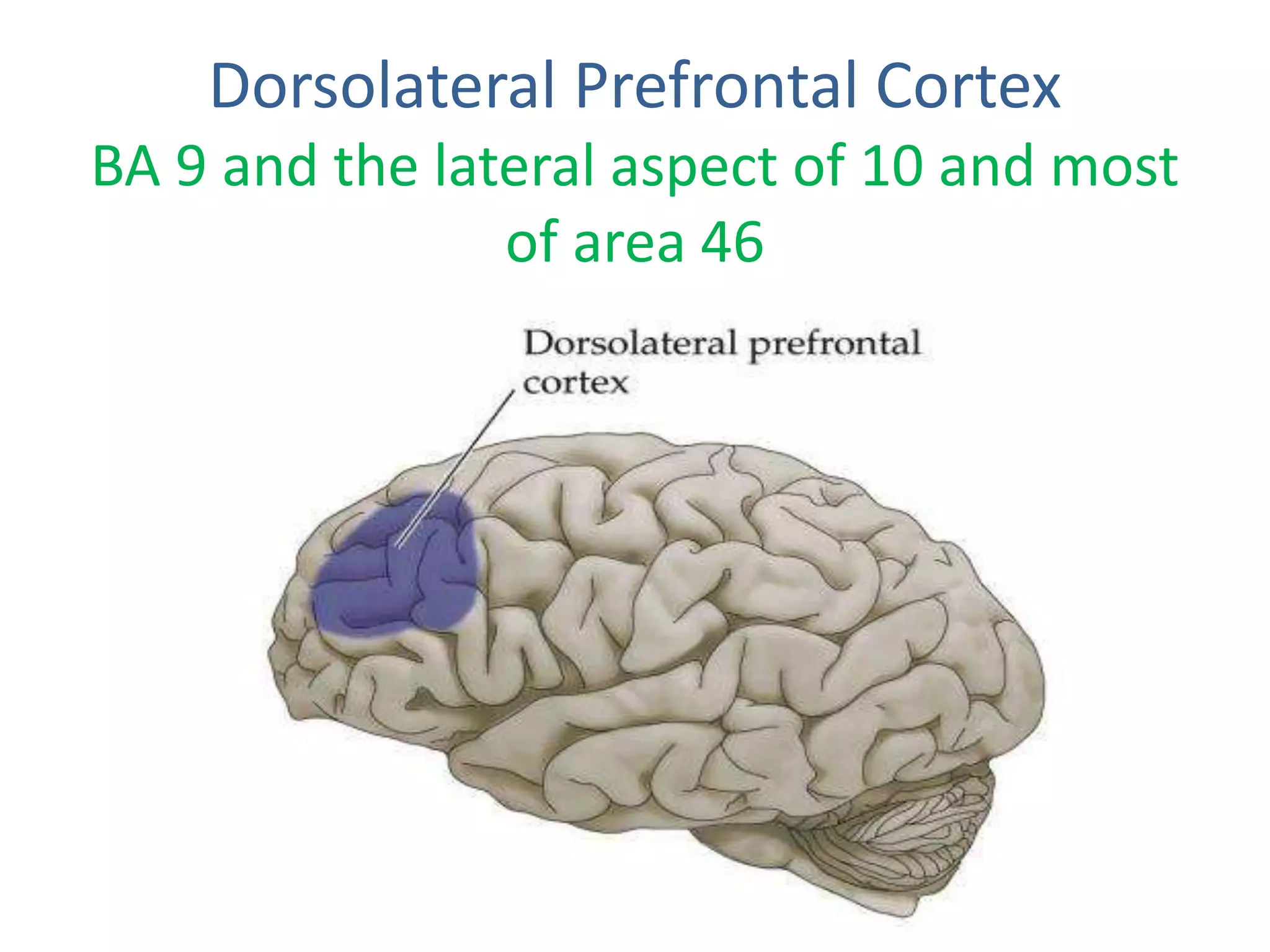
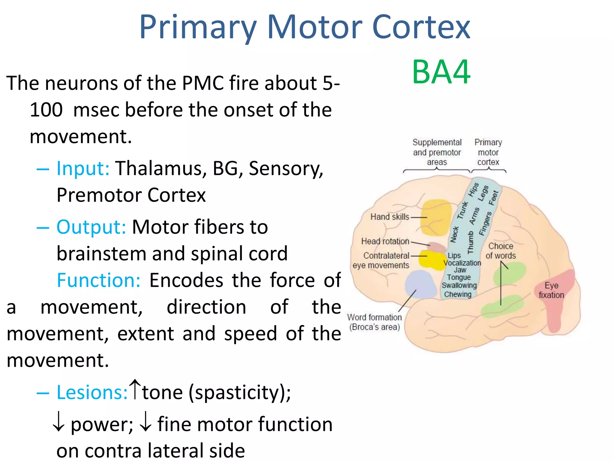
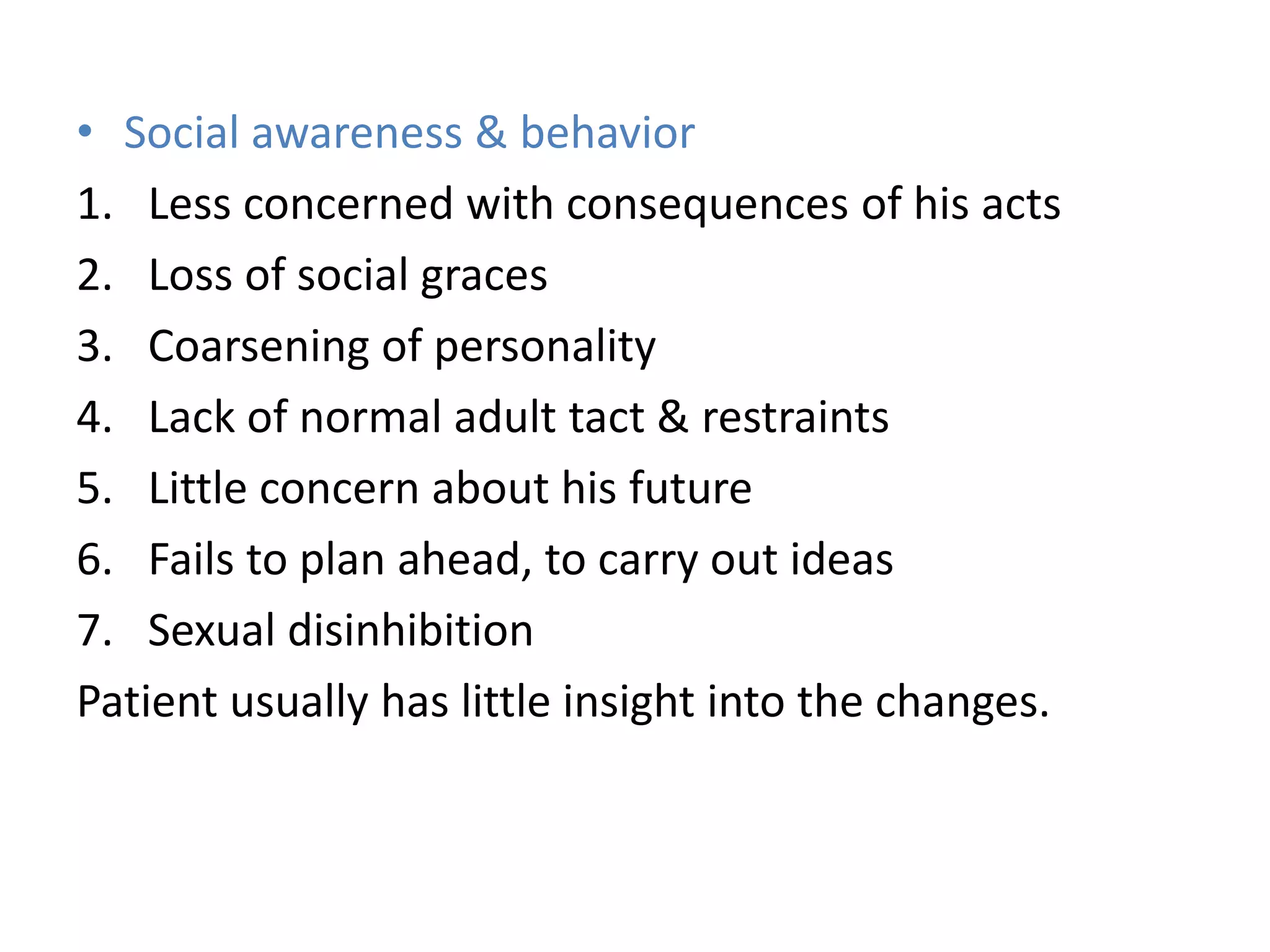
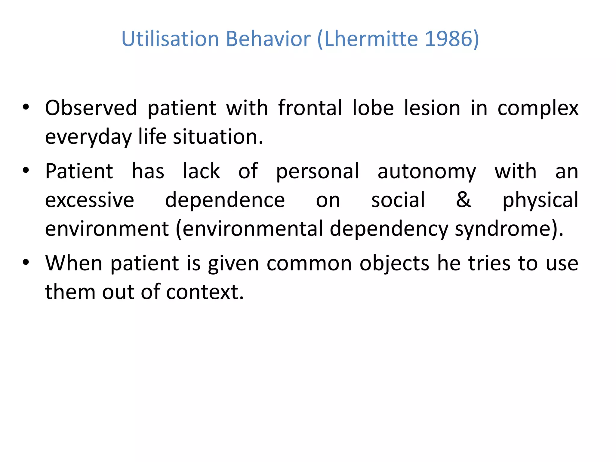
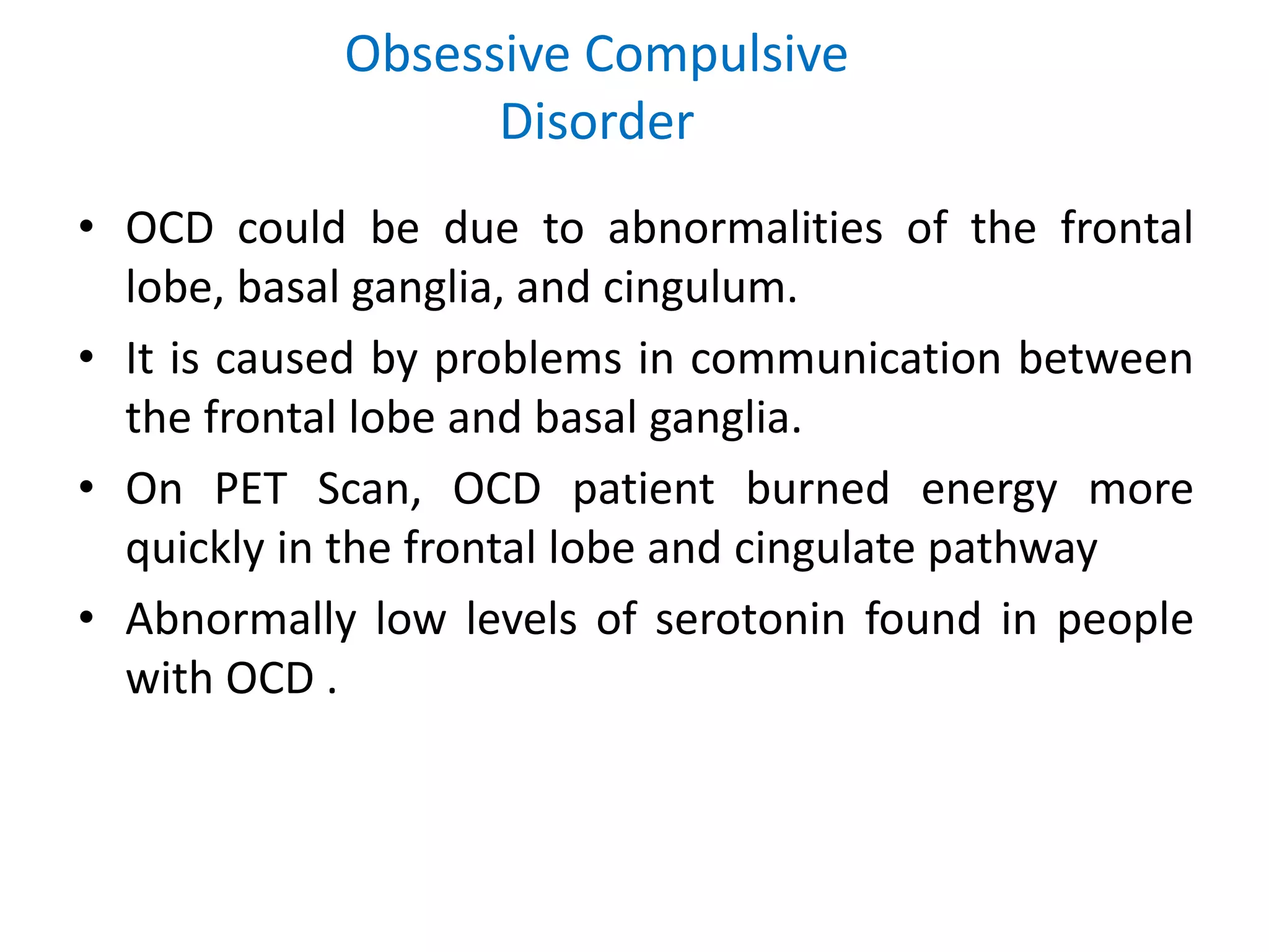
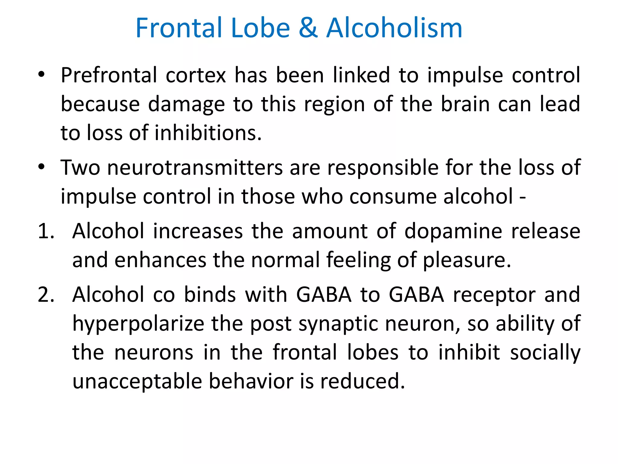
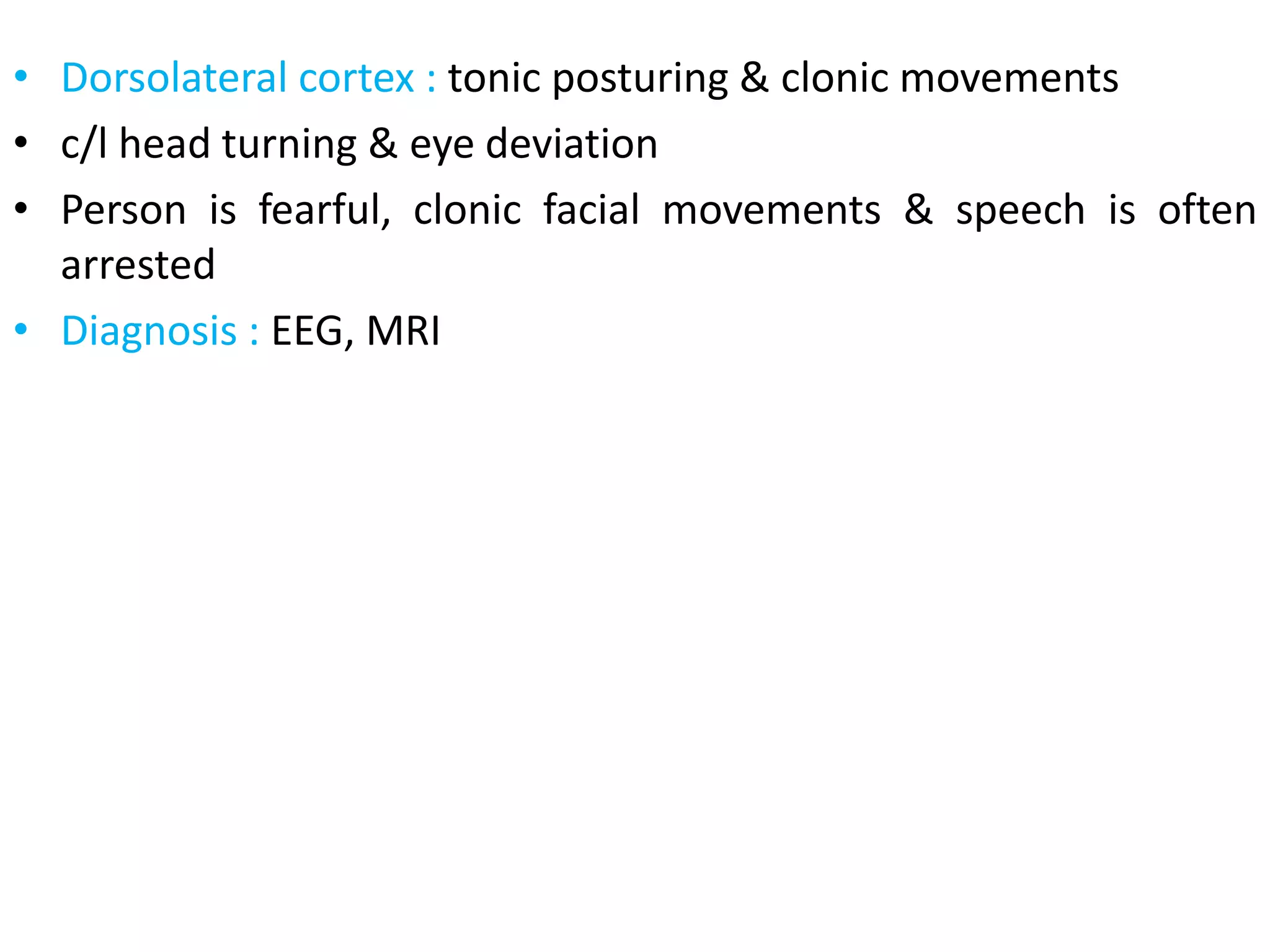
1. Presenter â Dr. Arpit Koolwal
Resident,
Department of Psychiatry
Subharti Medical College,
Meerut
FRONTAL LOBE
Neuroanatomy
And
Neuropsychiatric
Aspects
3. Cerebral Features
⢠Sulci â Small grooves dividing the gyri.
Central Sulcus â Divides the Frontal Lobe from the Parietal Lobe
⢠Fissures â Deep grooves, generally dividing large regions/lobes of the brain
Longitudinal Fissure â Divides the two Cerebral Hemispheres
Transverse Fissure â Separates the Cerebrum from the Cerebellum
Sylvian/Lateral Fissure â Divides the Temporal Lobe from the Frontal and
Parietal Lobes
⢠Gyri â Elevated ridges âwindingâ around the brain.
4. Frontal Lobe
⢠Phylogenetically youngest
part of brain.
⢠Located deep to the frontal
bone of the skull .
⢠Largest of all lobes
â SA: ~1/3 of hemisphere
⢠3 major areas in each lobe
â Dorsolateral aspect
â Medial aspect
â Inferior orbital aspect
8. Functional Anatomy of Frontal Lobe
Lateral sulcus/
Sylvian fissure
Central sulcus
Motor speech
area of Broca
Frontal eye field
B 44, 45
B 9, 10, 11, 12
B 8
Primary motor areaPremotor area
Prefrontal area
B6 B4
Supplementary
motor area
(medially)
Motor cortex
1. Primary
2. Premotor
3. Supplementar
y
4. Frontal eye
field
5. Brocaâs areaPrefrontal cortex
1. Dorsolateral
2. Medial
3. Orbitofrontal
11. ⢠Connections: Temporal, Parietal, Thalamus, GP,
Caudate, SN, Insula, Amygdala.
⢠Function: To mediate empathic, civil and socially
appropriate behavior, emotional input, arousal,
suppression of distracting signals, âdelayed
gratificationâ.
⢠Lesions: Emotional lability, disinhibition,
distractibility, increased risk taking.
Much of the personality changes described in cases of
frontal lobe injury are due to lesions in this area.
12. Tests:
1. Does the patient dress or behave in a way which suggests lack
of concern with the feelings of others or without concern to
accepted social customs.
2. Go/No-Go Test -
âtap once if I tap twice, donât tap if I tap onceâ
It measures a participant's capacity for sustained attention and
response control.
3. Iowa Gambling Task â Simulates real life decision making. Shows
increased risk taking behaviour.
15. ⢠Connections: Thalamus, GP, Caudate, SN
⢠Functions: âExecutive functioningâ including â
1. Working Memory
2. Planning
3. Shifting cognitive sets
4. Reasoning
5. Goal directed behavior
6. Sustaining and shifting attention
7. Abstraction
⢠Lesions: Executive function deficit, apathy, aspontaneity and
impoverished & stereotyped thought process
16. Tests:-
⢠Is the patient able to make an appointment and arrive on time?
⢠Is the patient able to give a coherent account of current problems.
⢠Attention â
1. Digit Repetition Test
2. Serial Sevens Subtraction Test
3. Repeat the days of a week backwards or name of months backwards.
⢠FAS verbal fluency test (Contolled oral word association test) -
produce as many words as possible, in one minute, starting with F,
then A, then S.
⢠Porteus Maze Test â Person is asked to work his way through a maze
and avoid blind alleys.
⢠Concrete thinking or lack of abstraction
â Proverb test
â Similarity test
17. ⢠Inability to plan & execute multistepped behavior is
hallmark of prefrontal lesions
⢠Can manage simple one or two step command
⢠Evaluated by asking the patient & spouse, do things get
started but not completed?
⢠Ask patient about planning a vacation, changing a tyre.
⢠Test of sequential motor & visual patterns
1. Reciprocal Coordination test
2. Fist-Palm-Side Test
3. Visual Pattern Completion Task
18. Wisconsin Card Sorting Test
âPlease sort the 60 cards
under the 4 samples
(stimulus cards).
I wonât tell you the rule, but I
will announce every
mistake.
The rule will change after
10 correct placements.â
Used to assess the following "frontal" lobe functions:
â˘strategic planning,
â˘organized searching,
â˘utilizing environmental feedback to shift cognitive sets,
â˘directing behavior toward achieving a goal
â˘modulating impulsive responding.
Frontal Lobe lesion ------------ Perseverative Errors!!!
19. Stroop Test
Demonstrates interference in the reaction time of a task.
The patient is asked to read the parts as quickly as possible.
Increased interference effect is found in disorders such as brain damage,
dementias, ADHD, Schizophrenia, depression, etc.
This test is considered to measure selective attention, cognitive
flexibility and processing speed, and it is used as a tool in the evaluation
of executive functions.
20. Trail Making Test
A
C1
2
7
3 D
5 B
4
6
It provides information about
â˘visual search speed
â˘scanning,
â˘speed of processing,
â˘mental flexibility, and executive functioning
21. Tower of London Tests
For the assessment of executive functioning, specifically to detect deficits in planning.
23. Primary Motor Cortex
BA4The neurons of the PMC fire about 5-
100 msec before the onset of the
movement.
â Input: Thalamus, BG, Sensory,
Premotor Cortex
â Output: Motor fibers to
brainstem and spinal cord
Function: Encodes the force of
a movement, direction of the
movement, extent and speed of the
movement.
â Lesions:ďtone (spasticity);
ďŻ power; ďŻ fine motor function
on contra lateral side
24. ⢠Tests
1. Motor strength of hand grip.
2. Motor speed on finger tapping.
26. Premotor Cortex
BA6
Input: Thalamus, Basal Ganglia, Sensory cortex
Output: Primary Motor Cortex
Function: Sensorimotor integration, stores motor
programs; controls coarse postural
movements.
Lesions: Moderate weakness in proximal muscles
on contralateral side, spasticity, grasp reflex,
buccofacial apraxia, inability to make use of sensory
feedback in the performance of smooth
movements.
27. ⢠Tests
1. Sensorimotor abilities are tested by asking the
patient touch each finger to the thumb in succession
as rapidly as possible (Watch for speed and dexterity)
2. Buccofacial apraxia can be tested by asking the
patient to "blow a kissâ.
Poor performance carries the diagnostic implications.
28. Supplementary motor area
medial aspect of BA 6
⢠Input: Cingulate Gyrus, Thalamus, Prefrontal Cortex
⢠Output: Premotor Cortex, Primary Motor Cortex
⢠Function: Involved in motivated behavior, initiation
and goal directed behavior, intentional preparation for
movement; procedural memory.
⢠Lesions: Transient Transcortical Motor Aphasia,
impairment in motor initiation ; impaired rapid
alternating movements, Limb Kinetic Apraxia, Alien
Hand Syndrome.
29. Frontal eye fields
BA 8 with some area of 9 & 6
â Input: Parietal / temporal (what is target);
posterior / parietal cortex (where is target)
â Output: Caudate; superior colliculus;
paramedian pontine reticular formation
â Function: Selects target and commands
movement (saccades) â Visually following an
object; locating an object visually.
â Lesion: Eyes deviate ipsilaterally with
destructive lesion and contralaterally with
irritating lesions.
30. ⢠Tests
1. Ask the patient to follow the movement of a finger
from left to right and up and down.
2. Ask the patient to look from left to right, up and
down (with no finger to follow).
Note inability to move or jerky movement.
3. Visual Field Search Test â Visually finding scattered
objects on a piece of paper.
31. Brocaâs speech area
BA 44, 45
â Input: Wernickeâs area
â Output: Primary motor cortex
â Function: Speech production (dominant hemisphere);
emotional melodic component of speech (non-dominant)
â Lesions: Motor aphasia; Dysprosody (monotonus speech)
Speech is sparse, slow, hesitant, disturbance of rhythm and
articulation, difficulty in word finding, wrong words are
chosen & often mispronounced, perseveration,
agrammatism (Brocaâs Aphasia or Expressive Aphasia).
Patient recognize his mistakes & tries to correct them but
becomes impatient.
Phrase length is small :- Telegraphic Language
Writing affected with speech, comprehension is preserved.
32. ⢠Arcuate Fasciculus is white matter tract that connects
Brocaâs Area and Wernickeâs Area through the temporal,
parietal and frontal lobes. Allows for coordinated
comprehensible speech. Damage may result in:
Conduction Aphasia â Auditory comprehension and
speech articulation are preserved but the patient finds it
difficult to repeat heard words.
34. Dopaminergic tracts
â Origin: Ventral tegmental area in midbrain
â Projections: Prefrontal cortex (mesocortical tract)
and to limbic system (mesolimbic tract)
â Function: Reward, motivation, spontaneity,
arousal
35. Norepinephrine tracts
â Origin: Locus ceruleus in brainstem and lateral
brainstem tegmentum
â Projections: Anterior cortex
â Functions: Alertness, arousal, cognitive processing
of somatosensory information
36. Serotonergic tracts
â Origin: Raphe nuclei in brainstem
â Projections: Number of forebrain structures
â Function: Minor role in prefrontal cortex; sleep,
mood, anxiety, feeding
37. Frontal lobe function
Motor Cognitive Behavior Arousal
Voluntary
movements
Working
Memory
Personality Attention
Planning,
Initiation
Problem
solving
Social and
sexual
behavior
Spontaneity Judgment Impulse
control
Language
Expression
Abstract
thinking
Mood and
affect
Eye
movements
39. Five âfrontal subcortical circuitsâ
1. Motor
2. Oculomotor
3. Dorsolateral prefrontal
4. Orbitofrontal
5. Anterior cingulate
Prefrontal cortex
Motor cortex
40. Frontal Subcortical Motor Circuit
⢠Neurophysiologic investigations of movement demonstrate
preparatory premovement activity, serial processing of movements
initiated in the cortex, and concurrent parallel processing in the
structures of the circuit.
SMA,
Premotor,Motor
Putamen
Globus PallidusThalamus
Hypo-thalamus
41. Frontal Oculomotor Circuit
⢠Voluntary scanning eye movement
⢠Independent of visual stimuli
Frontal
Eye field
Central
Caudate
Globus Pallidus
&
Substantia Nigra
Thalamus
42. Dorsolateral Prefrontal Circuit
⢠Executive functions: Planning, working memory, goal directed
behavior, attention span.
⢠Lesions lead to Executive deficits - Difficulty focusing and sustaining
attention, generating hypothesis, and maintaining or shifting sets.
⢠Depression has been associated with lesions of the dorsolateral
prefrontal cortex and caudate nucleus, particularly when the left
hemisphere is affected.
Dorsolateral Pre-
Frontal Cortex
Caudate
Globus Pallidus &
Substantia Nigra
Thalamus
43. Orbito-frontal Circuit
⢠Disinhibiton, inattention, distractibility, and increased motor
activity may be seen, and hypomania or mania is not
uncommon.
⢠PET scan in Huntington's disease and Parkinson's disease reveal
diminished metabolism in the orbitofrontal cortex in depressed
compared with non-depressed patients.
Orbito-frontal
Cortex
Caudate
Globus Pallidus &
Substantia Nigra
Thalamus
45. ⢠As the CNS matures, frontal lobe cells develop and
begin to inhibit the primitive reflexes which are present
in normal babies.
⢠These may reappear with brain damage or disease.
⢠However, they may also reappear with normal aging.
⢠Their significance is greater when they appear
unilaterally and in young individuals (Ross, 1985).
1. Grasp Reflex
2. Sucking Reflex (pout, snout)
3. Palmo-mental Reflex
4. Glabellar Reflex
Frontal Release Reflexes
46.
47. â˘Frontal lobe syndrome (FLS) reflects damage to
the prefrontal regions of the frontal lobe. It is
characterised by deterioration in behaviour and
personality in a previously normal individual.
Anterior Location of the Prefrontal region---------
------damage missed on neurological
examination!!.
48. ⢠A 45 year old man with b/l prefrontal strokes was
found to have
â Normal neurological examination
â Slightly flattened affect
â Lack of spontaneity, mental slowness
â Increased left sided motor tone
â Neuropsychological testing â normal intelligence &
memory
â Demoted at his managerial jobs d/t ineffective work habits
â Unable to adequately supervise children
â Often lost his temper
â Inattention
â Bad judgment
49. ⢠CLINICAL PICTURE : Varies among patiens.
Individual features depend on -----
1. Nature & time course of pathological process.
2. Lateralization
3. Localization
4. Extent of involvement among subcortical & callosal
fibers
5. Secondary effect of raised ICT
50. ⢠Kliest first suggested that components of frontal lobe syndrome
may be related to specific regional involvement ------
⢠Orbital lesions cause : Disinhibition, failure to appreciate
consequences of oneâs action, euphoria (effect on personality
& social behavior).
⢠Lesions of dorsolateral convexity cause : Apathy, aspontaneity,
impoverished & stereotyped thought process.
⢠Left prefrontal injury : loss of executive & planning function,
depression.
⢠When supplementary/ premotor area affected : transcortical
motor aphasia, impairment of rapid skilled manual
movements.
⢠Right prefrontal injury : left sided extinction & neglect,
blunted or labile affect, impersistence, disinhibition,
confabulation, alien hand syndrome.
51. ⢠Negative symptoms :
1. Lack of initiative & spontaneity
2. General diminution of motor activity (sluggish response)
3. Task are left unfinished
4. New initiatives are rarely undertaken
5. Capacity to function independently in life is affected
6. Cognition & intellect may remain unaffected
7. Yet when vigorously urged or constrained by structural
situation pt may function quite well
52. ⢠Other pts may show positive symptoms
1. Restless
2. Hyperactive yet lack of goal directed behavior
3. Mild euphoria
4. Tendency to joke/pun
5. State of excitement, pressured speech
6. Overfamilarity
7. Outburst of irritability
⢠Such changes are rarely sustained and when left to
themselves these patients become inert & apathetic.
53. ⢠Social awareness & behavior
1. Less concerned with consequences of his acts
2. Loss of social graces
3. Coarsening of personality
4. Lack of normal adult tact & restraints
5. Little concern about his future
6. Fails to plan ahead, to carry out ideas
7. Sexual disinhibition
Patient usually has little insight into the changes.
54. ⢠Abulia : Poverty of thought, action & emotion is
common with large midline and b/l dorsofrontal
lesion
⢠Abulia is characterized by loss of spontaneity & will
power.
⢠They comprehend the question, hesitate, delay
respond, seem to ignore or give yes â no answer.
⢠Severely abulic patient do not speak unless spoken, do
not move unless they are hungry or ready to void &
may be incontinent.
⢠âTidal wavesâ of emotional & motor behavior (brief
rage, irritability, hyperactivity) may emerge from
âtranquil seaâ of abulia (placid, apathetic,
disinterested).
55. Phineas Gage (1848)
On 13th Sept 1848 a railroad
worker who is a hard
working, diligent, reliable,
responsible, intelligent,
good humored, polite god
fearing, family oriented man
Following an explosion, an
iron bar drove into his
frontal lobe andâŚâŚ
56. He becomes....
1. Unreliable and fails to come to work and when present
he is "lazy."
2. Lost interest in going to church, constantly drinks
alcohol, gambles, and "whores about."
3. Accused of sexually molesting young children.
4. Ignores his wife and children and fails to meet his
financial and family obligations.
5. Lost his sense of humour.
6. Curses constantly and does so in inappropriate
circumstances.
It was said that âGage isnât Gage anymoreâ.
He died of status epilepticus in 1861.
57. Utilisation Behavior (Lhermitte 1986)
⢠Observed patient with frontal lobe lesion in complex
everyday life situation.
⢠Patient has lack of personal autonomy with an
excessive dependence on social & physical
environment (environmental dependency syndrome).
⢠When patient is given common objects he tries to use
them out of context.
58.
59. Schizophrenia
⢠Symptoms can be aggregated in 3 broad clusters----
1. Psychomotor poverty syndrome
Affecting speech & movement, blunting of affect
Decreased rCBF in left prefrontal & parietal cortex
60. 2. Reality distortion syndrome
Positive symptoms hallucinations & delusions
Increase rCBF in left parahippocampal gyrus & contiguous
area
3. Disorganization syndrome
Thought disorder & inappropriate affect
Increase resting rCBF in anterior cingulate region
61. Depression
⢠Right frontal lobe demonstrated increased activity in response
to negative moods whereas left frontal activity decreases.
⢠Injuries to the left frontal lobe have been consistently
associated with depression, psycho-motor retardation,
apathy, irritability, and blunted mental functioning.
⢠In severely depressed patients demonstrate insufficient
activation and a significant lower integrated amplitude of the
EEG evoked response over the left vs. right frontal lobe.
62. Attention Deficit Hyperactivity
Disorder
⢠ADHD suffers usually have deficits in the executive
functions.
⢠Right frontal lobe is smaller in children with ADHD.
⢠Problems in the circuit between three regions are
considered the underlying mechanisms that cause
ADHD symptoms -----
1. Prefrontal cortex
2. Caudate nucleus
3. Globus pallidus
63. Obsessive Compulsive
Disorder
⢠OCD could be due to abnormalities of the frontal
lobe, basal ganglia, and cingulum.
⢠It is caused by problems in communication between
the frontal lobe and basal ganglia.
⢠On PET Scan, OCD patient burned energy more
quickly in the frontal lobe and cingulate pathway
⢠Abnormally low levels of serotonin found in people
with OCD .
64. Frontal Lobe & Alcoholism
⢠Prefrontal cortex has been linked to impulse control
because damage to this region of the brain can lead
to loss of inhibitions.
⢠Two neurotransmitters are responsible for the loss of
impulse control in those who consume alcohol -
1. Alcohol increases the amount of dopamine release
and enhances the normal feeling of pleasure.
2. Alcohol co binds with GABA to GABA receptor and
hyperpolarize the post synaptic neuron, so ability of
the neurons in the frontal lobes to inhibit socially
unacceptable behavior is reduced.
65. Frontal Lobe Epilepsy
⢠2nd most common type of epilepsy.
⢠Brief recurring seizures often while the patient is sleeping,
⢠Two forms :-
⢠Simple partial seizures : does not affect awareness & memory.
⢠Complex partial seizures : affects awareness & memory.
⢠Symptoms :-
⢠Physical/emotional aura of tingling, numbness, tension. Fear
expressed on face.
⢠Tonic posturing & clonic movements.
⢠Often misdiagnosed as psychogenic seizures.
⢠More specific symptoms depends on area of frontal cortex
involved.
66. ⢠Supplementary motor area : Somatosensory aura precedes
tonic posturing which is u/l, asymmetrical.
⢠Motor symptoms :- facial grimacing, complex automatism like
kicking, pelvic thrusting.
⢠Vocal symptoms :- laughing, yelling or speech arrest.
⢠Primary motor cortex : Jacksonian seizures that spread to
adjacent area, often triggers to 2nd round of seizures
⢠Usually tonic, myoclonic movements with speech arrest.
⢠Medial frontal, Cingulate gyrus, Orbitofrontal, Frontopolar region :
⢠Short repetitive thrashing, pedaling, thrusting, laughing,
screaming, crying.
⢠Motor symptoms are accompanied by emotional feelings &
viscerosensory symptoms.
⢠Often misdiagnosed as psychological seizures.
67. ⢠Dorsolateral cortex : tonic posturing & clonic movements
⢠c/l head turning & eye deviation
⢠Person is fearful, clonic facial movements & speech is often
arrested
⢠Diagnosis : EEG, MRI
68. Frontal Lobe & Memory
⢠Focal frontal injury does not produce a severe
amnesic disorder.
⢠Often the patients with frontal lobe injury score
normal on standard memory tests.
⢠Memory deficits in form of an impairment in the
Working Memory.
⢠Prefrontal cortex appears to be crucial for the
monitoring and control of memory processes, both
at the time of encoding and at the time of retrieval.