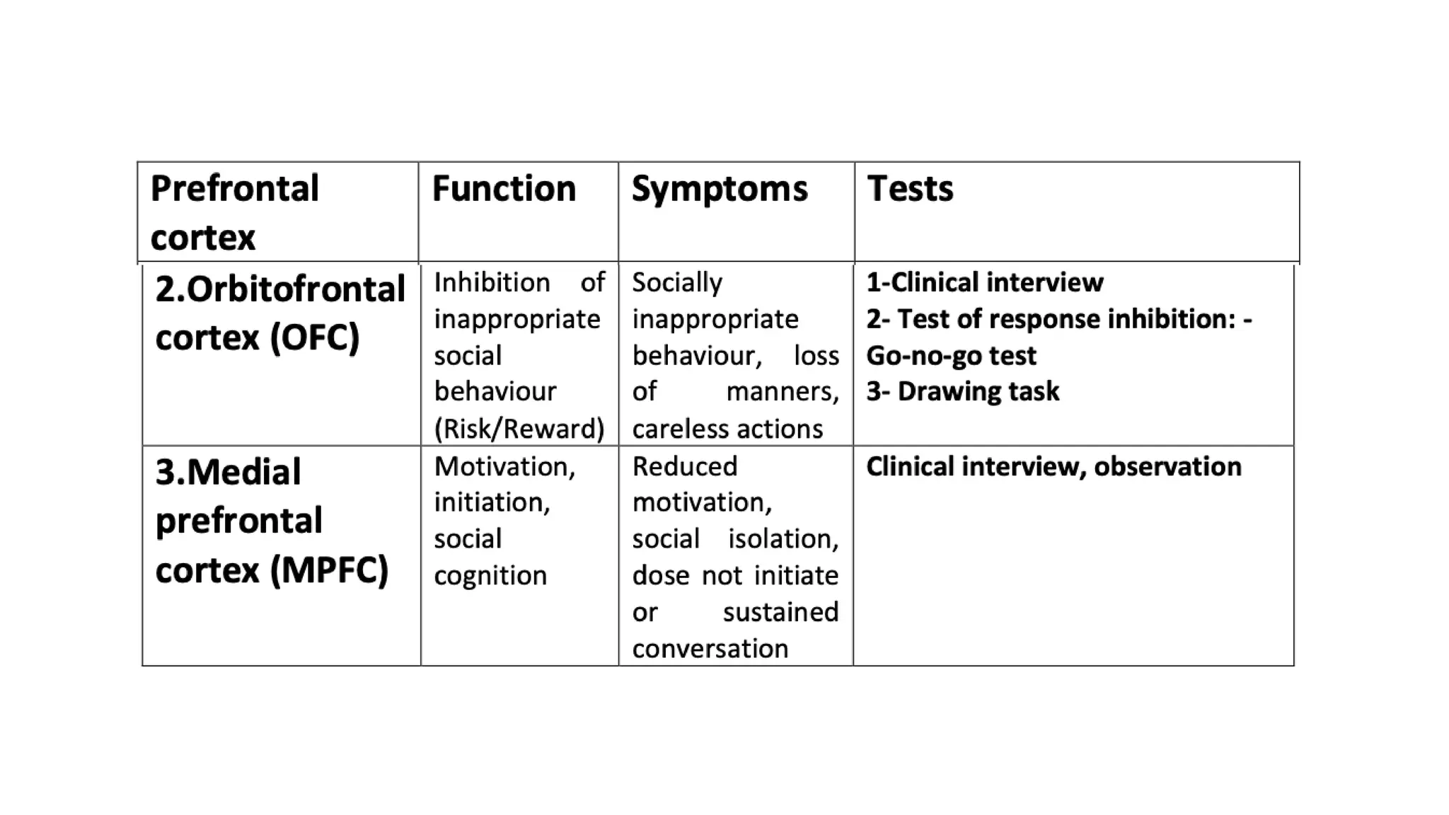
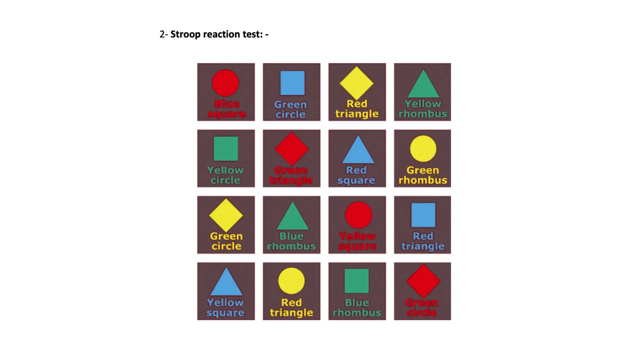
FRONTAL
LOBE
ANATOMY
AND SIGNS
PRESENTOR-DR CHARAN
KOLLANUR
MODERATOR- DR SUMIT
BANSAL
• Largest lobe of the brain
• Occupy 1/3rd
of Cerebral Hemisphere.
• larger in humans than in any other primates.
Location and Structure
• Largely in the anterior cranial fossa, lying on the orbital plate of
the frontal bone.
• Located at front of each cerebral hemisphere and
• positioned anterior to the parietal lobe &
• Antero-superior to temporal lobe.
• Extent:
• Central sulcus (posterior border)
• Lateral sulcus/ Sylvian fissure(Inferior border)
• Anterior most part- frontal pole
SURFACES
1. Lateral surface
2. Medial (Interhemispheric) surface
3. Inferior (Frontobasal/Orbital) surface
1. Lateral surface:
a. Pre-central gyrus
b. Superior frontal gyrus
c. Middle frontal gyrus
d. Inferior frontal gyrus
a. Precentral gyrus:
• Posterior most part of frontal lobe
• Between central and Precentral sulcus
• Contain : Primary motor cortex (Brodman’s area 4)
Premotor area/cortex (Brodman’s area 6)
Supplemental motor cortex
c. Inferior frontal gyrus:
Large part of antero-lateral prefrontal cortex
• Pars opercularis- Most dorsal vertically oriented
• Pars triangularis
• Pars orbitalis
Large part of antero-lateral prefrol
oriented
• Pars triangularis
• Pars orbitalis
2. Medial (Interhemispheric) surface:
a. Paracentral lobule
b. Medial extension of superior frontal gyrus
3. Inferior(Frontobasal/ Orbital) surface:
Lying on the floor of ACF
a. Straight gyrus (Gyrus recti)
b. Orbital gyrus-
a. Anterior& Posterior- Superiorly
b. Medial and lateral - Inferiorly
(separated by Olfactory sulcus
=olfactory tract and bulb)
Functional anatomy of frontal lobe
• The primary motor area.
• The premotor Area.
• The supplementary motor areas.
• The prefrontal region.
• The frontal eye fields.
• The motor speech areas.
Primary motor cortex :
• Integrate signals and modulate motor function
• Pyramidal cells from layer 5 -> Betz cells –> form corticospinal tract
and corticobulbar tracts.
• These are represented by Motor homunculus
oInput: Thalamus, Basal ganglia, sensory, premotor areas
oOutput: Motor fibers to brainstem and spinal cord
oFunction: Executes design into movement.
Pre motor cortex (Brodmann’s area 6):
Anterior part of Precentral gyrus (broader superiorly and narrows down
inferiorly).
Has no giant pyramidal (Betz) cells.
• Input: Thalamus, Basal ganglia, sensory cortex
• Output: Primary motor cortex
• Function: sensorimotor integration, stores motor programs
• Lesions: Inability to make use of sensory feedback in performance of smooth
movements and apraxia
Supplementary motor area:
• Located in medial surface of medial frontal gyrus on medial surface
anterior to paracentral lobule
• Input: Cingulate gyrus, Thalamus, sensory & prefrontal cortex
• Output: Premotor, primary motor cortex
• Function: Intentional preparation for movement; procedural memory: recall of
memorized motor sequences
• Lesions: Mutism, contralateral motor neglect, impairment of bi brachial
coordination.
Frontal eyefield (broadmann’s area 6, 8, 9):
• In middle frontal gyrus rostral to premotor region.
Input: Parietal / temporal (what is target & where is target)
Output: caudate; superior colliculus; paramedian pontine reticular
formation
Function: it is considered to control voluntary scanning(horizontal con
jugate) movements of the eye.
Lesion: Eyes deviate ipsilaterally with destructive lesion and
contralaterally with irritating lesions.
Broca’s Speech area (Broadmann’s area 44) :
• In inferior frontal gyrus on dominant hemisphere (triangular
and opercular parts).
Input: Wernicke’s area
Output: primary motor cortex
Function: speech production (dominant hemisphere); emotional,
melodic component of speech (non-dominant)
Lesions: motor aphasia.
Prefrontal area :
• Most anterior parts of frontal lobe (areas 9-12 & 45-47).
• Organ of civilization
• Receprocal connections to
• Anterior and dorsal nuclei of Thalamus
• Sensory processing areas of Posterior association
cortex.
• Limbic system (Hypothalamus, Amygdala and
Hippocanpus.
• Efferent to
• Subcortical structures: Basal ganglia, Cerebellum
and Thalamus
Pre frontal cortex divided into:
• Dorsolateral prefrontal cortex (DLPFC),
• Medial prefrontal cortex (MPC), and
• Orbitofrontal cortex (OFC)
Dorsolateral prefrontal cortex (DLPFC),
• Organization of self- ordered tasks.
• It plays a critical role in the working memory .
• The responsibility for executive function largely resides with the
DLPFC and its connections.
• Frontal lobe executive function is the ability to plan, carry out, and
monitor a series of actions intended to accomplish a goal.
• Lesions: executive function deficit; disinterest apathy, decreased
attention to relevant stimuli.
Medial prefrontal cortex (MPC)
Lesion causes:
• Paucity of spontaneous movements,
• sparse verbal output,
• lower extremity weakness and
• incontinence.
Orbitofrontal cortex (OFC)
• connections with the limbic system, including the amygdala.
• Lesion/dysfunction(mainly right sided): Disinhibition syndromes
 Mildly inappropriate social behavior to full-blown mania
Blood supply
• Medial parts of frontal lobe:
Anterior cerebral artery
• Convexity and deep regions:
Superior (rolandic) division of
MCA
• Underlying deep white
matter: series of small
penetrating arteries
(lenticulostriate) directly from
stem of MCA
Frontal subcortical circuits
Five FSCs have been described: Goldman Rakic
1.Motor
2.Oculomotor
3.Dorsolateral prefrontal
4.Lateral orbitofrontal
5.Anterior cingulate
• All FSC circuits have both a direct and an indirect pathway, which have
“opposite” or reciprocal functions.
Motor circuit
Oculomotor circuit
Dorso lateral prefrontal circuit
This circuit is responsible for executive functions.
•motor planning, deciding which stimuli to attend to, shifting cognitive sets
•Attention span and working memory
•Lesion: difficulty focusing and sustaining attention as well as reduced verbal fluency and
motor programming
Orbito frontal circuit
• This circuit mediates empathic, civil and socially
appropriate behavior.
• Lesion: Disinhibition and emotional liability
Anterior Cingulate circuit
• This circuit is involved in motivational mechanisms. •
• Lesions: Abulia, akinetic mutism.
LOBAR FUNCTION TESTS
A. Prefrontal cortex: -
3- Go- no- go test: -
“Tap once when I tap once”
“Do not tap when I tap twice.”
B. Primary motor cortex: -
• Motor power examination
C. Supplementary motor cortex / Cingulate cortex: -
Gait apraxia: - Loss of ability to properly use the lower limbs in the act of walking
which cannot be accounted for by demonstrable sensory impairment or motor
weakness.
• Dysfunction in the cingulate/SMA are associated with several uniquely bizarre
characteristics,
- apathy and akinetic mutism (reflecting a loss of drive
and motivation)
-complex attentional deficits and
-delayed habituation to external stimuli
D. Premotor cortex:
- Limb kinetic apraxia: - Loss of co-ordination between fine
skilled fingers movement.
➢ Ask patient to oppose their thumb to their index, middle,
ring and little fingers rapidly in succession
➢ Picking up a coin.
D. Frontal eye field: -
• Damage to the frontal eye fields will cause deficits in voluntary
eye movement to the contralateral visual field (leading to active
visual search deficits),
• but preserved passive eye movement (as in the following of a
moving object)
E. Frontal lobe release reflexes: -
a. Grasp reflex: -
Elicited by distally moving deep pressure stimulus over palmar aspect of
hand between thumb and index finger.
Two phases; Catching and holding phase.
Cingulate and SMA lesion are implicated in the aetiology of these responses.
b. Utilization behaviour: -
Tested by presenting the patient with a series of spectacle, which he
proceeds to put on his nose, one on top of themother. Can tested with
several shirts also.
c. Sucking reflex: -Place object (Spatula) in the mouth.
d. Tactile Rooting reflex: - Elicited by moving a finger or
handle of knee
hammer drawn across the lower or upper lips in a lateral
direction.
e. Snout reflex: -
Gentle pressure over the nasal philtrum; puckering of the lips
f. Pout reflex: -
Tapping around the mouth or the lips.  exaggerated myotactic
stretch reflex, not a true release reflex.
 Indicates damage to B/L corticobulbar fibres above 7th cranial
nerve nucleus.
g. Palmomental reflex: -
Slow contraction of I/L, C/L, B/L mentalis muscle in response to
stroking the thenar eminence from proximal to distally.
h. Glabellar tap reflex: -
Tapping the glabella from behind at a frequency of 2-3/sec.
Habituation usually occurs after 3rd tap. Positive glabella tap;
Failure of habituation.
i. Corneo-mandibular reflex: -
Horizontal movement of mandible to the C/L side on touching
the cornea of I/L eye.
Using a cutoff score of 12/18 can reasonably differentiate FTD from mild Alzheimer's (sensitivity of 77% and specificity of
87%)

Frontal lobe anatomy and clinical application.pptx

  • 1.
  • 2.
    • Largest lobeof the brain • Occupy 1/3rd of Cerebral Hemisphere. • larger in humans than in any other primates.
  • 3.
    Location and Structure •Largely in the anterior cranial fossa, lying on the orbital plate of the frontal bone. • Located at front of each cerebral hemisphere and • positioned anterior to the parietal lobe & • Antero-superior to temporal lobe. • Extent: • Central sulcus (posterior border) • Lateral sulcus/ Sylvian fissure(Inferior border) • Anterior most part- frontal pole
  • 4.
    SURFACES 1. Lateral surface 2.Medial (Interhemispheric) surface 3. Inferior (Frontobasal/Orbital) surface
  • 5.
    1. Lateral surface: a.Pre-central gyrus b. Superior frontal gyrus c. Middle frontal gyrus d. Inferior frontal gyrus
  • 7.
    a. Precentral gyrus: •Posterior most part of frontal lobe • Between central and Precentral sulcus • Contain : Primary motor cortex (Brodman’s area 4) Premotor area/cortex (Brodman’s area 6) Supplemental motor cortex
  • 8.
    c. Inferior frontalgyrus: Large part of antero-lateral prefrontal cortex • Pars opercularis- Most dorsal vertically oriented • Pars triangularis • Pars orbitalis Large part of antero-lateral prefrol oriented • Pars triangularis • Pars orbitalis
  • 9.
    2. Medial (Interhemispheric)surface: a. Paracentral lobule b. Medial extension of superior frontal gyrus
  • 11.
    3. Inferior(Frontobasal/ Orbital)surface: Lying on the floor of ACF a. Straight gyrus (Gyrus recti) b. Orbital gyrus- a. Anterior& Posterior- Superiorly b. Medial and lateral - Inferiorly (separated by Olfactory sulcus =olfactory tract and bulb)
  • 13.
    Functional anatomy offrontal lobe • The primary motor area. • The premotor Area. • The supplementary motor areas. • The prefrontal region. • The frontal eye fields. • The motor speech areas.
  • 14.
    Primary motor cortex: • Integrate signals and modulate motor function • Pyramidal cells from layer 5 -> Betz cells –> form corticospinal tract and corticobulbar tracts. • These are represented by Motor homunculus oInput: Thalamus, Basal ganglia, sensory, premotor areas oOutput: Motor fibers to brainstem and spinal cord oFunction: Executes design into movement.
  • 17.
    Pre motor cortex(Brodmann’s area 6): Anterior part of Precentral gyrus (broader superiorly and narrows down inferiorly). Has no giant pyramidal (Betz) cells. • Input: Thalamus, Basal ganglia, sensory cortex • Output: Primary motor cortex • Function: sensorimotor integration, stores motor programs • Lesions: Inability to make use of sensory feedback in performance of smooth movements and apraxia
  • 18.
    Supplementary motor area: •Located in medial surface of medial frontal gyrus on medial surface anterior to paracentral lobule • Input: Cingulate gyrus, Thalamus, sensory & prefrontal cortex • Output: Premotor, primary motor cortex • Function: Intentional preparation for movement; procedural memory: recall of memorized motor sequences • Lesions: Mutism, contralateral motor neglect, impairment of bi brachial coordination.
  • 19.
    Frontal eyefield (broadmann’sarea 6, 8, 9): • In middle frontal gyrus rostral to premotor region. Input: Parietal / temporal (what is target & where is target) Output: caudate; superior colliculus; paramedian pontine reticular formation Function: it is considered to control voluntary scanning(horizontal con jugate) movements of the eye. Lesion: Eyes deviate ipsilaterally with destructive lesion and contralaterally with irritating lesions.
  • 20.
    Broca’s Speech area(Broadmann’s area 44) : • In inferior frontal gyrus on dominant hemisphere (triangular and opercular parts). Input: Wernicke’s area Output: primary motor cortex Function: speech production (dominant hemisphere); emotional, melodic component of speech (non-dominant) Lesions: motor aphasia.
  • 21.
    Prefrontal area : •Most anterior parts of frontal lobe (areas 9-12 & 45-47). • Organ of civilization • Receprocal connections to • Anterior and dorsal nuclei of Thalamus • Sensory processing areas of Posterior association cortex. • Limbic system (Hypothalamus, Amygdala and Hippocanpus. • Efferent to • Subcortical structures: Basal ganglia, Cerebellum and Thalamus
  • 22.
    Pre frontal cortexdivided into: • Dorsolateral prefrontal cortex (DLPFC), • Medial prefrontal cortex (MPC), and • Orbitofrontal cortex (OFC)
  • 23.
    Dorsolateral prefrontal cortex(DLPFC), • Organization of self- ordered tasks. • It plays a critical role in the working memory . • The responsibility for executive function largely resides with the DLPFC and its connections. • Frontal lobe executive function is the ability to plan, carry out, and monitor a series of actions intended to accomplish a goal. • Lesions: executive function deficit; disinterest apathy, decreased attention to relevant stimuli.
  • 24.
    Medial prefrontal cortex(MPC) Lesion causes: • Paucity of spontaneous movements, • sparse verbal output, • lower extremity weakness and • incontinence.
  • 25.
    Orbitofrontal cortex (OFC) •connections with the limbic system, including the amygdala. • Lesion/dysfunction(mainly right sided): Disinhibition syndromes  Mildly inappropriate social behavior to full-blown mania
  • 26.
    Blood supply • Medialparts of frontal lobe: Anterior cerebral artery • Convexity and deep regions: Superior (rolandic) division of MCA • Underlying deep white matter: series of small penetrating arteries (lenticulostriate) directly from stem of MCA
  • 28.
    Frontal subcortical circuits FiveFSCs have been described: Goldman Rakic 1.Motor 2.Oculomotor 3.Dorsolateral prefrontal 4.Lateral orbitofrontal 5.Anterior cingulate • All FSC circuits have both a direct and an indirect pathway, which have “opposite” or reciprocal functions.
  • 30.
  • 32.
  • 33.
    Dorso lateral prefrontalcircuit This circuit is responsible for executive functions. •motor planning, deciding which stimuli to attend to, shifting cognitive sets •Attention span and working memory •Lesion: difficulty focusing and sustaining attention as well as reduced verbal fluency and motor programming
  • 34.
    Orbito frontal circuit •This circuit mediates empathic, civil and socially appropriate behavior. • Lesion: Disinhibition and emotional liability
  • 35.
    Anterior Cingulate circuit •This circuit is involved in motivational mechanisms. • • Lesions: Abulia, akinetic mutism.
  • 36.
  • 37.
  • 41.
    3- Go- no-go test: - “Tap once when I tap once” “Do not tap when I tap twice.”
  • 53.
    B. Primary motorcortex: - • Motor power examination
  • 54.
    C. Supplementary motorcortex / Cingulate cortex: - Gait apraxia: - Loss of ability to properly use the lower limbs in the act of walking which cannot be accounted for by demonstrable sensory impairment or motor weakness. • Dysfunction in the cingulate/SMA are associated with several uniquely bizarre characteristics, - apathy and akinetic mutism (reflecting a loss of drive and motivation) -complex attentional deficits and -delayed habituation to external stimuli
  • 55.
    D. Premotor cortex: -Limb kinetic apraxia: - Loss of co-ordination between fine skilled fingers movement. ➢ Ask patient to oppose their thumb to their index, middle, ring and little fingers rapidly in succession ➢ Picking up a coin.
  • 56.
    D. Frontal eyefield: - • Damage to the frontal eye fields will cause deficits in voluntary eye movement to the contralateral visual field (leading to active visual search deficits), • but preserved passive eye movement (as in the following of a moving object)
  • 57.
    E. Frontal loberelease reflexes: - a. Grasp reflex: - Elicited by distally moving deep pressure stimulus over palmar aspect of hand between thumb and index finger. Two phases; Catching and holding phase. Cingulate and SMA lesion are implicated in the aetiology of these responses. b. Utilization behaviour: - Tested by presenting the patient with a series of spectacle, which he proceeds to put on his nose, one on top of themother. Can tested with several shirts also.
  • 58.
    c. Sucking reflex:-Place object (Spatula) in the mouth. d. Tactile Rooting reflex: - Elicited by moving a finger or handle of knee hammer drawn across the lower or upper lips in a lateral direction.
  • 59.
    e. Snout reflex:- Gentle pressure over the nasal philtrum; puckering of the lips f. Pout reflex: - Tapping around the mouth or the lips.  exaggerated myotactic stretch reflex, not a true release reflex.  Indicates damage to B/L corticobulbar fibres above 7th cranial nerve nucleus. g. Palmomental reflex: - Slow contraction of I/L, C/L, B/L mentalis muscle in response to stroking the thenar eminence from proximal to distally.
  • 60.
    h. Glabellar tapreflex: - Tapping the glabella from behind at a frequency of 2-3/sec. Habituation usually occurs after 3rd tap. Positive glabella tap; Failure of habituation. i. Corneo-mandibular reflex: - Horizontal movement of mandible to the C/L side on touching the cornea of I/L eye.
  • 61.
    Using a cutoffscore of 12/18 can reasonably differentiate FTD from mild Alzheimer's (sensitivity of 77% and specificity of 87%)