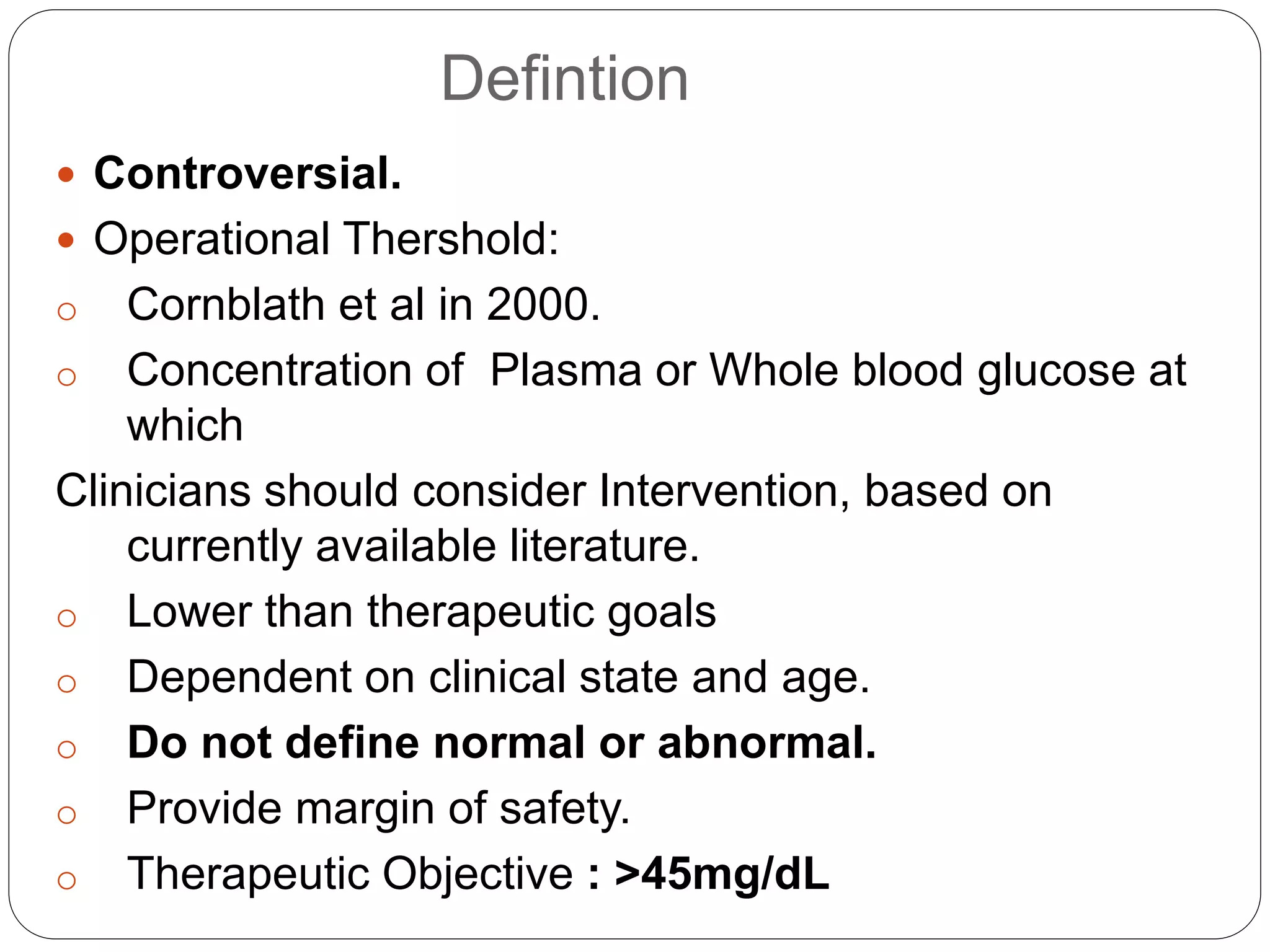
Neonatal Hypoglycemia
Defintion
 Controversial.
 Operational Thershold:
o Cornblath et al in 2000.
o Concentration of Plasma or Whole blood glucose at
which
Clinicians should consider Intervention, based on
currently available literature.
o Lower than therapeutic goals
o Dependent on clinical state and age.
o Do not define normal or abnormal.
o Provide margin of safety.
o Therapeutic Objective : >45mg/dL
OPERATIONAL THRESHOLDS:
Clinical condition Blood glucose level
1 a) Healthy <24hrs first episode
b) Healthy <24hrs persistent
30 to 35mg/dL
45mg/dL
2 Healthy > 24hrs 45 to 50mg/dL
3 Asymptomatic neonate with risk factors 36/dL
4 Neonate with abnormal signs and
symptoms
45mg/dL.
5 Any baby < 20 to 25mg/dL
 Whipple’s Triad:
1. Low Blood Glucose Level.
2. Symptoms of Hypoglycemia at the time of low
glucose level.
3. Symptom relief at the time of Hypoglycemia with
intervention.
Etiology
 Hyperinsulinemic hypoglycemia
 Large for gestational age infants.
 Decreased Production/Stores.
 Increased utilization/ Decreased production.
Hyperinsulinemic Hypoglycemia
 Maternal Hyperglycemia.
 Congenital Genetic: Mutation of genes- ABCC8,
KCNJ11, HNF4A gene.
 Secondary to following conditions:
1. Birth Asphyxia
2. Beckwith- Wiedemann syndrome
3. Abrupt cessation of high glucose infusion.
4. Insulin producing tumors.
5. Erythroblastosis(hyperplastic islets of
langerhans)
6. Malpositioned umblical artery.
Decreased Production/Stores
1. Prematurity.
2. Intrauterine growth restriction.
3. Inadequate caloric intake.
4. Delayed onset of feeding.
Increased utilization/ Decreased production
 Perinatal Stress:
Sepsis
Shock
Asphyxia
Hypothermia
Respiratory Distress
Postresuscitation.
 Defects in Carbohydrate metabolism:
Glycogen storage disease.
Frutose intolerance.
Galactosemia
 Polycythemia
 Maternal Therapy with BetaBlockers.
Increased utilization/ Decreased production
 Endocrine Deficiency:
Adrenal Insufficiency.
Hypothalamic Deficiency.
Congenital Hypopituitarism.
Glucagon Deficiency.
Epinephrine Deficiency.
 Defects in Amino acid metabolism:
Maple Syrup Urine disease.
Propionic acidemia.
Methylmalonic acidemia.
Tyrosinemia.
Glutaric acidemia Type 2.
Ethylmaloni adipic aciduria.
Indication for Routine Blood Glucose Screening
1. Infants <2000grams.
2. Infants <_35 weeks.
3. Small for Gestational Age.
4. Large for Gestational Age.
5. Infant of Diabetic Mother.
6. Infants with Rh Hemolytic disease.
7. Mothers receiving therapy with
Terbutaline/Propanolol/Lebatolol/Oral
Hypoglycemic agents
8. IUGR.
9. Any sick neonate (Perinatal
asphyxia/Polycythemia/Sepsis/Shock)
10. Infants on Parenteral Nutrition.
Schedule of Blood Glucose
Monitoring
Category of Infants Time Schedule
At Risk Infants( 1 to 8) 2, 6, 12, 24, 48 and 72
Hours
Sick Infants ( Infants with
Sepsis, Asphxia, Shock)
Every 6 to 8 hours.
Stable VLBW infants on
Parenteral Nutrition
Initial 72 hours: Every 6 to 8
hrs.
After 72 hours: Once a day.
Methods of Blood Glucose
Estimation
I. Point of Care Glucose Meter:
 Glucose Oxidase method.
 1st Generation: Change in colour by enzymatic
reaction on application of blood drop.
Read by Reflectance meters.
Affected by Hematocrit values, acidosis,
Bilirubin
 Newer Generation: Generate current on reaction of
glucose with enzymes such Glucose Oxidase or
Glucose Dehydrogenase
Amount of Current is proportional to
Amount of sugar in plasma
 Whole blood 10-15% < Plasma Value.
Methods of Glucose Estimation
II. Lab Diagnosis:
 Glucose Oxidase Method.
 Glucose Eletrode Method.
 Prompt analysis should be preformed –
Reduction of Glucose concentrate upto 18
mg/dL/hr.
III. Continous Glucose Monitoring:
 Detects using a Sensor.
 Detects many more episodes of low glucose
concentration than blood glucose
measurement.
Clinical Manifestations
 Asymptomatic.
 Neurogenic Symptoms:
Jitteriness/Tremors
Sweating.
Irritability.
Tachypnea.
Pallor.
 Neuroglycopenic Symptoms:
Poor suck/Poor feeding.
Weak or high pitched cry.
Change in Level of Consciousness.
Seizures.
Hypotonia.
 Apnea, Bradycardia, Cyanosis, Hypothermia.
Management of Neonatal
Hypoglycemia
Hypoglycemia
BGL < 40mg/dL
Asymptomatic Symptomatic
Asymptomatic Hypoglycemia
20 to
<40mg/dL
<20mg/dL
Follow Symptomatic
Hypoglycemia protocol
Trial of
Oral
Feeds
Monitor BGL after 1
hour.
> 40mg/dL <40mg/dL
Frequent Feeds
Monitor BGL
Stop after 48 hours
Ensure no feeding difficulty
before Discharge
Symptomatic including
Seizures
Bolus of 2ml/kg – 10%
Glucose
IV glucose infusion @ 6mg/kg/min.
Monitor hrly till euglycemia then
6hrly.
BGL >50mg/dL
Stable for 24 hrs on
IVF.
2 values > 50mg/dL
Weaning at 2mg/kg/min every 6
hrs
Increase feeds. Monitor 6 hrly.
Stop IVF when
4mg/kg/min and infant
stable
Stop monitoring when 2
values > 50mg/dL on full
feeds.
BGL < 50mg/dL
Increase glucose
@2mg/kg/min till
Euglycemia
Increase till GIR-
>12mg/kg/min
Refer to Specialist for
further evaluation
Hydrocortisone, Octreotide,
Diazoxide, Glucagon
Glucose Infusion Rate
 Neonatal bloody glucose concentration correlate
closely with Glucose infusion rates.
 Normal – 4 to 6 mg/kg/min
 Expressed in mg of glucose/kg body
weight/minute
 Infusion Rate(mg/kg/min) = IV rate(ml/kg/day) * % of
D
144
 % of Dextrose = GIR(mg/kg/min) X 144
IV rate(ml/kg/day)
 5X -25 = amount of 25% D to be added to 100ml of
5%D
 X is the % of Dextrose required.
Practical Cues
 Ensure continuous glucose infusion without any
interruption, stopping infusion abruptly - Rebound
Hypoglycemia.
 > 12.5% D – Use central line( Risk of
Thrombophlebitis)
 Persistent Hypoglycemia- check the IV line,
Infusion rate and intravenous fluid preparation.
Recurrent or Persistent
Hypoglycemia
 Recurrent episodes of Hypoglycemia or if he fails
to maintain normal BGL despite a GIR of
12mg/kg/min OR Requiring IV glucose for greater
than 7 days.
 Causes:
1. Congenital hypothyroidism
2. Adrenal insufficiency
3. Hyperinsulinemic states
4. Galactosemia
5. Glycogen storage disorders
6. Maple syrup urine disease
7. Mitochondrial disorders.
8. Fatty oxidation defect.
Recurrent or Persistent
Hypoglycemia
 Investigations to be done:
1. Serum insulin levels
2. Serum cortisol levels.
3. GH levels
4. Blood Ammonia
5. Blood lactate levels.
6. Urine ketones.
7. Free fatty acid levels.
8. Galatose 1 phosphate uridyl transferase levels.
Drugs used in Treatment of Hypoglycemia
Drug Dosage Mechanism of Action Issues
Hydrocorti
sone
5 mg/kg/day in
2 divided
doses.
Reduces peripheral
glucose utilization,
Increases
gluoneogenesis,
Increases effects of
glucagon
Cortisol levels to be
checked before
administration
Diazoxide 5 -8 mg/kg/day
in divided
doses; every 6-
8 hours
Inhibits release of
insulin by acting as a
specific ATP sensitive k
channel agonist in
normal pancreatic beta
cells
Can take upto 5 days
for positive effect to be
seen.
Octreotide 5-
20mcg/kg/day
sc or iv in
divided doses.
Every 6-8
Long acting
somatostatin analog
that inhibits insulin
secretion
Tachyphylaxis
Differential Diagnosis
 Sepsis
 CNS disease
 Toxic exposure
 Metabolic abnormalities:
1. Hypocalcemia
2. Hyponatremia or Hypernatremia.
3. Hypomagnesemia.
 Adrenal insufficieny
 Heart Failure
 Renal Failure
 Liver Failure
Neonatal hypoglycemia

Neonatal hypoglycemia

  • 1.
  • 2.
    Defintion  Controversial.  OperationalThershold: o Cornblath et al in 2000. o Concentration of Plasma or Whole blood glucose at which Clinicians should consider Intervention, based on currently available literature. o Lower than therapeutic goals o Dependent on clinical state and age. o Do not define normal or abnormal. o Provide margin of safety. o Therapeutic Objective : >45mg/dL
  • 3.
    OPERATIONAL THRESHOLDS: Clinical conditionBlood glucose level 1 a) Healthy <24hrs first episode b) Healthy <24hrs persistent 30 to 35mg/dL 45mg/dL 2 Healthy > 24hrs 45 to 50mg/dL 3 Asymptomatic neonate with risk factors 36/dL 4 Neonate with abnormal signs and symptoms 45mg/dL. 5 Any baby < 20 to 25mg/dL
  • 4.
     Whipple’s Triad: 1.Low Blood Glucose Level. 2. Symptoms of Hypoglycemia at the time of low glucose level. 3. Symptom relief at the time of Hypoglycemia with intervention.
  • 5.
    Etiology  Hyperinsulinemic hypoglycemia Large for gestational age infants.  Decreased Production/Stores.  Increased utilization/ Decreased production.
  • 6.
    Hyperinsulinemic Hypoglycemia  MaternalHyperglycemia.  Congenital Genetic: Mutation of genes- ABCC8, KCNJ11, HNF4A gene.  Secondary to following conditions: 1. Birth Asphyxia 2. Beckwith- Wiedemann syndrome 3. Abrupt cessation of high glucose infusion. 4. Insulin producing tumors. 5. Erythroblastosis(hyperplastic islets of langerhans) 6. Malpositioned umblical artery.
  • 7.
    Decreased Production/Stores 1. Prematurity. 2.Intrauterine growth restriction. 3. Inadequate caloric intake. 4. Delayed onset of feeding.
  • 8.
    Increased utilization/ Decreasedproduction  Perinatal Stress: Sepsis Shock Asphyxia Hypothermia Respiratory Distress Postresuscitation.  Defects in Carbohydrate metabolism: Glycogen storage disease. Frutose intolerance. Galactosemia  Polycythemia  Maternal Therapy with BetaBlockers.
  • 9.
    Increased utilization/ Decreasedproduction  Endocrine Deficiency: Adrenal Insufficiency. Hypothalamic Deficiency. Congenital Hypopituitarism. Glucagon Deficiency. Epinephrine Deficiency.  Defects in Amino acid metabolism: Maple Syrup Urine disease. Propionic acidemia. Methylmalonic acidemia. Tyrosinemia. Glutaric acidemia Type 2. Ethylmaloni adipic aciduria.
  • 10.
    Indication for RoutineBlood Glucose Screening 1. Infants <2000grams. 2. Infants <_35 weeks. 3. Small for Gestational Age. 4. Large for Gestational Age. 5. Infant of Diabetic Mother. 6. Infants with Rh Hemolytic disease. 7. Mothers receiving therapy with Terbutaline/Propanolol/Lebatolol/Oral Hypoglycemic agents 8. IUGR. 9. Any sick neonate (Perinatal asphyxia/Polycythemia/Sepsis/Shock) 10. Infants on Parenteral Nutrition.
  • 11.
    Schedule of BloodGlucose Monitoring Category of Infants Time Schedule At Risk Infants( 1 to 8) 2, 6, 12, 24, 48 and 72 Hours Sick Infants ( Infants with Sepsis, Asphxia, Shock) Every 6 to 8 hours. Stable VLBW infants on Parenteral Nutrition Initial 72 hours: Every 6 to 8 hrs. After 72 hours: Once a day.
  • 12.
    Methods of BloodGlucose Estimation I. Point of Care Glucose Meter:  Glucose Oxidase method.  1st Generation: Change in colour by enzymatic reaction on application of blood drop. Read by Reflectance meters. Affected by Hematocrit values, acidosis, Bilirubin  Newer Generation: Generate current on reaction of glucose with enzymes such Glucose Oxidase or Glucose Dehydrogenase Amount of Current is proportional to Amount of sugar in plasma  Whole blood 10-15% < Plasma Value.
  • 13.
    Methods of GlucoseEstimation II. Lab Diagnosis:  Glucose Oxidase Method.  Glucose Eletrode Method.  Prompt analysis should be preformed – Reduction of Glucose concentrate upto 18 mg/dL/hr. III. Continous Glucose Monitoring:  Detects using a Sensor.  Detects many more episodes of low glucose concentration than blood glucose measurement.
  • 14.
    Clinical Manifestations  Asymptomatic. Neurogenic Symptoms: Jitteriness/Tremors Sweating. Irritability. Tachypnea. Pallor.  Neuroglycopenic Symptoms: Poor suck/Poor feeding. Weak or high pitched cry. Change in Level of Consciousness. Seizures. Hypotonia.  Apnea, Bradycardia, Cyanosis, Hypothermia.
  • 15.
    Management of Neonatal Hypoglycemia Hypoglycemia BGL< 40mg/dL Asymptomatic Symptomatic
  • 16.
    Asymptomatic Hypoglycemia 20 to <40mg/dL <20mg/dL FollowSymptomatic Hypoglycemia protocol Trial of Oral Feeds Monitor BGL after 1 hour. > 40mg/dL <40mg/dL Frequent Feeds Monitor BGL Stop after 48 hours Ensure no feeding difficulty before Discharge
  • 17.
    Symptomatic including Seizures Bolus of2ml/kg – 10% Glucose IV glucose infusion @ 6mg/kg/min. Monitor hrly till euglycemia then 6hrly. BGL >50mg/dL Stable for 24 hrs on IVF. 2 values > 50mg/dL Weaning at 2mg/kg/min every 6 hrs Increase feeds. Monitor 6 hrly. Stop IVF when 4mg/kg/min and infant stable Stop monitoring when 2 values > 50mg/dL on full feeds. BGL < 50mg/dL Increase glucose @2mg/kg/min till Euglycemia Increase till GIR- >12mg/kg/min Refer to Specialist for further evaluation Hydrocortisone, Octreotide, Diazoxide, Glucagon
  • 18.
    Glucose Infusion Rate Neonatal bloody glucose concentration correlate closely with Glucose infusion rates.  Normal – 4 to 6 mg/kg/min  Expressed in mg of glucose/kg body weight/minute  Infusion Rate(mg/kg/min) = IV rate(ml/kg/day) * % of D 144  % of Dextrose = GIR(mg/kg/min) X 144 IV rate(ml/kg/day)  5X -25 = amount of 25% D to be added to 100ml of 5%D  X is the % of Dextrose required.
  • 19.
    Practical Cues  Ensurecontinuous glucose infusion without any interruption, stopping infusion abruptly - Rebound Hypoglycemia.  > 12.5% D – Use central line( Risk of Thrombophlebitis)  Persistent Hypoglycemia- check the IV line, Infusion rate and intravenous fluid preparation.
  • 20.
    Recurrent or Persistent Hypoglycemia Recurrent episodes of Hypoglycemia or if he fails to maintain normal BGL despite a GIR of 12mg/kg/min OR Requiring IV glucose for greater than 7 days.  Causes: 1. Congenital hypothyroidism 2. Adrenal insufficiency 3. Hyperinsulinemic states 4. Galactosemia 5. Glycogen storage disorders 6. Maple syrup urine disease 7. Mitochondrial disorders. 8. Fatty oxidation defect.
  • 21.
    Recurrent or Persistent Hypoglycemia Investigations to be done: 1. Serum insulin levels 2. Serum cortisol levels. 3. GH levels 4. Blood Ammonia 5. Blood lactate levels. 6. Urine ketones. 7. Free fatty acid levels. 8. Galatose 1 phosphate uridyl transferase levels.
  • 23.
    Drugs used inTreatment of Hypoglycemia Drug Dosage Mechanism of Action Issues Hydrocorti sone 5 mg/kg/day in 2 divided doses. Reduces peripheral glucose utilization, Increases gluoneogenesis, Increases effects of glucagon Cortisol levels to be checked before administration Diazoxide 5 -8 mg/kg/day in divided doses; every 6- 8 hours Inhibits release of insulin by acting as a specific ATP sensitive k channel agonist in normal pancreatic beta cells Can take upto 5 days for positive effect to be seen. Octreotide 5- 20mcg/kg/day sc or iv in divided doses. Every 6-8 Long acting somatostatin analog that inhibits insulin secretion Tachyphylaxis
  • 24.
    Differential Diagnosis  Sepsis CNS disease  Toxic exposure  Metabolic abnormalities: 1. Hypocalcemia 2. Hyponatremia or Hypernatremia. 3. Hypomagnesemia.  Adrenal insufficieny  Heart Failure  Renal Failure  Liver Failure

Editor's Notes

  • #3 1. Controversial because no concrete evidence to show the causation of adverse long term outcomes by a particular level or duration of Hypogycemia.
  • #7 Causes increased brain injury since it not only decreases serum blood glucose level but it also prevents the brain from using the secondary fuel by suppressing release of fatty acid and ketone body synthesis. Beckwith-Wiedemann syndrome: Macrosomia, mild microcephaly, macroglossia, Omphalocele, Hypoglycemia, Anterior linear ear lobe creases/posterior helical ear pits and Visceromegaly Malpositioned umblical artery into celia and mesenteri arteries will stimulate release of insulin from pancreas.
  • #9 Polycythemia: Due to higher glucose utilization by increased mass of red blood cells. Maternal Betablockers: Prevention of sympathetic stimuation of Glycogenolysis.
  • #14 II. Red cell Glycolysis. III. Harris DL et al. New Zealand. Journal of Pediatrics 2010.
  • #15 Neurogenic: Neural sympathetic discharges trigerred by hypoglycemia. Neuroglycopenic: Deficient glucose supply-- Impaired brain energy metabolism- Brain dysfunction.
  • #23 Persistently low BGL values for more than 48 hours of life. BOHB- Beta Hydroxy Butyrate