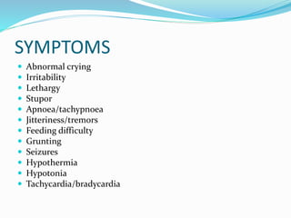
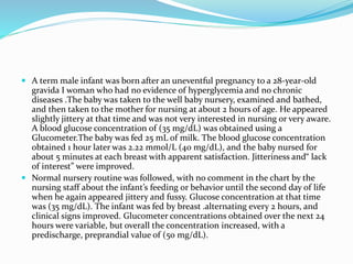
NEONATAL
HYPOGLYCEMIA
OBJECTIVES
Define hypoglycemia ?
Pathophysiology
Transient Vs Persistent
Lab
Therapeutics
Long term neurodevelopmental outcome of
hypoglycemia
NEONATAl HYPOGLYCEMIA
 WHY IT SHOULD BE AGGRESIVELY MANAGED
 Incidence is 1-5/1000 of live birth.8% in LGA and 15%
in preterms and IUGR upto 30 % in highrisk babies
 30 % incidence of neourodevelopmental sequlae if
hypogycemia was present for more than 3 days , even
ifintermittently and 40 % if for 5 days or more. ( lucas
BMJ)
 NH as the most common cause of remote symptomatic
infantile onset epilepsy( udani IP)
 Even moderate hypoglycemia ( < 47mg%) can have
significant outcome in PT babies.
NO DEFINITION
 CONTROVERSIAL:
 Current evidence does not support a specific concentration of
glucose that can discriminate euglycemia from hypoglycemia , or
can predict acute or chronic irreversible neurologic damage .
 No single blood value can be used to define physiologic hypoglycemia.
 No studies to date have established an absolute serum glucose
concentration at which short or long term organ dysfunction can occur.
 2 hr healthy newborn with 30 mg% might not demonstrate impaired
function but a stressed term may show s/s at 50mg%.
 Time of measurement. A value considered low normal or normal at 3
hrs of life might be termed hypoglycemic at 18 hrs
Cont.
Deft Definition
Definitions based on
 Clinical manifestations. (Along with Whipples triad)
 Epidemiologic and statistical; (less that 2 SD)
 Metabolic and hormonal mileu changes.
 Long term neurologic outcome.
 PRACTICAL DEFINITION:- levels at which Glucose
delivery or availability is inadequate to meet the
demand
Any value below 55 mg% be viewed cautiously and treated
aggressively
Cont.
WHIPPLES TRIAD
To attribute signs and symptoms to NH, Cornblath et al have
suggested that the Whipple triad be fulfilled:
1. Signs consistent with NH and
2. coincident with A low blood glucose concentration and
3. Resolution of signs and symptoms within min to hours after
restoring blood glucose concentrations to normal values.
though this glucose level may be quite deviant from the level and
duration that may be associated with long term damage.
Moreover this definition did not include asymptomatic
hypoglycemic babies.
Cont..
OPERATIONAL THRESHOLD
 Traditionally hypoglycemia has been defined blood
glucose < 40mg %( equi to plasma glucose of 45mg%)
 Cornblath suggested that OPERATIONAL
THRESHOLD should be established.
 It is defined as that level at which babies demonstrate
a unique response to abnormal mileu casused by
inadequate delivery of glucose to target organs
 Such operational thresholds are different from
therapeutic goals, and they do not define normal or
abnormal but provide a margin of safety for
interventions.
 There is no evidence to suggest differentiating critical
glucose levels in term and preterms.
Operational thresholds
 Term newborn:
 Healthy full term baby: no screening required
 Breast fed babies who lose wt may have low glucose but high ketone bodies as
an alternative substrate
 Symtomatic
 Intervention required ifIf value is < 45
 Newborn with risk factors of maladaptation
 In asymptomatic intervention if < 36 and not increasing with feeds.
 At < 25 start IV aimed at raising above 45.
 In PHHI goal is 60 mg%
 Preterm
 No data to support lower threshold for preterms.
 OT of 45mg%
 Newborn on parenteral nutrition
 Higher therapeutic levels needed
HIGH RISK BABIES(routine monitoring recommended)
 Preterm babies ( <37W)
 Large for gestational age (LGA) infants,
 small for gestational age (SGA) infants
 IDM Infants born to insulin-dependent mothers (1:1000 pregnant women) or
mothers with gestational diabetes (occurs in 2% of pregnant women)
 Sick Infants( sepsis ,asphyxia, resp. distress,hypothermia,polycythemia).
 Eythroblastosis fetalis and Post exchange blood transfusion
 Infants whose mothers have recd beta blockers, oral hypoglycemics,or
intrapartum dextrose infusion
 Newborns with symptoms suggestive of hypoglycemia, including jitteriness,
tachypnea, hypotonia, poor feeding, apnea, temperature instability, seizures,
and lethargy
 Additionally, consider hypoglycemia screening in infants with isolated
hepatomegaly (possible glycogen-storage disease), microcephaly, anterior
midline defects, gigantism, macroglossia or hemihypertrophy
(possible Beckwith- Wiedemann Syndrome), or any possibility of an inborn
error of metabolism.
 Transient : due to perinatal cond. Lower levels of GIR
needed for 24 to 48 hrs
 Refactory : Should be suspected if > 12 mg/kg/mt of
GIR is required for more than 24 hrs
 Mostly because of increased or in appropriate insulin or deficencey of one of the
glucose regulatory enzymes of liver
 Hyperinsulinemia, hypopituitarism, adrenal insufficiency, galactosemia, GSD,
organic acidenias and mitochondrila disorders.
 PHHI persistent hyperinsulinemia if insulin. 2 μU/ml in presence of glucose
<50mg/dl
 Prolonged : if blood glucose remains unstable beyond
5-7 days
 Famililial hyperinsulinemia
 Inborn errors of metabolism
Transient, Refractory and Prolonged hypoglycemia
Glucose homeostasis
 Clamping of cord cuts off maternal supply of glucose to newborn .
 Increased catecholamine ( immediately releases glucose)
 Decrease in insulin/glucagon ratio.( prolonged glucose release)
 This leads to inactivation of glycogen synthase and activation of glycogen
phosphorylase.
 Term infants have only enough hepatic glycogen to last for 10 hrs. even
less in preterms and IUGR.
 Basal glucose utilization rate is 4-6mg/kg/mt( twice as adult)
 During first 2-3 hours blood glucose level falls from fetal( 70% of
maternal level) to as low as 30mg% before compensatory homeostasis
sets in.
 Gluconeogenic enzymes may be slow to be induced in preterms and
cause prolonged hypoglycemia.
 Fetus does not produce any glucose .
PATHOPHYSIOLOGY
IDMs Vs PHHI
IDMs Hyperinsulism
 Increased insulin secretion in
response to antepartum
hyperglycemia
 Persists for 24 to 72 hours
 Increased Insulin/Glucagon
ratio leads to Decreased
glycogenolysis
 Increased peripheral glucose
utilisation
 Hyperinsulinism related to cong
defects
 Prolonged hypoglycemia(>7d)
 Rarely IUGRand HIE babies may
show prolonged
hyperinsulinism.
 AR K+ ATP channel
 AD hyperinsulinemia
 Congenital hyperinsulinemia
syndrome
 Beckwith Weidermann synd.
Erythroblastosis fetalis
 Increased levels of insulin and increased B cell .
 Glutathione released from hemolysed rbc
inactivates insulin in circulation leading to
increased secretion and up regulation of beta cells.
 Moreover ,exchange transfusion puts a load of ACD
dextrose leading to hyperinsulinism followed by
rebound hypoglycemia.
SYMPTOMS
 Abnormal crying
 Irritability
 Lethargy
 Stupor
 Apnoea/tachypnoea
 Jitteriness/tremors
 Feeding difficulty
 Grunting
 Seizures
 Hypothermia
 Hypotonia
 Tachycardia/bradycardia
LABORATORY
 Glucose levels in plasma (one of the components of blood) are generally 10%–15% higher than
glucose measurements in whole blood (and even more after eating). This is important because
home blood glucose meters measure the glucose in whole blood while most lab tests measure
the glucose in plasma. Currently, most meters on the market give results as "plasma
equivalent," even though they are measuring whole blood glucose. The plasma equivalent is
calculated from the whole blood glucose reading using an equation built into the glucose
meter.
 Factors affecting accuracy of various meters include calibration of meter, ambient temp,
pressure use to wipe off strip (if applicable), size and quality of blood sample, high levels of
certain substances (such as ascorbic acid in blood, hematocrit, dirt on meter, humidity, and
aging of test strips
 Most glucometers today use an electrochemical method. Test strips contain a capillary that
sucks up a reproducible amount of blood. The glucose in the blood reacts with an enzyme in
strip glucose oxidase (or dehydrogenase) and forms gluconic acid This reacts with ferricyanide
ion. And forms ferrocyanide which is read electronically to measure glucose.
 The colorimetric method is a technique where the total amount of charge generated by the
glucose oxidation reaction is measured over a period of time. The amperometric method is
used by some meters and measures the electric current generated at a specific point in time by
the glucose reaction. This is analogous to throwing a ball and using the speed at which it is
travelling at a point in time to estimate how hard it was thrown. The coulometric method can
allow for variable test times, whereas the test time on a meter using the amperometric method
is always fixed. Both methods give an estimation of the concentration of glucose in the initial
blood sample.`
 Arterial glucose > capillary > venous glucose.
 Glucose can fall @ 14-18 mg% /hr on standing sample. Can transport in SF vials.
 Recently subcutaneously inserted continuous glucose monitoring sensors are used in VLBW
babies.
Persistent hypoglycemia workup
 Serum GH , cortisol
•Congenital hypopituitarism Serum GH. ,Serum Cortisol
•Adrenal insufficiency Serum Cortisol
•Hyperinsulinemic states •Insulin glucose ratio>0.4
•Insulin .0.6 micu/ml
•Galactosemia Presence of non glucose reducing substances
in urine
Enzyme assay gal-1 pho uridyl
•Glycogen storage disorders Lactic acidosis
hyperuricemia
•MSUD Urinary aminpacids
•Mitochondrial disorders Lactic acidosis
SCREENING SCHEDULE
 Neonatal glucose concentrations decrease after birth, to as low as 30 mg/dL
during the first 1 to 2 hours after birth, and then increase to higher and
relatively more stable concentrations, generally above 45 mg/dL by 12
hours after birth. Data on the optimal timing and intervals for glucose
screening are limited. It is controversial whether to screen the
asymptomatic at-risk infant for NH during this normal physiologic nadir.
No studies have demonstrated harm from a few hours of asymptomatic
hypoglycemia during this normal postnatal period of establishing
“physiologic glucose homeostasis.” at-risk infants should be screened for
NH with a frequency and duration related to risk factors specific to the
individual infant. Screening the asymptomatic at-risk infant can be
performed within the first hours of birth and continued through multiple
feed-fast cycles.
 LGA And IDMs usually manifest as hypoglycemia early as compared
to preterms and IUGR.
 All Asymptomatic at risk neonates should be screened at 2 hours of
age and then 4-6 hrly until feeds are well established and glucose
levels are normalized( ususlly till 48 hrs)
Screening schedule
• 2-6-12-24-48+72 hr
At Risk
Neonates
• Every 6-8 hrs till stable
Sick
Neonates
• Every 6-8 hrs for 3 days
• Every 24 hrs after 3 days
Stable
VLBW
TREATMENT
Asymptomatic hypoglycemia
 Measured breast milk by spoon or gavage, or formula
milk, or dextrose gel.
 Check glucose after 30 to 60 min.
 If > 45 then 2-3 hrly feeds and 4-6 hrly testing upto 48
hrs.
 IV glucose in Asymptomatic baby if :-
1. Blood glucose < 25
2. Glucose < 40 despite feeding
3. enteral feeding CI
4. Baby becomes symptomatic
Symptomatic hypoglycemia
 Bolus of 2 ml/kg 10 % dextrose as mini bolus
 Followed by infusion @ 6 mg/kg/mt
Glucose rechecked after 15-30 min
 If > 45 increase testing interval
 If < 45 increase GIR @ 2 every 15 -30 min
 Tapering. Once BG stabilises over 45 for24 hrs then GIR
can be tapered @ 2 every 6 hrs.and can be stopped once
GIR reaches 4.
 In neonates who cant be fed orally GIR should be tapered
to min level at which euglycemia is maintained
 If GIR > 12 is recq then a diagnosis of resistant
hypoglycemia should be madeand managed accordingly
 ORAL feeds should be continued along with tapering of
infusion
Practice points
 Avoid using > 12.5 % GIR from peripheral veins.
 In addition to glucose infusion measures should be
taken to correct acidosis, maintain thermoneutral
environment and treatment of underlying conditions
 A continuous infusion should me maintained.
Rebound hypoglycemia may happen if infusion
stopped abruptly
 Intermittent boluses are not logical. May lead to labile
glycemic levels.
THERAPY
FEEDING
Dextrose
 Glucagon
 Glucocorticoids
 Diazoxide
 Octreotide
 Nifedipine
 A term male infant was born after an uneventful pregnancy to a 28-year-old
gravida I woman who had no evidence of hyperglycemia and no chronic
diseases .The baby was taken to the well baby nursery, examined and bathed,
and then taken to the mother for nursing at about 2 hours of age. He appeared
slightly jittery at that time and was not very interested in nursing or very aware.
A blood glucose concentration of (35 mg/dL) was obtained using a
Glucometer.The baby was fed 25 mL of milk. The blood glucose concentration
obtained 1 hour later was 2.22 mmol/L (40 mg/dL), and the baby nursed for
about 5 minutes at each breast with apparent satisfaction. Jitteriness and“ lack
of interest” were improved.
 Normal nursery routine was followed, with no comment in the chart by the
nursing staff about the infant’s feeding or behavior until the second day of life
when he again appeared jittery and fussy. Glucose concentration at that time
was (35 mg/dL). The infant was fed by breast .alternating every 2 hours, and
clinical signs improved. Glucometer concentrations obtained over the next 24
hours were variable, but overall the concentration increased, with a
predischarge, preprandial value of (50 mg/dL).
 At 2 weeks of life, the parents noted the infant to be very fussy and
jittery and to experience staring spells. At a local emergency
department, he was noted to have lost weight, appeared somnolent but
fussy when aroused, and started having tonic-clonic jerking
movements of all extremities. A “glucose concentration” was less than
(10 mg/dL). The infant was treated with intravenous glucose, and the
apparent seizure resolved. Over the next several weeks, the infant
returned to the emergency department several times with similar
episodes.
 When finally examined by the primary care physician, the infant had
gained 283.5 g and appeared“ puffy.” An “office glucose concentration”
was (35 mg/dL). The infant was referred to a pediatric endocrinologist,
who noted that the infant’s weight was approaching the 90th
percentile, there was definite hepatomegaly, and the infant appeared
“apathetic.” In the hospital, several serum glucose concentrations were
measured at less than (40 mg/dL), with plasma insulin concentrations
all greater than (20 mcU/mL).
Dextrose
 LOAD with 2 ml /kg of 10 % dext followed by 4 –
6mg/kg/mt ( Term) and 6-8mg/kg/mt ( preterm)
 Central line if conc > 12.5 mg%
 Increase @ 2 mg/kg.mt if still hypoglycemic.( give a
repeat bolus if symptomatic)
 If conc > 20 mg/kg.mt is required to maintain glucose
,consider additional therapeutics.
 Step Down gradually in interval of 4-6 hours @2
mg/kg/mt. or faster tapering at higher glucose levels
glucocorticoids
 Used as an adjunct to IV Dext if GIR > 15 is required
 Mech of action.↓ insulin secretion, ↑ insulin
resistance, ↑ gluconeogenesis, ↑ glyconeogenolysis
 Dexamethasone 0.25mg/kg bid,
 Hydrocortisone2.5mg/kg qid(freq req of addon)
 S/E :↓ growth , feed intol,GIT perforation, ↑ BP
Glucagon
 Used only when adeq glycogen stores are anticipated,and
awaiting iv access
 Effect is rapid but SHORT LASTING. Has to be followed by
IV Dext
 Used Esp in non PHHI ie term and preterm without
persistent hyperinsulinemia
 Continuous/intermittent( 30 mic/kg im dose )
 20-40 mcg/kg/hr for sick premies or flat 1 mg/day for term.
Dose 10 times may be required in high insulin levels.
 S/E Severe hyponatremia, Crystallization of glucagon,
thrombocytopenia
Diazoxide
 Used when hyperinsulinemic state leads to prolonged
and refractory hypoglycemia
 ßcell KATP channel opener leading to ↓insulin secretion
(therefore used in hyperinsulinemic states)
 10-15 mg/kg/day in 2 – 3 doses. ORAL
 S/E Hypertrichosis, Fluid retention
 Failure to respond makes way for octreotide which acts
distally.
octreotide
 Long acting Somatostatin analogue.
 5 mcg/kg/dose 4 doses or continuous inf
 S/E Growth hormone release block.
Dextrose Gel
 Dextrose gel contains dextrose, a simple carbohydrate, in concentrated aqueous solution,
which can be administered by direct application to mucosal surfaces of the mouth,
including buccal and lingual surfaces. Absorption from these sites may allow rapid access
to the circulation. Some proportion of the dose may be swallowed and absorbed from the
gastrointestinal tract.
 Commercial preparations of dextrose gel are widely available, as they are commonly used
for management of hypoglycaemia in patients with diabetes. Many preparations contain
preservatives and flavour additives as well as gelling agents, requiring individual
assessment for suitability in neonates. Dextrose gel can be manufactured by hospital
pharmacies with appropriate facilities. Costs for neonatal doses are low (a few dollars or
less per dose), and adverse effects have not been reported.
 In infants with hypoglycaemia, simple treatment with dextrose gel and potential
avoidance of more complex treatments, such as IV dextrose or complementary milks,
would provide an attractive option, if effective. Dextrose gel is typically available in 40
g/100 mL form (40%) and is administered at doses of 200 to 400 mg/kg. Two
observational studies suggest that it may be helpful for treatment of individuals with
hypoglycaemia, and these studies report no adverse effects . Treatment of the neonate
with hypoglycaemia usually involves additional feeding, often with formula milk, with
the potential for an adverse impact on the quality and duration of breast feeding. If
feeding is not effective, IV dextrose is usually administered, commonly requiring
admission to the NICU and resulting in separation of mother and infant, impaired
initiation of breast feeding and increased healthcare costs.
 Dextrose gel is inexpensive and simple to administer. It may be effective in treating
infants with neonatal hypoglycaemia without adverse effects. Further, dextrose gel can be
used in resource-poor settings in which higher levels of neonatal care are unavailable,
and it may prevent brain damage caused by untreated neonatal hypoglycaemia.
Neurologic outcome
HIE HGE
Serum lactic acid Increased Normal
cerebral Cortex Infarction in watershed zones Selective neuronal
necrosis
cerebellum involved spared
brainstem involved spared
Imaging
localisation
Non specific OCCIPITAL LOBE
Neurological outcome
 Outcome of hypoglycemia depends on
 Duration of hypoglycemia . The NHBI depends on the
duration of hypoglycemia rather than its severity
 Recurrent or protracted hypoglycemia seems to be a more sinister
risk factor rather than a single low value of blood sugar.
 Severity of hypoglycemia
 Cerebral blood flow
 Concomitant illness The neuronal injury is also influenced by the
existence of other comorbidities such as hypoxic ischemia, prematurity,
and SGA
Udani et al :neonatal hypoglycemia was the most common etiology for
remote symptomatic epilepsy with onset in the first 3 years of life, and
infantile spasms was the most common seizure type
FINDINGS
Pathological changes of NHBI
 Swelling of the neuronal and glial cells, necrosis, gyrus atrophy, and white
matter demyelination have been observed in association with NHBI.. NHBI
does not follow vascular territory and bilateral posterior parieto-occipital
regions are most commonly involved in term as well as preterm infants.91
 Diffuse white matter involvement particularly in the watershed areas is more
common with hypoxic ischemia, whereas cortical injury is more commonly
seen in NHBI.
Neuroimaging findings
 Cranial ultrasound and computed tomography scans of the brain lack
sensitivity and specificity, and have been replaced by MRI for diagnosing
NHBI.1 In order to detect acute manifestations, MRI should be performed
between 3 and 7 days after the hypoglycemic insult, Such images done within 7
days revealed marked parieto-occipital hyperintensity signals . Magnetic
resonance spectroscopy studies following the acute phase of hypoglycemia have
revealed increased lactate and free fatty acid peaks, reduced acetyl aspartic acid
peaks, and altered ATP/lactic acid ratio indicating altered brain function.108
Systemic review
Preventive practices
 Early exclusive BF with in 1st hr.
 Maintain thermoneutral environment
 Do not feed 5,10, 25 % dextrose as a substitute. It can
induce vomiting,decreased glucagon and rebound
hypoglycemia
 No interruption in IV infusion.
THANKS

Neonatal hypoglycemia

  • 1.
  • 2.
    OBJECTIVES Define hypoglycemia ? Pathophysiology TransientVs Persistent Lab Therapeutics Long term neurodevelopmental outcome of hypoglycemia
  • 3.
    NEONATAl HYPOGLYCEMIA  WHYIT SHOULD BE AGGRESIVELY MANAGED  Incidence is 1-5/1000 of live birth.8% in LGA and 15% in preterms and IUGR upto 30 % in highrisk babies  30 % incidence of neourodevelopmental sequlae if hypogycemia was present for more than 3 days , even ifintermittently and 40 % if for 5 days or more. ( lucas BMJ)  NH as the most common cause of remote symptomatic infantile onset epilepsy( udani IP)  Even moderate hypoglycemia ( < 47mg%) can have significant outcome in PT babies.
  • 4.
    NO DEFINITION  CONTROVERSIAL: Current evidence does not support a specific concentration of glucose that can discriminate euglycemia from hypoglycemia , or can predict acute or chronic irreversible neurologic damage .  No single blood value can be used to define physiologic hypoglycemia.  No studies to date have established an absolute serum glucose concentration at which short or long term organ dysfunction can occur.  2 hr healthy newborn with 30 mg% might not demonstrate impaired function but a stressed term may show s/s at 50mg%.  Time of measurement. A value considered low normal or normal at 3 hrs of life might be termed hypoglycemic at 18 hrs Cont.
  • 5.
    Deft Definition Definitions basedon  Clinical manifestations. (Along with Whipples triad)  Epidemiologic and statistical; (less that 2 SD)  Metabolic and hormonal mileu changes.  Long term neurologic outcome.  PRACTICAL DEFINITION:- levels at which Glucose delivery or availability is inadequate to meet the demand Any value below 55 mg% be viewed cautiously and treated aggressively Cont.
  • 6.
    WHIPPLES TRIAD To attributesigns and symptoms to NH, Cornblath et al have suggested that the Whipple triad be fulfilled: 1. Signs consistent with NH and 2. coincident with A low blood glucose concentration and 3. Resolution of signs and symptoms within min to hours after restoring blood glucose concentrations to normal values. though this glucose level may be quite deviant from the level and duration that may be associated with long term damage. Moreover this definition did not include asymptomatic hypoglycemic babies. Cont..
  • 7.
    OPERATIONAL THRESHOLD  Traditionallyhypoglycemia has been defined blood glucose < 40mg %( equi to plasma glucose of 45mg%)  Cornblath suggested that OPERATIONAL THRESHOLD should be established.  It is defined as that level at which babies demonstrate a unique response to abnormal mileu casused by inadequate delivery of glucose to target organs  Such operational thresholds are different from therapeutic goals, and they do not define normal or abnormal but provide a margin of safety for interventions.  There is no evidence to suggest differentiating critical glucose levels in term and preterms.
  • 8.
    Operational thresholds  Termnewborn:  Healthy full term baby: no screening required  Breast fed babies who lose wt may have low glucose but high ketone bodies as an alternative substrate  Symtomatic  Intervention required ifIf value is < 45  Newborn with risk factors of maladaptation  In asymptomatic intervention if < 36 and not increasing with feeds.  At < 25 start IV aimed at raising above 45.  In PHHI goal is 60 mg%  Preterm  No data to support lower threshold for preterms.  OT of 45mg%  Newborn on parenteral nutrition  Higher therapeutic levels needed
  • 9.
    HIGH RISK BABIES(routinemonitoring recommended)  Preterm babies ( <37W)  Large for gestational age (LGA) infants,  small for gestational age (SGA) infants  IDM Infants born to insulin-dependent mothers (1:1000 pregnant women) or mothers with gestational diabetes (occurs in 2% of pregnant women)  Sick Infants( sepsis ,asphyxia, resp. distress,hypothermia,polycythemia).  Eythroblastosis fetalis and Post exchange blood transfusion  Infants whose mothers have recd beta blockers, oral hypoglycemics,or intrapartum dextrose infusion  Newborns with symptoms suggestive of hypoglycemia, including jitteriness, tachypnea, hypotonia, poor feeding, apnea, temperature instability, seizures, and lethargy  Additionally, consider hypoglycemia screening in infants with isolated hepatomegaly (possible glycogen-storage disease), microcephaly, anterior midline defects, gigantism, macroglossia or hemihypertrophy (possible Beckwith- Wiedemann Syndrome), or any possibility of an inborn error of metabolism.
  • 10.
     Transient :due to perinatal cond. Lower levels of GIR needed for 24 to 48 hrs  Refactory : Should be suspected if > 12 mg/kg/mt of GIR is required for more than 24 hrs  Mostly because of increased or in appropriate insulin or deficencey of one of the glucose regulatory enzymes of liver  Hyperinsulinemia, hypopituitarism, adrenal insufficiency, galactosemia, GSD, organic acidenias and mitochondrila disorders.  PHHI persistent hyperinsulinemia if insulin. 2 μU/ml in presence of glucose <50mg/dl  Prolonged : if blood glucose remains unstable beyond 5-7 days  Famililial hyperinsulinemia  Inborn errors of metabolism Transient, Refractory and Prolonged hypoglycemia
  • 11.
    Glucose homeostasis  Clampingof cord cuts off maternal supply of glucose to newborn .  Increased catecholamine ( immediately releases glucose)  Decrease in insulin/glucagon ratio.( prolonged glucose release)  This leads to inactivation of glycogen synthase and activation of glycogen phosphorylase.  Term infants have only enough hepatic glycogen to last for 10 hrs. even less in preterms and IUGR.  Basal glucose utilization rate is 4-6mg/kg/mt( twice as adult)  During first 2-3 hours blood glucose level falls from fetal( 70% of maternal level) to as low as 30mg% before compensatory homeostasis sets in.  Gluconeogenic enzymes may be slow to be induced in preterms and cause prolonged hypoglycemia.  Fetus does not produce any glucose .
  • 12.
  • 14.
    IDMs Vs PHHI IDMsHyperinsulism  Increased insulin secretion in response to antepartum hyperglycemia  Persists for 24 to 72 hours  Increased Insulin/Glucagon ratio leads to Decreased glycogenolysis  Increased peripheral glucose utilisation  Hyperinsulinism related to cong defects  Prolonged hypoglycemia(>7d)  Rarely IUGRand HIE babies may show prolonged hyperinsulinism.  AR K+ ATP channel  AD hyperinsulinemia  Congenital hyperinsulinemia syndrome  Beckwith Weidermann synd.
  • 15.
    Erythroblastosis fetalis  Increasedlevels of insulin and increased B cell .  Glutathione released from hemolysed rbc inactivates insulin in circulation leading to increased secretion and up regulation of beta cells.  Moreover ,exchange transfusion puts a load of ACD dextrose leading to hyperinsulinism followed by rebound hypoglycemia.
  • 16.
    SYMPTOMS  Abnormal crying Irritability  Lethargy  Stupor  Apnoea/tachypnoea  Jitteriness/tremors  Feeding difficulty  Grunting  Seizures  Hypothermia  Hypotonia  Tachycardia/bradycardia
  • 17.
    LABORATORY  Glucose levelsin plasma (one of the components of blood) are generally 10%–15% higher than glucose measurements in whole blood (and even more after eating). This is important because home blood glucose meters measure the glucose in whole blood while most lab tests measure the glucose in plasma. Currently, most meters on the market give results as "plasma equivalent," even though they are measuring whole blood glucose. The plasma equivalent is calculated from the whole blood glucose reading using an equation built into the glucose meter.  Factors affecting accuracy of various meters include calibration of meter, ambient temp, pressure use to wipe off strip (if applicable), size and quality of blood sample, high levels of certain substances (such as ascorbic acid in blood, hematocrit, dirt on meter, humidity, and aging of test strips  Most glucometers today use an electrochemical method. Test strips contain a capillary that sucks up a reproducible amount of blood. The glucose in the blood reacts with an enzyme in strip glucose oxidase (or dehydrogenase) and forms gluconic acid This reacts with ferricyanide ion. And forms ferrocyanide which is read electronically to measure glucose.  The colorimetric method is a technique where the total amount of charge generated by the glucose oxidation reaction is measured over a period of time. The amperometric method is used by some meters and measures the electric current generated at a specific point in time by the glucose reaction. This is analogous to throwing a ball and using the speed at which it is travelling at a point in time to estimate how hard it was thrown. The coulometric method can allow for variable test times, whereas the test time on a meter using the amperometric method is always fixed. Both methods give an estimation of the concentration of glucose in the initial blood sample.`  Arterial glucose > capillary > venous glucose.  Glucose can fall @ 14-18 mg% /hr on standing sample. Can transport in SF vials.  Recently subcutaneously inserted continuous glucose monitoring sensors are used in VLBW babies.
  • 19.
    Persistent hypoglycemia workup Serum GH , cortisol •Congenital hypopituitarism Serum GH. ,Serum Cortisol •Adrenal insufficiency Serum Cortisol •Hyperinsulinemic states •Insulin glucose ratio>0.4 •Insulin .0.6 micu/ml •Galactosemia Presence of non glucose reducing substances in urine Enzyme assay gal-1 pho uridyl •Glycogen storage disorders Lactic acidosis hyperuricemia •MSUD Urinary aminpacids •Mitochondrial disorders Lactic acidosis
  • 20.
    SCREENING SCHEDULE  Neonatalglucose concentrations decrease after birth, to as low as 30 mg/dL during the first 1 to 2 hours after birth, and then increase to higher and relatively more stable concentrations, generally above 45 mg/dL by 12 hours after birth. Data on the optimal timing and intervals for glucose screening are limited. It is controversial whether to screen the asymptomatic at-risk infant for NH during this normal physiologic nadir. No studies have demonstrated harm from a few hours of asymptomatic hypoglycemia during this normal postnatal period of establishing “physiologic glucose homeostasis.” at-risk infants should be screened for NH with a frequency and duration related to risk factors specific to the individual infant. Screening the asymptomatic at-risk infant can be performed within the first hours of birth and continued through multiple feed-fast cycles.  LGA And IDMs usually manifest as hypoglycemia early as compared to preterms and IUGR.  All Asymptomatic at risk neonates should be screened at 2 hours of age and then 4-6 hrly until feeds are well established and glucose levels are normalized( ususlly till 48 hrs)
  • 21.
    Screening schedule • 2-6-12-24-48+72hr At Risk Neonates • Every 6-8 hrs till stable Sick Neonates • Every 6-8 hrs for 3 days • Every 24 hrs after 3 days Stable VLBW
  • 22.
  • 23.
    Asymptomatic hypoglycemia  Measuredbreast milk by spoon or gavage, or formula milk, or dextrose gel.  Check glucose after 30 to 60 min.  If > 45 then 2-3 hrly feeds and 4-6 hrly testing upto 48 hrs.  IV glucose in Asymptomatic baby if :- 1. Blood glucose < 25 2. Glucose < 40 despite feeding 3. enteral feeding CI 4. Baby becomes symptomatic
  • 24.
    Symptomatic hypoglycemia  Bolusof 2 ml/kg 10 % dextrose as mini bolus  Followed by infusion @ 6 mg/kg/mt Glucose rechecked after 15-30 min  If > 45 increase testing interval  If < 45 increase GIR @ 2 every 15 -30 min  Tapering. Once BG stabilises over 45 for24 hrs then GIR can be tapered @ 2 every 6 hrs.and can be stopped once GIR reaches 4.  In neonates who cant be fed orally GIR should be tapered to min level at which euglycemia is maintained  If GIR > 12 is recq then a diagnosis of resistant hypoglycemia should be madeand managed accordingly  ORAL feeds should be continued along with tapering of infusion
  • 25.
    Practice points  Avoidusing > 12.5 % GIR from peripheral veins.  In addition to glucose infusion measures should be taken to correct acidosis, maintain thermoneutral environment and treatment of underlying conditions  A continuous infusion should me maintained. Rebound hypoglycemia may happen if infusion stopped abruptly  Intermittent boluses are not logical. May lead to labile glycemic levels.
  • 28.
  • 29.
     A termmale infant was born after an uneventful pregnancy to a 28-year-old gravida I woman who had no evidence of hyperglycemia and no chronic diseases .The baby was taken to the well baby nursery, examined and bathed, and then taken to the mother for nursing at about 2 hours of age. He appeared slightly jittery at that time and was not very interested in nursing or very aware. A blood glucose concentration of (35 mg/dL) was obtained using a Glucometer.The baby was fed 25 mL of milk. The blood glucose concentration obtained 1 hour later was 2.22 mmol/L (40 mg/dL), and the baby nursed for about 5 minutes at each breast with apparent satisfaction. Jitteriness and“ lack of interest” were improved.  Normal nursery routine was followed, with no comment in the chart by the nursing staff about the infant’s feeding or behavior until the second day of life when he again appeared jittery and fussy. Glucose concentration at that time was (35 mg/dL). The infant was fed by breast .alternating every 2 hours, and clinical signs improved. Glucometer concentrations obtained over the next 24 hours were variable, but overall the concentration increased, with a predischarge, preprandial value of (50 mg/dL).
  • 30.
     At 2weeks of life, the parents noted the infant to be very fussy and jittery and to experience staring spells. At a local emergency department, he was noted to have lost weight, appeared somnolent but fussy when aroused, and started having tonic-clonic jerking movements of all extremities. A “glucose concentration” was less than (10 mg/dL). The infant was treated with intravenous glucose, and the apparent seizure resolved. Over the next several weeks, the infant returned to the emergency department several times with similar episodes.  When finally examined by the primary care physician, the infant had gained 283.5 g and appeared“ puffy.” An “office glucose concentration” was (35 mg/dL). The infant was referred to a pediatric endocrinologist, who noted that the infant’s weight was approaching the 90th percentile, there was definite hepatomegaly, and the infant appeared “apathetic.” In the hospital, several serum glucose concentrations were measured at less than (40 mg/dL), with plasma insulin concentrations all greater than (20 mcU/mL).
  • 31.
    Dextrose  LOAD with2 ml /kg of 10 % dext followed by 4 – 6mg/kg/mt ( Term) and 6-8mg/kg/mt ( preterm)  Central line if conc > 12.5 mg%  Increase @ 2 mg/kg.mt if still hypoglycemic.( give a repeat bolus if symptomatic)  If conc > 20 mg/kg.mt is required to maintain glucose ,consider additional therapeutics.  Step Down gradually in interval of 4-6 hours @2 mg/kg/mt. or faster tapering at higher glucose levels
  • 32.
    glucocorticoids  Used asan adjunct to IV Dext if GIR > 15 is required  Mech of action.↓ insulin secretion, ↑ insulin resistance, ↑ gluconeogenesis, ↑ glyconeogenolysis  Dexamethasone 0.25mg/kg bid,  Hydrocortisone2.5mg/kg qid(freq req of addon)  S/E :↓ growth , feed intol,GIT perforation, ↑ BP
  • 33.
    Glucagon  Used onlywhen adeq glycogen stores are anticipated,and awaiting iv access  Effect is rapid but SHORT LASTING. Has to be followed by IV Dext  Used Esp in non PHHI ie term and preterm without persistent hyperinsulinemia  Continuous/intermittent( 30 mic/kg im dose )  20-40 mcg/kg/hr for sick premies or flat 1 mg/day for term. Dose 10 times may be required in high insulin levels.  S/E Severe hyponatremia, Crystallization of glucagon, thrombocytopenia
  • 34.
    Diazoxide  Used whenhyperinsulinemic state leads to prolonged and refractory hypoglycemia  ßcell KATP channel opener leading to ↓insulin secretion (therefore used in hyperinsulinemic states)  10-15 mg/kg/day in 2 – 3 doses. ORAL  S/E Hypertrichosis, Fluid retention  Failure to respond makes way for octreotide which acts distally.
  • 35.
    octreotide  Long actingSomatostatin analogue.  5 mcg/kg/dose 4 doses or continuous inf  S/E Growth hormone release block.
  • 36.
    Dextrose Gel  Dextrosegel contains dextrose, a simple carbohydrate, in concentrated aqueous solution, which can be administered by direct application to mucosal surfaces of the mouth, including buccal and lingual surfaces. Absorption from these sites may allow rapid access to the circulation. Some proportion of the dose may be swallowed and absorbed from the gastrointestinal tract.  Commercial preparations of dextrose gel are widely available, as they are commonly used for management of hypoglycaemia in patients with diabetes. Many preparations contain preservatives and flavour additives as well as gelling agents, requiring individual assessment for suitability in neonates. Dextrose gel can be manufactured by hospital pharmacies with appropriate facilities. Costs for neonatal doses are low (a few dollars or less per dose), and adverse effects have not been reported.  In infants with hypoglycaemia, simple treatment with dextrose gel and potential avoidance of more complex treatments, such as IV dextrose or complementary milks, would provide an attractive option, if effective. Dextrose gel is typically available in 40 g/100 mL form (40%) and is administered at doses of 200 to 400 mg/kg. Two observational studies suggest that it may be helpful for treatment of individuals with hypoglycaemia, and these studies report no adverse effects . Treatment of the neonate with hypoglycaemia usually involves additional feeding, often with formula milk, with the potential for an adverse impact on the quality and duration of breast feeding. If feeding is not effective, IV dextrose is usually administered, commonly requiring admission to the NICU and resulting in separation of mother and infant, impaired initiation of breast feeding and increased healthcare costs.  Dextrose gel is inexpensive and simple to administer. It may be effective in treating infants with neonatal hypoglycaemia without adverse effects. Further, dextrose gel can be used in resource-poor settings in which higher levels of neonatal care are unavailable, and it may prevent brain damage caused by untreated neonatal hypoglycaemia.
  • 37.
    Neurologic outcome HIE HGE Serumlactic acid Increased Normal cerebral Cortex Infarction in watershed zones Selective neuronal necrosis cerebellum involved spared brainstem involved spared Imaging localisation Non specific OCCIPITAL LOBE
  • 38.
    Neurological outcome  Outcomeof hypoglycemia depends on  Duration of hypoglycemia . The NHBI depends on the duration of hypoglycemia rather than its severity  Recurrent or protracted hypoglycemia seems to be a more sinister risk factor rather than a single low value of blood sugar.  Severity of hypoglycemia  Cerebral blood flow  Concomitant illness The neuronal injury is also influenced by the existence of other comorbidities such as hypoxic ischemia, prematurity, and SGA Udani et al :neonatal hypoglycemia was the most common etiology for remote symptomatic epilepsy with onset in the first 3 years of life, and infantile spasms was the most common seizure type
  • 39.
    FINDINGS Pathological changes ofNHBI  Swelling of the neuronal and glial cells, necrosis, gyrus atrophy, and white matter demyelination have been observed in association with NHBI.. NHBI does not follow vascular territory and bilateral posterior parieto-occipital regions are most commonly involved in term as well as preterm infants.91  Diffuse white matter involvement particularly in the watershed areas is more common with hypoxic ischemia, whereas cortical injury is more commonly seen in NHBI. Neuroimaging findings  Cranial ultrasound and computed tomography scans of the brain lack sensitivity and specificity, and have been replaced by MRI for diagnosing NHBI.1 In order to detect acute manifestations, MRI should be performed between 3 and 7 days after the hypoglycemic insult, Such images done within 7 days revealed marked parieto-occipital hyperintensity signals . Magnetic resonance spectroscopy studies following the acute phase of hypoglycemia have revealed increased lactate and free fatty acid peaks, reduced acetyl aspartic acid peaks, and altered ATP/lactic acid ratio indicating altered brain function.108
  • 40.
  • 41.
    Preventive practices  Earlyexclusive BF with in 1st hr.  Maintain thermoneutral environment  Do not feed 5,10, 25 % dextrose as a substitute. It can induce vomiting,decreased glucagon and rebound hypoglycemia  No interruption in IV infusion.
  • 42.