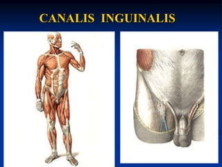
4. The walls of the inguinal canal
Anterior wall is formed by the aponeurosis of the
external oblique muscle;
Posterior wall is formed by the transverse fascia;
Superior wall is formed by the free inferior
borders of the internal oblique and transverse
muscles.
Inferior wall is formed by the inguinal ligament
(medial 1/3);
6. SUPERFICIAL INGUINAL RING
Superficial inguinal ring (anulus
inguinalis superficialis) is in the anterior
wall of inguinal canal and limited
1. From above by a medial crus of an
inguinal ligament;
2. From below by a lateral crus of an
inguinal ligament;
3. medially by a ligamentum reflexum;
4. laterally by intercrural fibres;
8. CANALIS INGUINALIS
Deep inguinal ring (anulus inguinalis
profundus) is in the posterior wall of
inguinal canal formed by the fascia
transversalis and is projected on the
lateral inguinal fossa.
10. REGIO SUBINGUINALE
The Iliopectineal arch divides the space
below the inguinal ligament into two
parts:
1. A lateral part, the lacuna
musculorum, containing iliopsoas
muscle and femoral nerve;
2. A medial part, the lacuna vasorum,
transmitting the femoral artery and
vein;
12. Pelvic region
1. The piriform muscle passes through the
greater sciatic foramen above and below
which openings (foramen suprapiriforme
and foramen infrapiriforme) remain and
transmit the gluteal vessels and nerves.
2. Obturator membrane closes most of the
obturator foramen leaving obturator groove
and thus converts the groove to an
obturator canal.
13. TRIGONUM FEMORALE
The femoral triangle is
bounded
1. Above by the inguinal
ligament;
2. Laterally by the sartorius
muscle;
3. Medially by the adductor
longus muscle.
Two grooves (iliopectineal
groove and anterior
femoral groove) are in
femoral triangle.
15. CANALIS ADDUCTORIUS
Adductor canal (femoropopliteal, or
Hunter’s canal) connects the anterior
region of the thigh with the popliteal
fossa. It has 3 walls and 3 openings.
Medial wall is formed by the adductor
magnus muscle. Lateral wall is formed
by the vastus medialis of quadriceps
muscle, and anteriorly it is bounded
by the lamina vastoadductoria.
17. CANALIS ADDUCTORIUS
The openings of canalis adductorius:
1. Proximal foramen is located at the apex
(inferior angle) of femoral triangle;
2. distal foramen is called hiatus adductorius
is formed by the diverging fibres of the
adductor magnus muscle. It opens into
popliteal fossa (at the upper corner of
fossa);
3. anterior opening is in the center of the
lamina vastoadductoria.
19. FOSSA POPLITEA
Fossa poplitea is a rhomboidal region posterior to
the knee joint. The superior angle is formed
(laterally) by the biceps femoris muscle and
(medially) by the semimembranous and
semitendinous muscles. The inferior angle is
bounded by the medial and lateral heads of the
gastrocnemius muscle.
The floor of fossa is formed by the popliteal muscle,
popliteal surface of the femur, posterior surface of
the articular capsule of knee joint.
Popliteal fossa contains fatty tissue with the
popliteal lymph nodes, popliteal artery and vein,
tibial nerve that pass from the superior angle to
the inferior angle of the fossa.
21. CANALIS CRUROPOPLITEUS
The popliteal fossa is continues with the
cruropopliteal canal. It is located between the
superficial and deep layers of the posterior leg
muscles. It transmits the tibial nerve and the
posterior tibial artery and vein.
Openings of canal:
1. Superior opening is located at the inferior angle
of popliteal fossa;
2. Inferior opening is located at lower 1/3 of the
leg, medially to calcaneal (Achillis) tendon;
3. Anterior opening is on the interosseous
membrane of the leg.
22.
23. Superior and inferior musculoperoneal canals are
located in the leg.
Inferior musculoperoneal canal branches off from
cruropopliteal canal. It transmits the peroneal
artery.
Superior musculoperoneal canal is independent
canal, it transmits the superficial peroneal
nerve.
24. Medial and lateral plantar
grooves are found on
the sole along the
edges of the flexor
digitorum brevis
muscle.
28. Sheath of the rectus abdominis muscle
Above umbilicus anterior wall is formed by the
aponeurosis of obliquus externus abdominis and
anterior leaf of aponeurosis of obliquus internus
abdominis. Posterior wall is formed by posterior
leaf of aponeurosis of obliquus internus
abdominis and aponeurosis of transversus
abdominis.
29. Sheath of the rectus abdominis muscle
At about 4-5 cm below the umbilicus anterior
wall is formed by the aponeuroses of the
three abdominal muscles (external oblique,
internal oblique and transverse muscles).
Posterior wall is formed by fascia
transversalis.