3. • Coronary sulcus
– Atrioventricular sulcus
• Circumvents the heart
• Interventricular sulcus
– Anterior
– Posterior
Sulci of the heart
4.
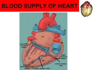
5. Coronary arteries
• The coronary arteries & their branches form a circle
& loop around the heart.
• Coronary= encircling like a crown.
• The heart is supplied by two coronary arteries (right
& left) which arise from the ascending aorta.
• The right coronary artery arises from the anterior
aortic sinus, while the left coronary artery arises from
the left posterior aortic sinus.
6. Arterial supply
of Heart
Rt. & Lt. Coronary arteries
Origin
- from Ascending aorta
Rt.coronary
-Rt. (or ) Anterior Aortic sinus
Lt.coronary
- Left posterior Aortic sinus
Pulmon.
Tr.
SVC.
R A.
L A.
R V. L V.
Ascending
Aorta
7. RIGHT CORONARY ARTERY
• ORIGIN- RCA from anterior aortic sinus, smaller than left
coronary artery(LCA).
• After arising from ascending aorta runs between pulmonary
trunk & right auricle.
• Then runs downwards in right anterior coronary sulcus upto
junction of right & inferior border of heart
• It winds around inferior border to reach diaphragmatic surface.
Here it runs backwards & to left in right posterior coronary
sulcus to reach posterior Interventricular groove.
• TERMINATION- terminates by anastomosing with circumflex
branch of Left coronary artery.
8.
9. Branches & distribution
• Right conus artery- supply base of pulmonary trunk
• Right Atrial branch - supply atria & SA node through NODAL
BRANCH (in 60% cases) .
• Right Ant. Ventricular branch- supply anterior surface of right
ventricle.
• Marginal artery- runs along inferior border to supply both ventricles.
• Right Post. Ventricular branch- supply diaphragmatic surface of right
ventricle.
• Post. Interventricular artery- runs in posterior Interventricular groove
& supplies posterior 1/3rd of interventricular septum, AV node in 60%
cases & Right & Left ventricles.
10.
11. AREA OF DISTRIBUTION OF RIGHT
CORONARY ARTERY
• Right Atrium
• Ventricles
Greater part of right Ventricle except the area adjoining anterior
Interventricular septum
Small part of left Ventricle adjoining posterior Interventricular
groove
• Post 1/3rd part of interventricular septum
• Whole conducting system of heart except a part of AV bundle
SA node (supplied by Left Coronary artery In about 40%)
12.
13. LEFT CORONARY ARTERY
• ORIGIN- from left posterior aortic sinus.
• After arising from ascending aorta runs forward & to left between
pulmonary trunk & left auricle.
• Then divides into :
1) Anterior Interventricular
branch/left anterior descending
artery/LAD
2) Circumflex artery.
14. Branches & distribution
• Anterior Interventricular Artery / Left Anterior Descending Artery
(LAD) – runs downwards in ant. Interventricular groove winding
around apex to enter posterior Interventricular groove & terminating by
anastomosing with posterior Interventricular artery ( br.of RCA) .
• BRANCHEs:
1) Right & left ventricular branch: supply ventricles
2) Left conus branch: anastomoses with right conus(RCA)
3)Diagonal artery- one of large branch of left anterior ventricular
branches or may arise directly from trunk of LCA.
4)Anterior septal branch: supply ant. 2/3rd of interventricular septum.
15. Branches & distribution
Circumflex artery- continuation of LCA.
Circumflex a. winds around Left margin of heart & continues in posterior
Coronary sulcus. Near, posterior Interventricular groove it terminates
by anastomosing with RCA.
BRANCHES:
1) Left marginal artery that supplies left margin of left ventricle upto
apex of heart.
2) Ventricular branch to inferior surface of left ventricle.
3) left Atrial branch supply left atrium.
16.
17. AREA OF DISTRIBUTION OF LCA
• Left Atrium
• Ventricles
Greater part of left Ventricle except the area adjoining post.
Interventricular septum
Small part of right Ventricle adjoining anterior Interventricular
groove
• Anterior 2/3rd part of interventricular septum
• Part of left branch of AV bundle
SA node(40% of cases)
18. Cardiac Dominance
• The artery giving the posterior interventricular branch is
the dominant artery.
• Majority of people possess ‘ Right Coronary dominance’
means RCA gives post. Interventricular a.
• 10% population possess ‘ Left Coronary dominance’ where
circumflex a. continuation of LCA provides post.
Interventricular br. as well as to AV node .
19. CARDIAC ANASTOMOSIS
• In normal healthy heart, the coronary
arteries are TRUE END ARTERIES as
their anastomoses are inadequate to
maintain circulation in sudden occlusion.
20. CLINICAL ANATOMY
• Angina pectoris
narrowing of coronary
artery leads to
ischaemia of cardiac
muscles on excertion
causing pain in chest
(radiating to Lt
shoulder & arm)
21. CLINICAL
ANATOMY
• Myocardial Ischaemia (MI)– sudden
block of one of larger br. of coronary
a. lead to myocardial ischaemia
followed by necrosis (myocardial
infarction) , part of heart suffering
from MI stops functioning & often
causes death called heart attack
commonest site of coronary a.
occlusion is LAD, RCA, circumflex
br. LCA
MI mostly occurs at rest & angina on
exertion
23. CLINICAL
ANATOMY
• Angioplasty–
Done for increasing
diameter of narrowed
coronary artery.
A balloon catheter is
passed & balloon is
inflated at the site of
block. Then stent is
place in artery.
24. CLINICAL
ANATOMY
• Coronary artery
bypass graft(CABG)
• surgical procedure where
patients own vessels are
used for graft.
Internal mammary is
commonly used for graft.
Great saphenous
vein,radial artery also
used.
27. Venous drainage of heart
• Venous blood from
heart is drained into
Right atrium by
Coronary sinus
Anterior Cardiac vein.
Venae cordis minimae
(thebesian v.)
28. Coronary sinus
• Largest vein of heart
• Lies in posterior part of
Coronary sulcus.
• BEGINS as continuation of
great cardiac vein at left end
of coronary sulcus & opens
into right atrium
• Guarded by small
valves(THEBESIAN
VALVES)
29. Tributaries of coronary
sinus
1. Great cardiac vein
2. Middle cardiac vein
3. Small cardiac vein
4. Posterior Vein of Left ventricle
5. Oblique vein of Left atrium ( vein of marshall)
6. Right marginal vein
30.
31. Tributaries of coronary sinus
1. Great cardiac vein– begins at apex, ascends in anterior
interventricular groove & enter left end of sinus, accompanies anterior
Interventricular & circumflex artery.
2. Middle cardiac vein– accompanies posterior Interventricular artery &
joins middle part of sinus.
3. Small cardiac vein– follows right coronary artery & open into right end
of coronary sinus. Right marginal vein may drain into it.
4. Posterior Vein of Left ventricle- runs on diaphragmatic surface of left
ventricle & ens in middle if sinus.
5. Oblique vein of marshall- small vein on posterior of left atrium &
terminate in left end of sinus
6. Right marginal vein- accompanies marginal br. of RCA.may drain into
small cardiac vein or directly into right atrium.
32. VENOUS DRAINAGE OF HEART
• Anterior cardiac veins.– these
are series of 3 or 4 small veins,
open directly into Right atrium
• Venae cordis minimae
(thebesian v.)– numerous small
veins in walls of all four chambers
of heart that open directly into
respective chambers. (Most
numerous in Rightt atrium)