Radio immunoassay (ria)
•Download as PPTX, PDF•
9 likes•534 views

Radioimmunoassay is the technique in which radioisotopes is used as a tag or label radioisotopes is covalently linked with Ag & Ab for the detection of ( Ag & Abs) complex

Recommended
More Related Content
What's hot
What's hot (20)
Similar to Radio immunoassay (ria)
Similar to Radio immunoassay (ria) (20)
More from Vipin Shukla
More from Vipin Shukla (20)
Recently uploaded
Recently uploaded (20)
Radio immunoassay (ria)
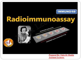
- 1. Prepared By: Vipin Kr Shukla Assistant Lecturer.
- 2. Radio-Immunoassay: Radioisotope is defined as a technique In which Radio-Isotope is used as a tag or label, (i.e.) Radioisotope is covalently linked to Antigen or Antibody for the detection of (AG & AB) complex. Radioimmunoassay (RIA) is a very sensitive in vitro assay technique used to measure concentrations of antigens (for example, hormone levels in the blood) by use of antibodies.
- 3. Continued….. RIA technique is extremely sensitive and extremely specific, requiring specialized equipment, it remains the least expensive method to perform such tests.
- 4. History of RIA: This technique was developed in the year (1959) by Endocrinologist (Rosalyn Yalow and Solomon Berson), to measure the concentration of an Ag & Ab in the sample. In the year (1977), Rosalyn Yalow was awarded Nobel prize for medicine for the development of RIA. But Rosalyn Yalow could not shared the Nobel prize because of her certain death.
- 5. Continued…… The technique was introduced in 1960 by Berson and Yalow as an assay for the concentration of insulin in plasma. It represented the first time that hormone levels in the blood could be detected by an in vitro assay . The technique of radioimmunoassay has revolutionized research and clinical practice in many areas, e.g., Blood banking Diagnosis of allergies Endocrinology
- 6. Continued…… In RIA, Radioisotopes are covalently attached to Ag & Ab. We know that Radioisotopes emit the radiations, which can be measured by using instruments such as Gamma Counter. Most popular Radioisotopes – I- 125.
- 8. Principle of RIA: The technique is based on the ability of an unlabelled form of the substance to inhibit competitively the binding of a radioactively labelled substance by specific antibodies .
- 9. PRINCIPLE OF RADIOIMMUNO ASSAY: It involves combination of three principles. An immune reaction i.e. antigen, antibody binding. A competitive binding or competitive displacement reaction. (It gives specificity) Measurement of radio emission. (It gives sensitivity)
- 10. IMMUNE REACTION: When a foreign biological substance enters into body blood stream through non oral route, body recognizes the specific chemistry on surface of foreign substance as antigen and produces specific antibodies against the antigen so as nullify the effects and keep the body safe. The antibodies are produced by body immune system so, it is an immune reaction. Here the antibodies or antigens bind move due to chemical influence. This is different to principle of electrophoresis where proteins are separated due to charge.
- 11. COMPETITIVE BINDING OR COMPETITIVE DISPLACEMENT REACTION: This is a phenomenon wherein when there are two antigens which can bind to same antibody, the antigen with more concentration binds extensively with the limited antibody displacing other. Radiolabelled antigen is allowed to bind to high affinity antibody. Then when patient serum is added unlabelled antigens in it start binding to the antibody displacing the labelled antigen.
- 12. MEASUREMENT OF RADIO EMISSION: Once the incubation is over, then washings are done to remove any unbound antigens. Then radio emission of the antigen antibody complex is taken, the gamma rays from radio labelled antigen are measured. The target antigen is labelled radioactively and bound to its specific antibodies (a limited and known amount of the specific antibody has to be added).
- 13. Continued…… A sample, for e.g. blood-serum, is added in order to initiate a competitive reaction of the labelled antigens from the preparation, and the unlabelled antigens from the serum- sample, with the specific antibodies. The competition for the antibodies will release a certain amount of labelled antigen. This amount is proportional to the ratio of labelled to unlabelled antigen. A binding curve can then be generated which allows the amount of antigen in the patient’s serum to be derived.
- 14. Continued…… That means as the concentration of unlabelled antigen is increased, more of it binds to the antibody, displacing the labelled variant. The bound antigens are then separated from the unbound ones, and the radioactivity of the free antigens remaining in the supernatant is measured.
- 15. Continued……. Antigen–antibody complexes are precipitated either by cross-linking with a second antibody or by means of the addition of reagents that promote the precipitation of antigen–antibody complexes. Counting radioactivity in the precipitates allows the determination of the amount of Radiolabelled antigen precipitated with the antibody.
- 16. Continued……. A standard curve is constructed by plotting the percentage of antibody-bound Radiolabelled antigen against known concentrations of a standardized unlabelled antigen, and the concentrations of antigen in patient samples are extrapolated from that curve. The extremely high sensitivity of RIA is its major advantage.
- 18. Process of RIA: In RIA, The Antigen & Antibody are labelled with Radioisotopes. We know that the radioisotopes emit the radiations, these radiations can be detected and measured by specialized instruments such as Gamma Counter. Most popular Radioisotope used as a tag is 125-I ( iodine). RIA, determines the concentration of an Antigen, in a sample based on Competitive binding between Radiolabelled ( Ag*) & unlabelled (Ag) for its specific high affinity Antibody.
- 19. Continued……. We add specific Radiolabelled Antigen, to this well, the amount of these Antigen, are such that they saturate all the Antigen binding sites, present in the well. Then this well rinsed to remove the unbound Radiolabelled Antigen. At this point if we measure, the Radioactivity of the well it will be maximum (100%). Now, In a second well again we took the same amount of immobilized Antibody, but this time we add unlabelled Antigen.
- 20. Continued……. The Antibody, present in the well has same affinity for both labelled and unlabelled Ag, Since both the (Ag) same, therefore these antigens will complete with each other for the Antigen binding sites. Now, some of the antigens binding sites will be occupied by the unlabelled Antigens. Again after binding takes place the well is rinsed to remove any unbound antigens. Now, measure the radioactivity of the well, it will not be (100%). It will decrease because the unlabelled (Ag) have replaced some of the Radiolabelled antigens and decreasing of the radioactivity tell us that the (Ag) is present in the sample.
- 21. RIA as a major clinical tool: It is used to assay plasma levels of most of our hormones. Digitoxin or digoxin in patients receiving these drugs Certain abused drugs. For the presence of hepatitis B surface antigen (hbsag) in donated blood. Anti-dna antibodies in systemic lupus erythematosus (SLE).
- 22. Continued…. Narcotics (drug) detection Blood bank screening for the hepatitis (a highly contagious condition) virus Early cancer detection. Measurement of growth hormone levels tracking of the leukemia virus. Diagnosis and treatment of peptic ulcers research with brain chemicals called neurotransmitters.