obturation
•
10 likes•292 views

obturation criteria for ideal obturating materials endodontic sealers obturation techniques criteria for the evaluation of the obturation's quality

Recommended
More Related Content
What's hot
What's hot (20)
Similar to obturation
Similar to obturation (20)
More from Toleen Mazloum
Recently uploaded
Recently uploaded (20)
obturation
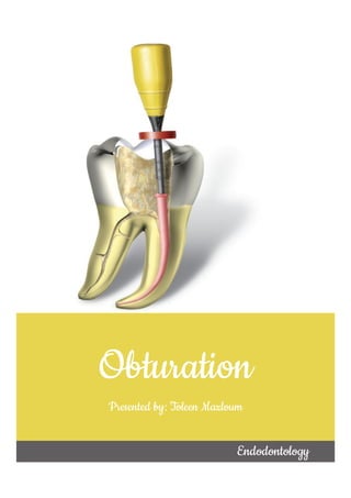
- 1. Obturation Presented by: Toleen Mazloum Endodontology
- 2. Outline: "criteria for ideal obturating materials" • > "criteria for ideal sealers" • >"techniques for placing sealers" "endodontic sealers" • >"lateral condensation of cold gutta-percha" • >"vertical compaction of warm gutta-percha" • >"thermomechanical compaction" • >"custom cone technique" "obturation technqiues" "citeria for the evaluation of the obturation's quality"
- 3. "Criteria for ideal obturating materials"i "easily introduced into the canal" "shouldn't stain tooth structure" "bacteriostatic (at least don't allow bacterial growth)" "easily removed from the root canal (when necessary)" "sterile (or easily & quickly sterilized prior to insertion )" "imprevious to moisture" "seal the canal laterally & apically" "shouldn't shrink after insertion" "semi-solid upon insertion & solid afterward" "radiopaque " "not irritant to periapical tissues"
- 4. "Endodontic sealers" "Criteria for ideal sealer"ii "shouldn't stain tooth structure" "bacteriostatic (at least don't allow bacterial growth)" "soluble in a common solvent >> can be removed (when necessary)" "sets slowly >> provide adequate working time" "tissue tolerant (doesn't irritate periapical tissues)" "insoluble in oral & tissue fluids" "shouldn't shrink upon setting" "consist of very fine powder particles >> optimal mix" "radiopaque " "able to make hermetic seal" "tacky when mixed >> provide good adhesion with canal walls" "shouldn't provoke an immune reponse in periradicular tissues" "non- carcinogenic & non- mutagenic"
- 5. "techniques for placing sealers"iii, iv "lentulo spiral" "manual" "rotary" "endodontic files" "gutta- percha cone" "ultrasonic file" "direct placement through intraoral tips"
- 6. "Obturation techniques:" "Lateral condensation of cold gutta-percha" ֍ "Technique:"v • "dry the canal using paper points" Definition "in this method, the root canal is filled by condensing gutta- percha points laterally against one canal wall using spreaders" • > "select a proper master cone (ideally same size as MAF)" • > "try the MC in a wet canal" • > "it should have tug-back at a point 0.5 - 1mm short of the radiographic apex" 1- "Cone fitting" • > "dry the canal using paper points" • > "mix the sealer & coat the canal walls" • > "coat the master cone (apical part) with sealer & insert into the canal" • > "insert the spreader next to the master cone against 1 wall (to within 1mm of the WL)" • > "this spreader compacts GP>> create space for accessory cones" 2- "lateral condensation" • > "remove the spreader" • > "insert the proper accessory cone coated with sealer" • > "place the spreader 1mm shorter than the previous one, remove it, then place the coated accessory cone" • > "repreat until spreader can no longer be insterted >> canal is full" • > "take an X-ray to make sure everything is alright" • > "remove the excess GP at the canal orifice using a heated instrument" • > "condense the top of GP vertically with a heated plugger" • > "clean the pulp chamber" 3- "accessory cones placement"
- 7. "Summary"
- 8. ֍ "Indications:" – "This technique is the most commonly used among other techniques" – " it is used in almost all situations except:" ֍ "Advantages:" ֍ "Disadvantages:" "severely curved canals" "abnormally shaped canals" "canals with gross irregularities (ex: internal resorption)" "simple" "requires simple equipment" "length control" "ease of retreatment" "adaptation to the canal wall" "ability to prepare post space" "positive dimensional stability" "minimized apical leakage" "time consuming" "not suitable in certain cases such as internal resorption, severe curvature..." "does not produce a homogeneous mass"
- 9. "Vertical compaction of warm gutta-percha" "In general, we'll need 3 pluggers:" ֍ "Technique:"vi Definition "this method was introduced by Schilder in which warmed & softened GP is adapted to irregularities & accessory and lateral canals within the root canal system (by vertical condensation)" "the canal should be a continuous tapered funnel & the apex should be kept as small as possible" "the widest plugger" "for the coronal 1/3" "narrower plugger" "for the middle 1/3" "the narrowest plugger" "for the apical 1/3"
- 10. An alternative method of backpacking may be done by injecting plasticized GP such as Obtura II ֍ "Indications:" – "This method is an alternative to the cold lateral compaction method" – "It's used in cases where the fitting of master cone to the apical part is impossible" – Example: "cases where there is "ledge formation" "perforation" "unusual canal curvature" "internal resorption" "large lateral canals"
- 11. ֍ "Advantages:" ֍ "Disadvantages:" "Thermomechanical compaction" "filling the canal irregularities" "preparation of post space" "excellent sealing of the canal apically, laterally & obturation of lateral and accessory canals" " lack of length control" "increased risk of vertical root fracture" "time consuming" "overfilling of canals with GP or sealer that can't be retrieved from periradicular tissues" " warming process >> generates temeprature within the canal" "difficult in curved canals" Definition "this method was introduced by Dr. John McSpadden" "it consists of a compactor which resembles a reverse H-file (H-file with blades toward the tip) & placed on a hand-piece (8,000-10,000 rpm)" " frictional heat from the compactor >> GP is plasticized & forced 1mm ahead & lateral to the compactor shaft""
- 12. ֍ "Technique:"vi ֍ "Indications:" ֍ "Disadvantages:" "Custom cone technique " • > "fit a master cone 1.5 mm shorter than the radiographic apex (since later the compactor will push GP 1mm apically)" • > "coat the master cone with sealer & introduce it into the canal" 1- "fit the master cone" • > "select the proper compactor (ideally same size as the largest file used within 1.5 of the apical stop)" • > "insert the compactor until a slight resistance is felt" • > "rotate the compactor to maximum speed" • > "after 1 second >> advance the compactor apically to the determined length" • > "remove the compactor slowly while it's still rotating at maximum speed" • > "don't withdraw the compactor quickly >> otherwise, voids will occur" 2- "compaction" "in cases where other techniques are difficult (ex: internal resorption)" "high rotational speed" "increased risk of instrument fracture" "heat generation" "risk of fracture around curves" "definition" "in this technique, a GP cone is customized specifically to fit the root canal." "chloroform dip technique" "solvents such as chloroform, eucalyptol or halothane are used to soften the outer surface of the cone as if making an impression of the apical portion of the canal." "Rolled technique" "in this technique, several large GP cones are heated & rolled between 2 glass slabs >>> single large cone"
- 13. ֍ Chloroform Dip Technique:" vii ֍ Clinical tips:" ֍ "Indications:" "mark the cone for orientation (to place it in the same position each time)" "never leave the cone in the canal while it's soft (its tip may separate while removing the master cone)" "wet with the canal with irrigants >> to prevent sticking of softened point to the canal's wall" "canal walls are not coated with sealer (only the apical 1/3 of the master cone)" "more sealer is added on accessory cones before placement" "choose a large standardized master cone that stops 2-4mm shorter than WL" "dip the master cone tip in chloroform for 3-4 seconds to soften it" "pack the cone apically in the canal & repeat several times" "grasp the cone at the reference point & measure it" "repeat softening & packing till the cone reaches the WL" "the cone tip should take an impression of the apical portion" "after reaching the WL, remove the cone & leave it to dry for 2-3 minutes" "immerse the tip of the cone in sealer & insert into canal" "complete obturation with lateral compaction technique" "cases of open apex (apical stop is lacking)" "cases where the apical portion is irregular or very large"
- 14. ֍ "Disadvantages:" ֍ Advantages: ֍ "Rolled Technique:" viii ֍ Indications:" "chemical solvents are irritant to periapical tissues" "chemically softened GP is dimesionally unstable" "solvents evaporate >> shrinkage of the root filling" "exact impression of the apical portion is taken >> improve the resultant seal" "arrange several large GP cones tip to butt" "heat them together & roll them between 2 glass slabs" "a single large cone is obtained" "repeat heating & rolling till the size of the obtained GP be similar to that of the canal" "cill the Tailor-made GP cone with ethyl chloride spray or water" "try it in the canal" "it should fit 1-2mm from the radiographic apex" "this cone is used as master cone" "continue obturation with lateral condensation technique" "when the canal is very wide & no single canal can be adjusted to fit (even when dipped in solvent)" "when the root canal is larger than the biggest standardized GP"
- 15. "Criteria for the evaluation of the obturation's quality" 1- "Radiographic evaluation:" "evaluation criteria" "radiographic evaluation" "clinical evaluation" "histological evaluation" •> "voids within the body or at the interface of obturation material & dentin wall >>> incomplete obturation" "Radiolucencies" •> "material should be of uniform density from coronal to apical part" •> "margins of GP should be sharp & distinct with no fuzziness >> indicates close adaptation" "Density" •> "the length of an ideal fill should be from the canal's apical minor constriction to the canal orifice (unless a post is planned)" "Length" •> "the restoration (whether permanent or temporary) should be contacting enough dentin surface to ensure a coronal seal" "Restoration" •> "the GP should reflect the canal shape (tapered from coronal to apical region)" "Taper"
- 16. 2-"Clinical evaluation:" "After a successful endodontic treatment, the following should be achieved:" 3- "Histological evaluation:" "after a successful endodontic treatment, the histological section should show" "Success" •> "normal PDL thickness & lamina dura when compared to adjacent teeth" •> "no evidence of resorption" •> "hermetic filling of root canal space" •> "dissappearance of radiolucency (if present) within 1 year" "Questionable" •> "radiolucency neither significantly improved nor deteriorated" •> "after endodontic surgery >> periapical scarring occurs" "Failure" •> "radiolucency has developed, persisted, or enlarged" "3 conditions" "no pain or swelling" "disappearance of fistula" "no loss of function" "no evidence of soft tissue distruction" "no tenderness to percussion or palpation" "no inflammation" •> "regeneration of PDL" •> "evidence of osseous repair with healthy osteoblast surrounding newly formed bone" "reconstitution of periapical structures"
- 17. i "Dr. Pradnya V.Bansode "Obturating Materials Present and Past: A Review” IOSR Journal of Dental and Medical Sciences (IOSR-JDMS), vol. 17, no. 3, 2018, pp. 27-33" ii " Garg N, Garg A. Textbook of endodontics. New Delhi: Jaypee Bros. Medical Publishers; 2010." iii "Hoen M, LaBounty G, Keller D. Ultrasonic endodontic sealer placement. Journal of Endodontics. 1988;14(4):169-174." iv "Sheetal M, Narayan R, Dhamali D, Singh A, Thakur A, Patil A. Effect of Placement Techniques on Sealing Ability of Root Canal Sealers. International Journal of Oral Care & Research. 2016;4(3):201-203." v"http://ccnmtl.columbia.edu/projects/virtechs2006/pdfs/endolateralcondens ationtechnique.pdf" vi "http://www.uobabylon.edu.iq/eprints/publication_4_472_1726.pdf" vii "https://pocketdentistry.com/root-canal-obturation-6/" viii "https://pocketdentistry.com/root-canal-obturation-6/"