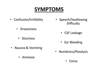
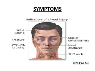
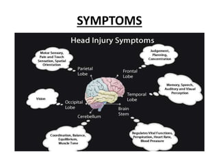
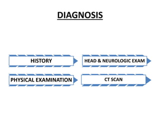
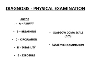
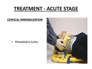
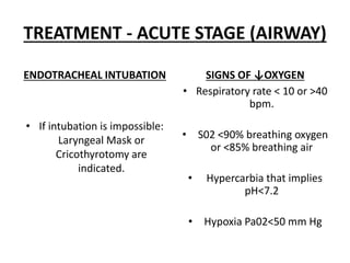
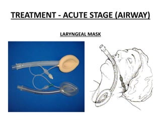
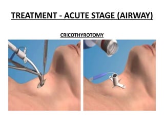
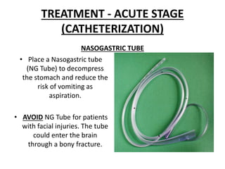
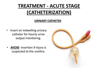
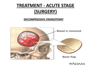
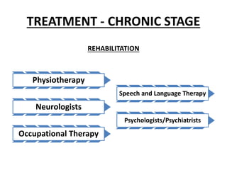
CROSS-SECTION
HEAD INJURY - DEFINITION
• Any injury that results
in trauma to the SCALP,
SKULL or BRAIN.
• TRAUMATIC BRAIN
INJURY and HEAD
INJURY are often used
interchangeably.
HEAD INJURY - TYPES
OPEN HEAD INJURY:
There is penetration to the skull.
CLOSED HEAD INJURY
There is NO penetration to the skull.
COUP-CONTRECOUP INJURIES
• Damage may occur
directly under the site
of impact (COUP), or it
may occur on the side
opposite the impact
(CONTRECOUP).
HEAD INJURY - MECHANISMS
PRIMARY INTRACRANIAL INJURY
• It is the initial neuronal
damage that occurs
IMMEDIATELY as result of
trauma.
SECONDARY INTRACRANIAL
INJURY
• Secondary injuries are the
result of the
neurophysiological and
anatomic changes, which
occur from MINUTES to
DAYS after the original
trauma.
HEAD INJURY - MECHANISMS
PRIMARY INTRACRANIAL INJURY
• Cerebral Laceration
• Cerebral Contusion
• Epidural Hematoma
• Subdural Hematoma
• Subarachnoid Hematoma
• Intracerebral Hematoma
• Diffuse Axonal Injury
SECONDARY INTRACRANIAL
INJURY
• Edema
• Impaired Metabolism
• Altered Cerebral Blood Flow
• Free Radical Formation
• Excitotoxicity
SCALP INJURIES
LACERATIONS SUBGALEAL HEMATOMA
SKULL INJURIES
CLOSED FRACTURES
• A closed fracture has a
significant chance of
associated intracranial
haematoma.
OPEN FRACTURES
• Open fractures have
potential for serious
infection.
• Any foreign matter impaled
in the skull should be left in
place for removal by the
neurosurgeons.
• Cover it lightly with a sterile
dressing that has been
moistened with a sterile
saline.
SKULL INJURIES
CT SCAN OT
SKULL INJURIES
DEPRESSED FRACTURES/COMPOUND
DEPRESSED FRACTURES
NON-DEPRESSED LINEAL
FRACTURES
SKULL INJURIES - BASILAR SKULL
FRACTURE
SKULL INJURIES - BASILAR SKULL
FRACTURE
RACCOON EYE
SKULL INJURIES - BASILAR SKULL
FRACTURE
BATTLE’S SIGN
SKULL INJURIES - BASILAR SKULL
FRACTURE
BLEEDING FROM THE EAR CANAL
CSF LEAKAGE FROM THE EAR OR
NOSE
BRAIN INJURIES
DIFFUSE
• Concussion
• Diffuse Axonal Injury
FOCAL
• Contusion
• Brain Lacerations
• Epidural haematoma
• Subdural haematoma
• Subarachnoid haemorrhage
• Parenchymal haematoma
HEAD INJURY (DIFFUSE) -
CONCUSSION
• Brain injury that does
not result in any
evidence of structural
alteration.
• Return of consciousness
moments or minutes
after impact.
• There may be brief
confusion,
disorientation,
headache, dizziness,
amnesia.
• CT scan is normal.
HEAD INJURY (DIFFUSE) - DIFFUSE
AXONAL INJURY
BRAIN CONTUSION
EPIDURAL HEMATOMA
SCHEMATIC CT SCAN
SUBDURAL HEMATOMA
SCHEMATIC CT SCAN
SUBARACHNOID HEMATOMA
SCHEMATIC CT SCAN
INTRACEREBRAL HEMATOMA
SCHEMATIC CT SCAN
HEMATOMAS
CEREBRAL EDEMA
NORMAL CT SCAN CEREBRAL EDEMA
SIGNS
A sign of ↑ICP
(INTRACRANIAL PRESSURE)
CUSHING REFLEX
↑ Blood Pressure
↓ Pulse Rate
↓ Respiratory Rate
SIGNS
• A UNILATERAL , FIXED
DILATED PUPIL indicates
neurologic deterioration
may be secondary to
hypoxia, hypovolaemia or
hypoglycaemia, due to
↑ICP, and compression of
the 3rd Cranial Nerve
(OCULOMOTOR NERVE).
DILATED PUPIL
SIGNS
SIGNS
DECORTICATE POSTURING
• Arms Flexed
• Arms bent inward on the
chest
• Hands clenched into fists
• Legs Extended
• Feet turned Inward
• Score of 3 in the Motor
section of the Glasgow
Coma Scale
SIGNS
DECEREBRATE POSTURING
• Head is arched back
• Arms Extended by the sides
• Legs Extended
• Patient is rigid with the
teeth clenched.
• Score of 2 in the Motor
section of the Glasgow
Coma Scale
SYMPTOMS
• Confusion/Irritibility
• Drowsiness
• Dizziness
• Nausea & Vomiting
• Amnesia
• Speech/Swallowing
Difficulty
• CSF Leakage
• Ear Bleeding
• Numbness/Paralysis
• Coma
SYMPTOMS
SYMPTOMS
DIAGNOSIS
HISTORY
PHYSICAL EXAMINATION
HEAD & NEUROLOGIC EXAM
CT SCAN
DIAGNOSIS - HISTORY
PATIENT
PEOPLE
DIAGNOSIS - PHYSICAL EXAMINATION
ABCDE
• A = AIRWAY
• B = BREATHING
• C = CIRCULATION
• D = DISABILITY
• E = EXPOSURE
• GLASGOW COMA SCALE
(GCS)
• SYSTEMIC EXAMINATION
GLASGOW COMA SCALE
MINIMUM=3/15 MAXIMUM=15/15 INTUBATION <8/15
GLASGOW COMA SCALE (GCS)
SEVERITY SCORE
13-15
9-12
3-8
MILD
MODERATE
SEVERE
GLASGOW COMA SCALE (GCS)
SEVERITY LOSS OF CONSCIOUSNESS
0-30 mins
>30 mins to <24 hrs
>24 hrs
MILD
MODERATE
SEVERE
DIAGNOSIS - HEAD AND NEUROLOGIC
EXAM
HEAD EXAM
• Hematoma
• Contusion
• Fracture e.g. Basilar Skull
Fracture
• Laceration
NEUROLOGIC EXAM
• Cranial Nerves
• Muscle Tone
• Muscle Power
• Sensations
• Walking Gait
DIAGNOSIS - OTHERS
X-RAYS / MRI
ANGIOGRAPHY
EEG
TRANSCRANIAL DOPPLER
TREATMENT
ACUTE STAGE
CHRONIC STAGE
TREATMENT - ACUTE STAGE
CERVICAL IMMOBILIZATION
• Philadelphia Collar
TREATMENT - ACUTE STAGE (AIRWAY)
ENDOTRACHEAL INTUBATION
• If intubation is impossible:
Laryngeal Mask or
Cricothyrotomy are
indicated.
SIGNS OF ↓OXYGEN
• Respiratory rate < 10 or >40
bpm.
• S02 <90% breathing oxygen
or <85% breathing air
• Hypercarbia that implies
pH<7.2
• Hypoxia Pa02<50 mm Hg
TREATMENT - ACUTE STAGE (AIRWAY)
LARYNGEAL MASK
TREATMENT - ACUTE STAGE (AIRWAY)
CRICOTHYROTOMY
TREATMENT - ACUTE STAGE (AIRWAY)
ENDOTRACHEAL INTUBATION
• Rapid sequence intubation
is performed, using sedative
agents and muscle
relaxants.
MECHANICAL VENTILATION
STANDARD PARAMETERS
• Tidal Volume: 8-10 ml/kg
• Rate: 12-15 bpm
• Pressure: 15-20 cm H20
• Fi02: 1
TREATMENT - ACUTE STAGE
(BREATHING)
• Start high-flow oxygen
administration (10-12 l/min)
TREATMENT - ACUTE STAGE
(CIRCULATION)
• Establish IV access with
two large-bore(14- or16
gauge) IV cannulas.
• IV infusion of Normal
Saline (NS).
• IV Norepinephrine
• AVOID giving 5% Dextrose
unless hypoglycaemia is
present.
• Dextrose ↑cerebral
oedema
• If BP is normal AVOID
giving excessive volumes
of fluids that may
↑cerebral oedema.
TREATMENT - ACUTE STAGE
(DISABILITY)
TREATMENT FOR ↑ICP
• IV Mannitol (Osmotic
Diuretic)
• IV Furosemide
• Hyperventilation
TREATMENT - ACUTE STAGE
(DISABILITY)
TREATMENT FOR ↑ICP
• If there are no counter-
indications (hypovolaemia,
spine injury) place the
patient in
“Reverse-Trendelenburg”
position
REVERSE-TRENDELENBURG
TREATMENT - ACUTE STAGE
(DISABILITY)
• If significant agitation and after excluding
hypoxia, hypovolaemia or pain, as the cause of
agitation: IV Midazolam
TREATMENT - ACUTE STAGE
(EXPOSURE)
• AVOID ↓Body
Temperature
• ↑Body Temperature:
Cooling measures and
IV Paracetamol
• Pain medication: IV
Fentanyl
• Anti-Emetics
• Post-Traumatic
Seizures: IV Diazepam
TREATMENT - ACUTE STAGE
(PARAMETERS)
MONITOR
• Blood Pressure
• Heart Rate
• Respiratory Rate
• S02, Etc02
• ECG
BLOOD SAMPLES
• Serum Electrolytes
• Arterial Blood Gas
• Hyper/Hypoglycaemia
TREATMENT - ACUTE STAGE
(CATHETERIZATION)
NASOGASTRIC TUBE
• Place a Nasogastric tube
(NG Tube) to decompress
the stomach and reduce the
risk of vomiting as
aspiration.
• AVOID NG Tube for patients
with facial injuries. The tube
could enter the brain
through a bony fracture.
TREATMENT - ACUTE STAGE
(CATHETERIZATION)
URINARY CATHETER
• Insert an indwelling urinary
catheter for hourly urine
output monitoring.
• AVOID insertion if injury is
suspected to the urethra.
TREATMENT - ACUTE STAGE
(SURGERY)
DECOMPRESSIVE CRANIOTOMY
TREATMENT - CHRONIC STAGE
REHABILITATION
Physiotherapy
Neurologists
Occupational Therapy
Speech and Language Therapy
Psychologists/Psychiatrists
COMPLICATIONS
• Personality Changes
• Hypopituitarism e.g. DI
• Post-Traumatic Seizures
• Infections e.g. Meningitis
• Vasospasm, Aneurysm
• Coma, Brain Death
LONG-TERM EFFECTS
• Parkinson’s
• Alzheimer’s Dementia
PREVENTION
HELMETS
SEAT BELTS
FALLS IN THE ELDERLY
RESTRICTING ALCOHOL USE

Head injuries Overview

  • 2.
  • 3.
    HEAD INJURY -DEFINITION • Any injury that results in trauma to the SCALP, SKULL or BRAIN. • TRAUMATIC BRAIN INJURY and HEAD INJURY are often used interchangeably.
  • 4.
    HEAD INJURY -TYPES OPEN HEAD INJURY: There is penetration to the skull. CLOSED HEAD INJURY There is NO penetration to the skull.
  • 5.
    COUP-CONTRECOUP INJURIES • Damagemay occur directly under the site of impact (COUP), or it may occur on the side opposite the impact (CONTRECOUP).
  • 6.
    HEAD INJURY -MECHANISMS PRIMARY INTRACRANIAL INJURY • It is the initial neuronal damage that occurs IMMEDIATELY as result of trauma. SECONDARY INTRACRANIAL INJURY • Secondary injuries are the result of the neurophysiological and anatomic changes, which occur from MINUTES to DAYS after the original trauma.
  • 7.
    HEAD INJURY -MECHANISMS PRIMARY INTRACRANIAL INJURY • Cerebral Laceration • Cerebral Contusion • Epidural Hematoma • Subdural Hematoma • Subarachnoid Hematoma • Intracerebral Hematoma • Diffuse Axonal Injury SECONDARY INTRACRANIAL INJURY • Edema • Impaired Metabolism • Altered Cerebral Blood Flow • Free Radical Formation • Excitotoxicity
  • 8.
  • 9.
    SKULL INJURIES CLOSED FRACTURES •A closed fracture has a significant chance of associated intracranial haematoma. OPEN FRACTURES • Open fractures have potential for serious infection. • Any foreign matter impaled in the skull should be left in place for removal by the neurosurgeons. • Cover it lightly with a sterile dressing that has been moistened with a sterile saline.
  • 10.
  • 11.
    SKULL INJURIES DEPRESSED FRACTURES/COMPOUND DEPRESSEDFRACTURES NON-DEPRESSED LINEAL FRACTURES
  • 12.
    SKULL INJURIES -BASILAR SKULL FRACTURE
  • 13.
    SKULL INJURIES -BASILAR SKULL FRACTURE RACCOON EYE
  • 14.
    SKULL INJURIES -BASILAR SKULL FRACTURE BATTLE’S SIGN
  • 15.
    SKULL INJURIES -BASILAR SKULL FRACTURE BLEEDING FROM THE EAR CANAL CSF LEAKAGE FROM THE EAR OR NOSE
  • 16.
    BRAIN INJURIES DIFFUSE • Concussion •Diffuse Axonal Injury FOCAL • Contusion • Brain Lacerations • Epidural haematoma • Subdural haematoma • Subarachnoid haemorrhage • Parenchymal haematoma
  • 17.
    HEAD INJURY (DIFFUSE)- CONCUSSION • Brain injury that does not result in any evidence of structural alteration. • Return of consciousness moments or minutes after impact. • There may be brief confusion, disorientation, headache, dizziness, amnesia. • CT scan is normal.
  • 18.
    HEAD INJURY (DIFFUSE)- DIFFUSE AXONAL INJURY
  • 19.
  • 20.
  • 21.
  • 22.
  • 23.
  • 24.
  • 25.
    CEREBRAL EDEMA NORMAL CTSCAN CEREBRAL EDEMA
  • 26.
    SIGNS A sign of↑ICP (INTRACRANIAL PRESSURE) CUSHING REFLEX ↑ Blood Pressure ↓ Pulse Rate ↓ Respiratory Rate
  • 27.
    SIGNS • A UNILATERAL, FIXED DILATED PUPIL indicates neurologic deterioration may be secondary to hypoxia, hypovolaemia or hypoglycaemia, due to ↑ICP, and compression of the 3rd Cranial Nerve (OCULOMOTOR NERVE). DILATED PUPIL
  • 28.
  • 29.
    SIGNS DECORTICATE POSTURING • ArmsFlexed • Arms bent inward on the chest • Hands clenched into fists • Legs Extended • Feet turned Inward • Score of 3 in the Motor section of the Glasgow Coma Scale
  • 30.
    SIGNS DECEREBRATE POSTURING • Headis arched back • Arms Extended by the sides • Legs Extended • Patient is rigid with the teeth clenched. • Score of 2 in the Motor section of the Glasgow Coma Scale
  • 31.
    SYMPTOMS • Confusion/Irritibility • Drowsiness •Dizziness • Nausea & Vomiting • Amnesia • Speech/Swallowing Difficulty • CSF Leakage • Ear Bleeding • Numbness/Paralysis • Coma
  • 32.
  • 33.
  • 34.
  • 35.
  • 36.
    DIAGNOSIS - PHYSICALEXAMINATION ABCDE • A = AIRWAY • B = BREATHING • C = CIRCULATION • D = DISABILITY • E = EXPOSURE • GLASGOW COMA SCALE (GCS) • SYSTEMIC EXAMINATION
  • 37.
    GLASGOW COMA SCALE MINIMUM=3/15MAXIMUM=15/15 INTUBATION <8/15
  • 38.
    GLASGOW COMA SCALE(GCS) SEVERITY SCORE 13-15 9-12 3-8 MILD MODERATE SEVERE
  • 39.
    GLASGOW COMA SCALE(GCS) SEVERITY LOSS OF CONSCIOUSNESS 0-30 mins >30 mins to <24 hrs >24 hrs MILD MODERATE SEVERE
  • 40.
    DIAGNOSIS - HEADAND NEUROLOGIC EXAM HEAD EXAM • Hematoma • Contusion • Fracture e.g. Basilar Skull Fracture • Laceration NEUROLOGIC EXAM • Cranial Nerves • Muscle Tone • Muscle Power • Sensations • Walking Gait
  • 41.
    DIAGNOSIS - OTHERS X-RAYS/ MRI ANGIOGRAPHY EEG TRANSCRANIAL DOPPLER
  • 42.
  • 43.
    TREATMENT - ACUTESTAGE CERVICAL IMMOBILIZATION • Philadelphia Collar
  • 44.
    TREATMENT - ACUTESTAGE (AIRWAY) ENDOTRACHEAL INTUBATION • If intubation is impossible: Laryngeal Mask or Cricothyrotomy are indicated. SIGNS OF ↓OXYGEN • Respiratory rate < 10 or >40 bpm. • S02 <90% breathing oxygen or <85% breathing air • Hypercarbia that implies pH<7.2 • Hypoxia Pa02<50 mm Hg
  • 45.
    TREATMENT - ACUTESTAGE (AIRWAY) LARYNGEAL MASK
  • 46.
    TREATMENT - ACUTESTAGE (AIRWAY) CRICOTHYROTOMY
  • 47.
    TREATMENT - ACUTESTAGE (AIRWAY) ENDOTRACHEAL INTUBATION • Rapid sequence intubation is performed, using sedative agents and muscle relaxants. MECHANICAL VENTILATION STANDARD PARAMETERS • Tidal Volume: 8-10 ml/kg • Rate: 12-15 bpm • Pressure: 15-20 cm H20 • Fi02: 1
  • 48.
    TREATMENT - ACUTESTAGE (BREATHING) • Start high-flow oxygen administration (10-12 l/min)
  • 49.
    TREATMENT - ACUTESTAGE (CIRCULATION) • Establish IV access with two large-bore(14- or16 gauge) IV cannulas. • IV infusion of Normal Saline (NS). • IV Norepinephrine • AVOID giving 5% Dextrose unless hypoglycaemia is present. • Dextrose ↑cerebral oedema • If BP is normal AVOID giving excessive volumes of fluids that may ↑cerebral oedema.
  • 50.
    TREATMENT - ACUTESTAGE (DISABILITY) TREATMENT FOR ↑ICP • IV Mannitol (Osmotic Diuretic) • IV Furosemide • Hyperventilation
  • 51.
    TREATMENT - ACUTESTAGE (DISABILITY) TREATMENT FOR ↑ICP • If there are no counter- indications (hypovolaemia, spine injury) place the patient in “Reverse-Trendelenburg” position REVERSE-TRENDELENBURG
  • 52.
    TREATMENT - ACUTESTAGE (DISABILITY) • If significant agitation and after excluding hypoxia, hypovolaemia or pain, as the cause of agitation: IV Midazolam
  • 53.
    TREATMENT - ACUTESTAGE (EXPOSURE) • AVOID ↓Body Temperature • ↑Body Temperature: Cooling measures and IV Paracetamol • Pain medication: IV Fentanyl • Anti-Emetics • Post-Traumatic Seizures: IV Diazepam
  • 54.
    TREATMENT - ACUTESTAGE (PARAMETERS) MONITOR • Blood Pressure • Heart Rate • Respiratory Rate • S02, Etc02 • ECG BLOOD SAMPLES • Serum Electrolytes • Arterial Blood Gas • Hyper/Hypoglycaemia
  • 55.
    TREATMENT - ACUTESTAGE (CATHETERIZATION) NASOGASTRIC TUBE • Place a Nasogastric tube (NG Tube) to decompress the stomach and reduce the risk of vomiting as aspiration. • AVOID NG Tube for patients with facial injuries. The tube could enter the brain through a bony fracture.
  • 56.
    TREATMENT - ACUTESTAGE (CATHETERIZATION) URINARY CATHETER • Insert an indwelling urinary catheter for hourly urine output monitoring. • AVOID insertion if injury is suspected to the urethra.
  • 57.
    TREATMENT - ACUTESTAGE (SURGERY) DECOMPRESSIVE CRANIOTOMY
  • 58.
    TREATMENT - CHRONICSTAGE REHABILITATION Physiotherapy Neurologists Occupational Therapy Speech and Language Therapy Psychologists/Psychiatrists
  • 59.
    COMPLICATIONS • Personality Changes •Hypopituitarism e.g. DI • Post-Traumatic Seizures • Infections e.g. Meningitis • Vasospasm, Aneurysm • Coma, Brain Death LONG-TERM EFFECTS • Parkinson’s • Alzheimer’s Dementia
  • 60.
    PREVENTION HELMETS SEAT BELTS FALLS INTHE ELDERLY RESTRICTING ALCOHOL USE