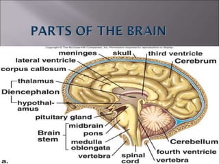
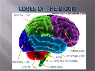
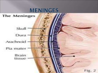
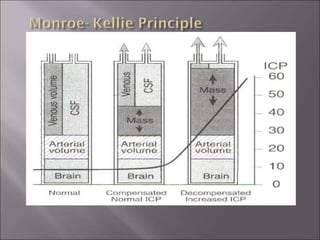
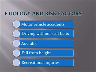
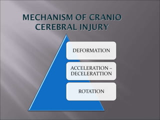
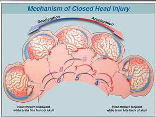
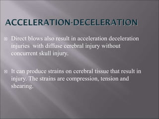
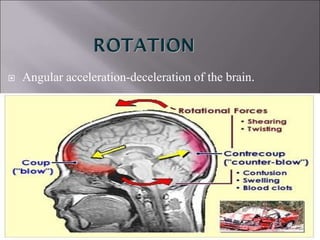
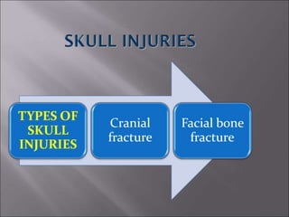
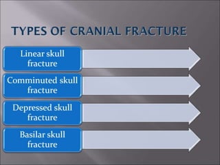
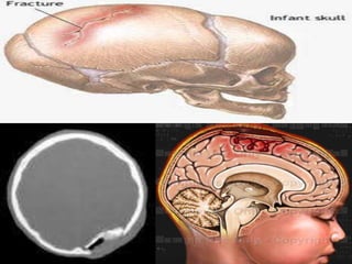
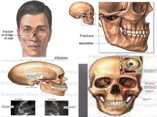
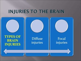
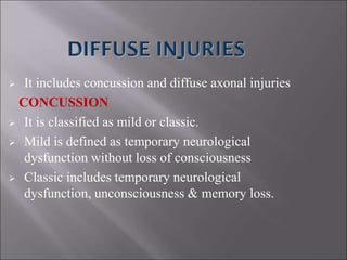
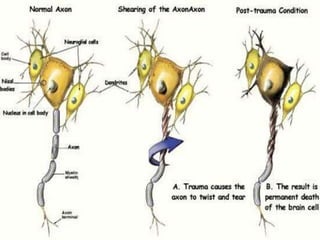
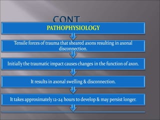
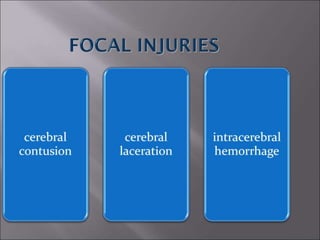
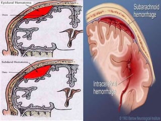
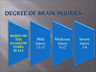
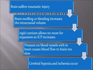
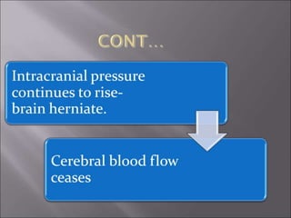
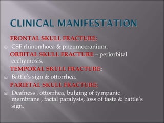
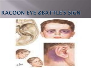
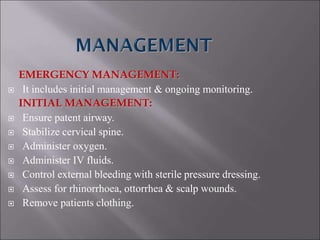
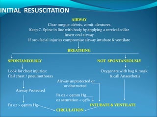
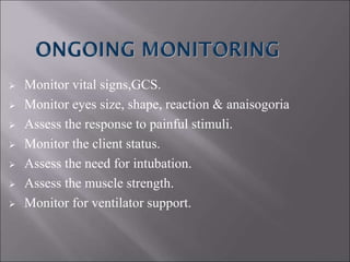
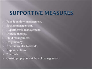
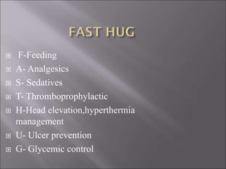
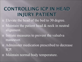
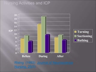
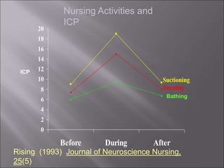
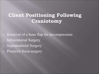
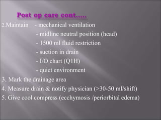
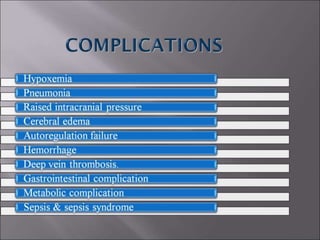
LOBES OF THE BRAIN
ACCELERATION-DECELERATION:-
The rapid changes in velocity of the brain with
in the cranial vault along a straight line from forward
movement to an abrupt stop.
ROTATION:-
Angular acceleration-deceleration of the brain.
TERMINOLOGY
COUP INJURY:-
A focal cerebral injury directly under the
area of impact is called a coup injury.
COUNTERCOUP INJURY:-
A focal cerebral injury sustained to the
opposite pole of direct impact
HALO SIGN:-
The appearance of blood encircled by a
yellowish stain on the dressing or bed linen is called
halo sign.
CEREBRAL CONCUSSION:-
It is defined as a transient temporary
,neurogenic dysfunction caused by mechanical force
to the brain
CEREBRAL CONTUSION:-
A cerebral contusion is a bruising of
the surface of the brain.
 Head injury refers to any injury to the scalp, skull
or brain.
 The terms head injury and cranio cerebral trauma is
an insult to the brain that is capable of producing
physical, intellectual emotional ,social and
vocational changes.
 Direct blows also result in acceleration deceleration
injuries with diffuse cerebral injury without
concurrent skull injury.
 It can produce strains on cerebral tissue that result in
injury. The strains are compression, tension and
shearing.
 Angular acceleration-deceleration of the brain.
 It includes concussion and diffuse axonal injuries
CONCUSSION
 It is classified as mild or classic.
 Mild is defined as temporary neurological
dysfunction without loss of consciousness
 Classic includes temporary neurological
dysfunction, unconsciousness & memory loss.
DEFINITION
 It is a widespread axonal damage occuring after a
mild, moderate or severe traumatic brain injury. The
damage occurs primarily around the axons in
subcortical white matter of cerebral hemispheres,
basal ganglia, thalamus & brainstem.
 DEFINITION
 It is a bruising of the surface of the brain. It may
occur from blunt trauma , penetrating wounds or
acceleration- deceleration closed injuries.
 It may be coup or counter coup depending on the
degree of acceleration – deceleration.
 It refers to be a traumatic tearing of the cortical
surface of the brain.
 Usually it is similar like a contusion
DEFINITION
 Hemorrhage can occur beneath the fracture or from
an acceleration – deceleration injury in which there
is shearing of the bridging veins or cortical artery.
FRONTAL SKULL FRACTURE:
 CSF rhinorrhoea & pneumocranium.
ORBITAL SKULL FRACTURE – periorbital
ecchymosis.
TEMPORAL SKULL FRACTURE:
 Battle’s sign & ottorrhea.
PARIETAL SKULL FRACTURE:
 Deafness , ottorrhea, bulging of tympanic
membrane , facial paralysis, loss of taste & battle’s
sign.
BASILLAR SKULL FRACTURE:
 CSF or brain ottorrhea, tinnitus or hearing
difficulties, facial paralysis, vertigo & halo sign.
CONCUSSION:
 Headache, dizziness, lethargy, irritability, inability
to concentrate, memory loss & vomiting.
TEMPORAL LOBE CONTUSION:
 Agitation, confusion & remain alert.
 FRONTAL LOBE CONTUSION:
 Alert & hemiparesis.
 BRAIN STEM CONTUSION:
 Unresponsive or partially comatose, rapid
respiration, loss of normal eye movement and small
, equal & reactive pupils.
 INCREASED ICP:
 Early findings:
 Deterioration in the level of consciousness.
 Pupillary dysfunction.
 motor weakness
 sensory deficit
 headache
 possible seizure
later findings:
 Cushing triad
 possible vomiting
 possible papilledema
 hemiplegia, decortication or decerebration
 changes in vital signs
EMERGENCY MANAGEMENT:
 It includes initial management & ongoing monitoring.
INITIAL MANAGEMENT:
 Ensure patent airway.
 Stabilize cervical spine.
 Administer oxygen.
 Administer IV fluids.
 Control external bleeding with sterile pressure dressing.
 Assess for rhinorrhoea, ottorrhea & scalp wounds.
 Remove patients clothing.
INITIAL RESUSCITATION
AIRWAY
Clear tongue, debris, vomit, dentures
Keep C. Spine in line with body by applying a cervical collar
Insert oral airway
If oro–facial injuries compromise airway intubate & ventilate
BREATHING
SPONTANEOUSLY NOT SPONTANEOUSLY
Look for chest injuries: Oxygenate witb bag & mask
flail chest / pneumothorax & call Anaesthetis
Airway unptotected or
or obstructed
Airway Protected
Pa 02 < 95mm Hg
02 saturation < 95%
Pa 02 > 95mm Hg INTUBATE & VENTILATE
CIRCULATION
 Monitor vital signs,GCS.
 Monitor eyes size, shape, reaction & anaisogoria
 Assess the response to painful stimuli.
 Monitor the client status.
 Assess the need for intubation.
 Assess the muscle strength.
 Monitor for ventilator support.
 Pain & anxiety management.
 Seizure management.
 Hyperthermia management.
 Diuretic therapy.
 Fluid management.
 Drug therapy.
 Neuromuscular blockade.
 Hyperventilation
 ?Steroids.
 Gastric prophylaxis & bowel management.
 F-Feeding
 A- Analgesics
 S- Sedatives
 T- Thromboprophylactic
 H-Head elevation,hyperthermia
management
 U- Ulcer prevention
 G- Glycemic control
 Elevate the head of the bed to 30 degree.
 Maintain the patient head & neck in neutral
alignment.
 Initiate measures to prevent the valsalva
maneuver.
 Administer medication prescribed to decrease
ICP.
 Maintain normal body temperature.
Keep neck mid-line and elevate head of bed …. To what degree?
Dicarlo in ALL-NET Pediatric Critical Care Textbook
www.med.ub.es/All-
Net/english/neuropage/protect/icp-tx-3.htm
Feldman et al.
(1992) Journal
of
Neurosurgery,
76
March et al.
(1990) Journal
of Neuroscience
Nursing, 22(6)
Parsons &
Wilson (1984)
Nursing
Research, 33(2)
0
2
4
6
8
10
12
14
16
18
20
Before During After
Turning
Suctioning
Bathing
Nursing Activities and ICP
Rising (1993) Journal of Neuroscience
Nursing, 25(5)
ICP
0
2
4
6
8
10
12
14
16
18
20
Before During After
Suctioning
Turning
Nursing Activities and
ICP
Rising (1993) Journal of Neuroscience Nursing,
25(5)
ICP
Bathing
Family Contact and ICP
Bruya (1981) Journal of Neuroscience Nursing, 13
Hendrickson (1987) Journal of Neuroscience Nursing, 19(1)
Mitchell (1985) Nursing Administration Quarterly, 9(4)
Treolar (1991) Journal of Neuroscience Nursing, 23(5)
Presence, touch and voice of family / significant
others...
• Does not significantly increase ICP
• Has been demonstrated to decrease ICP
 Hyperventilate the patient on mechanical
ventilation- administer oxygen.
 Maintain fluid restriction.
 Avoid noxious stimuli.
 Administer sedation to reduce metabolic
demands.
 Maintain the blood pressure – 140/90 mmhg
 Avoid physical restraint
 Indications
 Hemorrhage
 EDH,SDH
 Cerebral aneurysm
 Brain abscess
 Damage to tissues covering the brain
 Pre operative nursing care
Client Positioning Following
Craniotomy
1. Removal of a bone flap for decompression
2. Infratentorial Surgery
3. Supratentorial Surgery
4. Posterior fossa surgery
1. Monitor - vital signs & neurological status
- ↓sed LOC ,motor weakness
- aphasia ,visual changes
- personality changes
- ↑sed ICP
- head dressing (drainage sign)
- drain
- electrolyte levels
- dysrhythmias
2.Maintain - mechanical ventilation
- midline neutral position (head)
- 1500 ml fluid restriction
- suction in drain
- I/O chart (Q1H)
- quiet environment
3. Mark the drainage area
4. Measure drain & notify physician (>30-50 ml/shift)
5. Give cool compress (ecchymosis /periorbital edema)
New head injury nursing

New head injury nursing

  • 5.
  • 8.
    ACCELERATION-DECELERATION:- The rapid changesin velocity of the brain with in the cranial vault along a straight line from forward movement to an abrupt stop. ROTATION:- Angular acceleration-deceleration of the brain. TERMINOLOGY
  • 9.
    COUP INJURY:- A focalcerebral injury directly under the area of impact is called a coup injury. COUNTERCOUP INJURY:- A focal cerebral injury sustained to the opposite pole of direct impact
  • 10.
    HALO SIGN:- The appearanceof blood encircled by a yellowish stain on the dressing or bed linen is called halo sign. CEREBRAL CONCUSSION:- It is defined as a transient temporary ,neurogenic dysfunction caused by mechanical force to the brain
  • 11.
    CEREBRAL CONTUSION:- A cerebralcontusion is a bruising of the surface of the brain.
  • 12.
     Head injuryrefers to any injury to the scalp, skull or brain.  The terms head injury and cranio cerebral trauma is an insult to the brain that is capable of producing physical, intellectual emotional ,social and vocational changes.
  • 17.
     Direct blowsalso result in acceleration deceleration injuries with diffuse cerebral injury without concurrent skull injury.  It can produce strains on cerebral tissue that result in injury. The strains are compression, tension and shearing.
  • 18.
  • 26.
     It includesconcussion and diffuse axonal injuries CONCUSSION  It is classified as mild or classic.  Mild is defined as temporary neurological dysfunction without loss of consciousness  Classic includes temporary neurological dysfunction, unconsciousness & memory loss.
  • 27.
    DEFINITION  It isa widespread axonal damage occuring after a mild, moderate or severe traumatic brain injury. The damage occurs primarily around the axons in subcortical white matter of cerebral hemispheres, basal ganglia, thalamus & brainstem.
  • 31.
     DEFINITION  Itis a bruising of the surface of the brain. It may occur from blunt trauma , penetrating wounds or acceleration- deceleration closed injuries.  It may be coup or counter coup depending on the degree of acceleration – deceleration.
  • 33.
     It refersto be a traumatic tearing of the cortical surface of the brain.  Usually it is similar like a contusion
  • 34.
    DEFINITION  Hemorrhage canoccur beneath the fracture or from an acceleration – deceleration injury in which there is shearing of the bridging veins or cortical artery.
  • 41.
    FRONTAL SKULL FRACTURE: CSF rhinorrhoea & pneumocranium. ORBITAL SKULL FRACTURE – periorbital ecchymosis. TEMPORAL SKULL FRACTURE:  Battle’s sign & ottorrhea. PARIETAL SKULL FRACTURE:  Deafness , ottorrhea, bulging of tympanic membrane , facial paralysis, loss of taste & battle’s sign.
  • 43.
    BASILLAR SKULL FRACTURE: CSF or brain ottorrhea, tinnitus or hearing difficulties, facial paralysis, vertigo & halo sign. CONCUSSION:  Headache, dizziness, lethargy, irritability, inability to concentrate, memory loss & vomiting. TEMPORAL LOBE CONTUSION:  Agitation, confusion & remain alert.
  • 44.
     FRONTAL LOBECONTUSION:  Alert & hemiparesis.  BRAIN STEM CONTUSION:  Unresponsive or partially comatose, rapid respiration, loss of normal eye movement and small , equal & reactive pupils.  INCREASED ICP:  Early findings:  Deterioration in the level of consciousness.  Pupillary dysfunction.
  • 45.
     motor weakness sensory deficit  headache  possible seizure later findings:  Cushing triad  possible vomiting  possible papilledema  hemiplegia, decortication or decerebration  changes in vital signs
  • 47.
    EMERGENCY MANAGEMENT:  Itincludes initial management & ongoing monitoring. INITIAL MANAGEMENT:  Ensure patent airway.  Stabilize cervical spine.  Administer oxygen.  Administer IV fluids.  Control external bleeding with sterile pressure dressing.  Assess for rhinorrhoea, ottorrhea & scalp wounds.  Remove patients clothing.
  • 49.
    INITIAL RESUSCITATION AIRWAY Clear tongue,debris, vomit, dentures Keep C. Spine in line with body by applying a cervical collar Insert oral airway If oro–facial injuries compromise airway intubate & ventilate BREATHING SPONTANEOUSLY NOT SPONTANEOUSLY Look for chest injuries: Oxygenate witb bag & mask flail chest / pneumothorax & call Anaesthetis Airway unptotected or or obstructed Airway Protected Pa 02 < 95mm Hg 02 saturation < 95% Pa 02 > 95mm Hg INTUBATE & VENTILATE CIRCULATION
  • 50.
     Monitor vitalsigns,GCS.  Monitor eyes size, shape, reaction & anaisogoria  Assess the response to painful stimuli.  Monitor the client status.  Assess the need for intubation.  Assess the muscle strength.  Monitor for ventilator support.
  • 51.
     Pain &anxiety management.  Seizure management.  Hyperthermia management.  Diuretic therapy.  Fluid management.  Drug therapy.  Neuromuscular blockade.  Hyperventilation  ?Steroids.  Gastric prophylaxis & bowel management.
  • 52.
     F-Feeding  A-Analgesics  S- Sedatives  T- Thromboprophylactic  H-Head elevation,hyperthermia management  U- Ulcer prevention  G- Glycemic control
  • 53.
     Elevate thehead of the bed to 30 degree.  Maintain the patient head & neck in neutral alignment.  Initiate measures to prevent the valsalva maneuver.  Administer medication prescribed to decrease ICP.  Maintain normal body temperature.
  • 54.
    Keep neck mid-lineand elevate head of bed …. To what degree? Dicarlo in ALL-NET Pediatric Critical Care Textbook www.med.ub.es/All- Net/english/neuropage/protect/icp-tx-3.htm Feldman et al. (1992) Journal of Neurosurgery, 76 March et al. (1990) Journal of Neuroscience Nursing, 22(6) Parsons & Wilson (1984) Nursing Research, 33(2)
  • 55.
    0 2 4 6 8 10 12 14 16 18 20 Before During After Turning Suctioning Bathing NursingActivities and ICP Rising (1993) Journal of Neuroscience Nursing, 25(5) ICP
  • 56.
    0 2 4 6 8 10 12 14 16 18 20 Before During After Suctioning Turning NursingActivities and ICP Rising (1993) Journal of Neuroscience Nursing, 25(5) ICP Bathing
  • 57.
    Family Contact andICP Bruya (1981) Journal of Neuroscience Nursing, 13 Hendrickson (1987) Journal of Neuroscience Nursing, 19(1) Mitchell (1985) Nursing Administration Quarterly, 9(4) Treolar (1991) Journal of Neuroscience Nursing, 23(5) Presence, touch and voice of family / significant others... • Does not significantly increase ICP • Has been demonstrated to decrease ICP
  • 58.
     Hyperventilate thepatient on mechanical ventilation- administer oxygen.  Maintain fluid restriction.  Avoid noxious stimuli.  Administer sedation to reduce metabolic demands.  Maintain the blood pressure – 140/90 mmhg  Avoid physical restraint
  • 60.
     Indications  Hemorrhage EDH,SDH  Cerebral aneurysm  Brain abscess  Damage to tissues covering the brain  Pre operative nursing care
  • 61.
    Client Positioning Following Craniotomy 1.Removal of a bone flap for decompression 2. Infratentorial Surgery 3. Supratentorial Surgery 4. Posterior fossa surgery
  • 64.
    1. Monitor -vital signs & neurological status - ↓sed LOC ,motor weakness - aphasia ,visual changes - personality changes - ↑sed ICP - head dressing (drainage sign) - drain - electrolyte levels - dysrhythmias
  • 65.
    2.Maintain - mechanicalventilation - midline neutral position (head) - 1500 ml fluid restriction - suction in drain - I/O chart (Q1H) - quiet environment 3. Mark the drainage area 4. Measure drain & notify physician (>30-50 ml/shift) 5. Give cool compress (ecchymosis /periorbital edema)