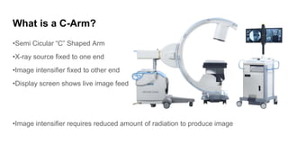
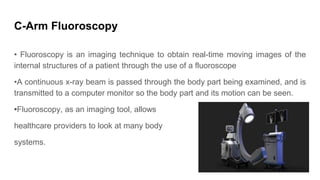
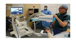
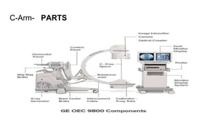
All about C-Arm system and how it workds
Copy this link to know more about Carm system -https://thetrend4u.blogspot.com/2022/10/what-is-c-arm-system-download-power.html
1. Introduction
•A C-Arm machine is an advanced medical imaging device based on X-ray
technology
•The name derives from the C-shaped arm used to connect the x-ray source and
x-ray detector to one another. C-arms have radiographic capabilities, though they
are used primarily for fluoroscopic intraoperative imaging during surgical,
orthopedic and emergency care procedures.
2. What is a C-Arm?
•Semi Cicular “C” Shaped Arm
•X-ray source fixed to one end
•Image intensifier fixed to other end
•Display screen shows live image feed
•Image intensifier requires reduced amount of radiation to produce image
3. What is an X-ray image intensifier ?
•As the name implies, this intensifier creates images with higher intensity.
•It converts X-rays into clear visible light better than the ordinary flourescent
screens.
•With this intensifying effect, low-intensity x-rays are conveniently seen in a bright
manner, helping the physicians to view the X-rayed object more clearly
4. C-Arm Fluoroscopy
• Fluoroscopy is an imaging technique to obtain real-time moving images of the
internal structures of a patient through the use of a fluoroscope
•A continuous x-ray beam is passed through the body part being examined, and is
transmitted to a computer monitor so the body part and its motion can be seen.
•Fluoroscopy, as an imaging tool, allows
healthcare providers to look at many body
systems.
6. Working
•A c-arm’s general makeup is a generator and an image intensifier or flat-panel detector. The C-
shaped connecting allows movement in all directions so that images of the patient can be
produced from almost any angle.
•The generator emits X-rays that penetrate the patient’s body and the image intensifier or detector
catches the x-ray and converts it into a visible image that is displayed on the C-arm monitor. The
physician can identify and check details on the image such as bone structure, blood flow and other
various images often captured with x-ray.
7. Uses
•C-Arm systems are used in the places where greater flexibility is required.
•They are being used in Orthopedic procedures, cardiac and angiography studies,
and in therapeutic studies including stents and line placements.
•Since it is an overhead x-ray image intensifier, the physician can get a real-time
clear-cut view of anatomic structures of the patients
•C-Arms have radiographic capabilities, though they are used primarily for
fluoroscopic intra operative imaging during surgical, orthopedic, and emergency
care procedures.
8. C-Arm Principle
•Fluoroscopy uses a continuous X-ray beam with a special machine called a C-
Arm that rotates around your body to look at different angles. During the
procedure, a series of x-ray images are projected onto a screen.
11. C arm part
X-ray tube
Image intensifier
Collimator
Display monitor
Function
Source of X-ray beam
Captures the X-ray beams and converts them
into an image that is displayed on the monitor
Determines the size and shape of the X-ray
beam
Displays the X-ray
12. Applications
•If there is a need to place needles or stents during a complicated surgery, C-Arm will be a
handy choice.
•During surgeries, the real-time view of the gallbladder, liver, bone and several structures can
be obtained.
•Multiple views of the same part are possible
•C-Arm systems can also guide a needle placement procedure mainly while injecting anesthetic
medicines.
•They can identify the joints and medication can be inserted perfectly onto the required
shoulders and knees without damaging the other structures.
13. Advantage
•Ability to move around the patient(Mobile C-Arm)
•Good resolution, heat capacity
•Large image intensifier
•Better perception among IR’s (interventional radiography)
•Lesstime for procedure
15. Conclusion
When it comes to medical devices, accuracy is the key, and in the C-arm X-ray machine, the
same has been considered; the device has been customized in a way it both offers a
solution in terms of flexibility and quality images for better decisions in surgical procedures.