SlideShare a Scribd company logo
Introduction
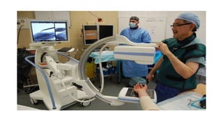
•A C-Arm machine is an advanced medical imaging device based on X-ray
technology
•The name derives from the C-shaped arm used to connect the x-ray source and
x-ray detector to one another. C-arms have radiographic capabilities, though they
are used primarily for fluoroscopic intraoperative imaging during surgical,
orthopedic and emergency care procedures.
What is a C-Arm?
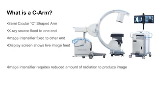
•Semi Cicular “C” Shaped Arm
•X-ray source fixed to one end
•Image intensifier fixed to other end
•Display screen shows live image feed
•Image intensifier requires reduced amount of radiation to produce image
What is an X-ray image intensifier ?
•As the name implies, this intensifier creates images with higher intensity.
•It converts X-rays into clear visible light better than the ordinary flourescent
screens.
•With this intensifying effect, low-intensity x-rays are conveniently seen in a bright
manner, helping the physicians to view the X-rayed object more clearly
C-Arm Fluoroscopy
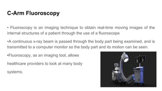
• Fluoroscopy is an imaging technique to obtain real-time moving images of the
internal structures of a patient through the use of a fluoroscope
•A continuous x-ray beam is passed through the body part being examined, and is
transmitted to a computer monitor so the body part and its motion can be seen.
•Fluoroscopy, as an imaging tool, allows
healthcare providers to look at many body
systems.
.
Working
•A c-arm’s general makeup is a generator and an image intensifier or flat-panel detector. The C-
shaped connecting allows movement in all directions so that images of the patient can be
produced from almost any angle.
•The generator emits X-rays that penetrate the patient’s body and the image intensifier or detector
catches the x-ray and converts it into a visible image that is displayed on the C-arm monitor. The
physician can identify and check details on the image such as bone structure, blood flow and other
various images often captured with x-ray.
Uses
•C-Arm systems are used in the places where greater flexibility is required.
•They are being used in Orthopedic procedures, cardiac and angiography studies,
and in therapeutic studies including stents and line placements.
•Since it is an overhead x-ray image intensifier, the physician can get a real-time
clear-cut view of anatomic structures of the patients
•C-Arms have radiographic capabilities, though they are used primarily for
fluoroscopic intra operative imaging during surgical, orthopedic, and emergency
care procedures.
C-Arm Principle
•Fluoroscopy uses a continuous X-ray beam with a special machine called a C-
Arm that rotates around your body to look at different angles. During the
procedure, a series of x-ray images are projected onto a screen.
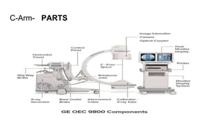
C-Arm- PARTS
…
C arm part
X-ray tube
Image intensifier
Collimator
Display monitor
Function
Source of X-ray beam
Captures the X-ray beams and converts them
into an image that is displayed on the monitor
Determines the size and shape of the X-ray
beam
Displays the X-ray
Applications
•If there is a need to place needles or stents during a complicated surgery, C-Arm will be a
handy choice.
•During surgeries, the real-time view of the gallbladder, liver, bone and several structures can
be obtained.
•Multiple views of the same part are possible
•C-Arm systems can also guide a needle placement procedure mainly while injecting anesthetic
medicines.
•They can identify the joints and medication can be inserted perfectly onto the required
shoulders and knees without damaging the other structures.
Advantage
•Ability to move around the patient(Mobile C-Arm)
•Good resolution, heat capacity
•Large image intensifier
•Better perception among IR’s (interventional radiography)
•Lesstime for procedure
Disadvantages
•exposure to c-arm x-ray radiation might lead to cancer and other side effects and health complications.
Conclusion
When it comes to medical devices, accuracy is the key, and in the C-arm X-ray machine, the
same has been considered; the device has been customized in a way it both offers a
solution in terms of flexibility and quality images for better decisions in surgical procedures.
THANK YOU.

More Related Content

What's hot

PTBD Percutaneous transhepatic biliary drainage
PTBD Percutaneous transhepatic biliary drainagePTBD Percutaneous transhepatic biliary drainage
PTBD Percutaneous transhepatic biliary drainage
Shiva Prakash
 
Ptc )percutaneous transhepatic cholangiography
Ptc )percutaneous transhepatic cholangiographyPtc )percutaneous transhepatic cholangiography
Ptc )percutaneous transhepatic cholangiography
Yashawant Yadav
 
Emegency drugs in radiology department
Emegency drugs in radiology department Emegency drugs in radiology department
Emegency drugs in radiology department
AbubakarMustaphaAman
 
Responsibility of radiographer
Responsibility of radiographerResponsibility of radiographer
Responsibility of radiographer
mohammed althaf
 
OT theatre radiography.ppt
OT theatre radiography.pptOT theatre radiography.ppt
OT theatre radiography.ppt
drparamesh93
 
Peripheral angiography
Peripheral angiographyPeripheral angiography
Peripheral angiography
InosRagan
 
Arthrography of the Shoulder, Ankle and Wrist.pptx
Arthrography of the Shoulder, Ankle and Wrist.pptxArthrography of the Shoulder, Ankle and Wrist.pptx
Arthrography of the Shoulder, Ankle and Wrist.pptx
Deneicer Guy
 
Digital subtraction angiography
Digital subtraction angiographyDigital subtraction angiography
Digital subtraction angiography
Jaya Yadav
 
SELDINGER TECHNIQUE & INTERVENTIONAL RADIOLOGY
SELDINGER TECHNIQUE & INTERVENTIONAL RADIOLOGYSELDINGER TECHNIQUE & INTERVENTIONAL RADIOLOGY
SELDINGER TECHNIQUE & INTERVENTIONAL RADIOLOGY
Riyas M K
 
Ercp
ErcpErcp
Flouroscopy ppt
Flouroscopy pptFlouroscopy ppt
Flouroscopy ppt
Syed Hammad .
 
Tools in interventional radiology
Tools in interventional radiologyTools in interventional radiology
Tools in interventional radiology
Anjan Dangal
 
Ct and mri preparation
Ct and mri preparationCt and mri preparation
Ct and mri preparation
Fadzlina Zabri
 
Barium Meal
Barium MealBarium Meal
Barium Meal
Anjan Dangal
 
Mobile & portable radiography
Mobile & portable radiographyMobile & portable radiography
Mobile & portable radiography
Priyanka Parimala
 
Intravenous pyelogram
Intravenous pyelogram Intravenous pyelogram
Intravenous pyelogram
Ramayya Pramila
 
Patient preparation for radiological procedure
Patient preparation for radiological procedurePatient preparation for radiological procedure
Patient preparation for radiological procedure
farranajwa
 

What's hot (20)

PTBD Percutaneous transhepatic biliary drainage
PTBD Percutaneous transhepatic biliary drainagePTBD Percutaneous transhepatic biliary drainage
PTBD Percutaneous transhepatic biliary drainage
 
Ptc )percutaneous transhepatic cholangiography
Ptc )percutaneous transhepatic cholangiographyPtc )percutaneous transhepatic cholangiography
Ptc )percutaneous transhepatic cholangiography
 
Emegency drugs in radiology department
Emegency drugs in radiology department Emegency drugs in radiology department
Emegency drugs in radiology department
 
Responsibility of radiographer
Responsibility of radiographerResponsibility of radiographer
Responsibility of radiographer
 
PTBD
PTBDPTBD
PTBD
 
OT theatre radiography.ppt
OT theatre radiography.pptOT theatre radiography.ppt
OT theatre radiography.ppt
 
Peripheral angiography
Peripheral angiographyPeripheral angiography
Peripheral angiography
 
Arthrography of the Shoulder, Ankle and Wrist.pptx
Arthrography of the Shoulder, Ankle and Wrist.pptxArthrography of the Shoulder, Ankle and Wrist.pptx
Arthrography of the Shoulder, Ankle and Wrist.pptx
 
Digital subtraction angiography
Digital subtraction angiographyDigital subtraction angiography
Digital subtraction angiography
 
SELDINGER TECHNIQUE & INTERVENTIONAL RADIOLOGY
SELDINGER TECHNIQUE & INTERVENTIONAL RADIOLOGYSELDINGER TECHNIQUE & INTERVENTIONAL RADIOLOGY
SELDINGER TECHNIQUE & INTERVENTIONAL RADIOLOGY
 
Angiogram
AngiogramAngiogram
Angiogram
 
Ercp
ErcpErcp
Ercp
 
Ercp
ErcpErcp
Ercp
 
Flouroscopy ppt
Flouroscopy pptFlouroscopy ppt
Flouroscopy ppt
 
Tools in interventional radiology
Tools in interventional radiologyTools in interventional radiology
Tools in interventional radiology
 
Ct and mri preparation
Ct and mri preparationCt and mri preparation
Ct and mri preparation
 
Barium Meal
Barium MealBarium Meal
Barium Meal
 
Mobile & portable radiography
Mobile & portable radiographyMobile & portable radiography
Mobile & portable radiography
 
Intravenous pyelogram
Intravenous pyelogram Intravenous pyelogram
Intravenous pyelogram
 
Patient preparation for radiological procedure
Patient preparation for radiological procedurePatient preparation for radiological procedure
Patient preparation for radiological procedure
 

Similar to C-arm system Presentation

Radiology_Equipment_Lec-1_Dr. Emad Taleb.pdf
Radiology_Equipment_Lec-1_Dr. Emad Taleb.pdfRadiology_Equipment_Lec-1_Dr. Emad Taleb.pdf
Radiology_Equipment_Lec-1_Dr. Emad Taleb.pdf
EmadTaleb1
 
Cone Beam Computed Tomography.pptx
Cone Beam Computed Tomography.pptxCone Beam Computed Tomography.pptx
Cone Beam Computed Tomography.pptx
DrAnand22
 
Radiographic aids in dental implants
Radiographic aids in dental implantsRadiographic aids in dental implants
Radiographic aids in dental implants
Thaslim Fathima
 
Cyberknife
Cyberknife Cyberknife
Cyberknife
Aaditya Sinha
 
Multidimentional imaging in spinal fractures
Multidimentional imaging in spinal fracturesMultidimentional imaging in spinal fractures
Multidimentional imaging in spinal fractures
Sumit2018
 
Principles of beam direction and use of simulators
Principles of beam direction and use of simulators Principles of beam direction and use of simulators
Principles of beam direction and use of simulators
Anil Gupta
 
Robotic surgery in ENT
Robotic surgery in ENTRobotic surgery in ENT
Robotic surgery in ENT
Jinu Iype
 
CAT
CATCAT
Portable ct mobile ct
Portable ct mobile ctPortable ct mobile ct
Portable ct mobile ct
anilayyakutty
 
7200 35328
7200 353287200 35328
Radiographic Considerations in Implants
Radiographic Considerations in ImplantsRadiographic Considerations in Implants
Radiographic Considerations in Implants
Dr.Richa Sahai
 
CBCT.pptx
CBCT.pptxCBCT.pptx
CBCT.pptx
AyushiGautam41
 
Advanced radiographic techniques in pedodontics
Advanced radiographic techniques in pedodonticsAdvanced radiographic techniques in pedodontics
Advanced radiographic techniques in pedodontics
Dr. Deepashree Paul
 
Immobilization techniques in SRS and SBRT
Immobilization techniques in SRS and SBRTImmobilization techniques in SRS and SBRT
Immobilization techniques in SRS and SBRT
Shreya Singh
 
X-ray machines - HPD
X-ray machines - HPDX-ray machines - HPD
X-ray machines - HPD
Shubham388431
 
White Paper: The Benefits of Mobile X-rays in Thoracic and Cardiac Care
White Paper: The Benefits of Mobile X-rays in Thoracic and Cardiac CareWhite Paper: The Benefits of Mobile X-rays in Thoracic and Cardiac Care
White Paper: The Benefits of Mobile X-rays in Thoracic and Cardiac Care
Carestream
 
Role of Image Guidance in Radiotherapy
Role of Image Guidance in RadiotherapyRole of Image Guidance in Radiotherapy
Role of Image Guidance in Radiotherapy
Susan Rochelle
 
Image guided radiation therapy
Image guided radiation therapyImage guided radiation therapy
Image guided radiation therapy
Swarnita Sahu
 

Similar to C-arm system Presentation (20)

O arm
O  armO  arm
O arm
 
Radiology_Equipment_Lec-1_Dr. Emad Taleb.pdf
Radiology_Equipment_Lec-1_Dr. Emad Taleb.pdfRadiology_Equipment_Lec-1_Dr. Emad Taleb.pdf
Radiology_Equipment_Lec-1_Dr. Emad Taleb.pdf
 
Cone Beam Computed Tomography.pptx
Cone Beam Computed Tomography.pptxCone Beam Computed Tomography.pptx
Cone Beam Computed Tomography.pptx
 
Radiographic aids in dental implants
Radiographic aids in dental implantsRadiographic aids in dental implants
Radiographic aids in dental implants
 
Cyberknife
Cyberknife Cyberknife
Cyberknife
 
Multidimentional imaging in spinal fractures
Multidimentional imaging in spinal fracturesMultidimentional imaging in spinal fractures
Multidimentional imaging in spinal fractures
 
Principles of beam direction and use of simulators
Principles of beam direction and use of simulators Principles of beam direction and use of simulators
Principles of beam direction and use of simulators
 
Robotic surgery in ENT
Robotic surgery in ENTRobotic surgery in ENT
Robotic surgery in ENT
 
CAT
CATCAT
CAT
 
Portable ct mobile ct
Portable ct mobile ctPortable ct mobile ct
Portable ct mobile ct
 
7200 35328
7200 353287200 35328
7200 35328
 
Radiographic Considerations in Implants
Radiographic Considerations in ImplantsRadiographic Considerations in Implants
Radiographic Considerations in Implants
 
Ct scan
Ct scanCt scan
Ct scan
 
CBCT.pptx
CBCT.pptxCBCT.pptx
CBCT.pptx
 
Advanced radiographic techniques in pedodontics
Advanced radiographic techniques in pedodonticsAdvanced radiographic techniques in pedodontics
Advanced radiographic techniques in pedodontics
 
Immobilization techniques in SRS and SBRT
Immobilization techniques in SRS and SBRTImmobilization techniques in SRS and SBRT
Immobilization techniques in SRS and SBRT
 
X-ray machines - HPD
X-ray machines - HPDX-ray machines - HPD
X-ray machines - HPD
 
White Paper: The Benefits of Mobile X-rays in Thoracic and Cardiac Care
White Paper: The Benefits of Mobile X-rays in Thoracic and Cardiac CareWhite Paper: The Benefits of Mobile X-rays in Thoracic and Cardiac Care
White Paper: The Benefits of Mobile X-rays in Thoracic and Cardiac Care
 
Role of Image Guidance in Radiotherapy
Role of Image Guidance in RadiotherapyRole of Image Guidance in Radiotherapy
Role of Image Guidance in Radiotherapy
 
Image guided radiation therapy
Image guided radiation therapyImage guided radiation therapy
Image guided radiation therapy
 

Recently uploaded

一比一原版(UMich毕业证)密歇根大学|安娜堡分校毕业证如何办理
一比一原版(UMich毕业证)密歇根大学|安娜堡分校毕业证如何办理一比一原版(UMich毕业证)密歇根大学|安娜堡分校毕业证如何办理
一比一原版(UMich毕业证)密歇根大学|安娜堡分校毕业证如何办理
peuce
 
一比一原版(IIT毕业证)伊利诺伊理工大学毕业证如何办理
一比一原版(IIT毕业证)伊利诺伊理工大学毕业证如何办理一比一原版(IIT毕业证)伊利诺伊理工大学毕业证如何办理
一比一原版(IIT毕业证)伊利诺伊理工大学毕业证如何办理
aozcue
 
一比一原版(UCSB毕业证)圣塔芭芭拉社区大学毕业证如何办理
一比一原版(UCSB毕业证)圣塔芭芭拉社区大学毕业证如何办理一比一原版(UCSB毕业证)圣塔芭芭拉社区大学毕业证如何办理
一比一原版(UCSB毕业证)圣塔芭芭拉社区大学毕业证如何办理
aozcue
 
Building a Raspberry Pi Robot with Dot NET 8, Blazor and SignalR - Slides Onl...
Building a Raspberry Pi Robot with Dot NET 8, Blazor and SignalR - Slides Onl...Building a Raspberry Pi Robot with Dot NET 8, Blazor and SignalR - Slides Onl...
Building a Raspberry Pi Robot with Dot NET 8, Blazor and SignalR - Slides Onl...
Peter Gallagher
 
Schematic Diagram MSI MS-7309 - REV 1.0 PDF .pdf
Schematic Diagram MSI MS-7309 - REV 1.0 PDF .pdfSchematic Diagram MSI MS-7309 - REV 1.0 PDF .pdf
Schematic Diagram MSI MS-7309 - REV 1.0 PDF .pdf
nikoloco007
 
欧洲杯冠军-欧洲杯冠军网站-欧洲杯冠军|【​网址​🎉ac123.net🎉​】领先全球的买球投注平台
欧洲杯冠军-欧洲杯冠军网站-欧洲杯冠军|【​网址​🎉ac123.net🎉​】领先全球的买球投注平台欧洲杯冠军-欧洲杯冠军网站-欧洲杯冠军|【​网址​🎉ac123.net🎉​】领先全球的买球投注平台
欧洲杯冠军-欧洲杯冠军网站-欧洲杯冠军|【​网址​🎉ac123.net🎉​】领先全球的买球投注平台
andreassenrolf537
 
天博体育下载-可靠的网络天博体育下载-网络天博体育下载|【​网址​🎉ac123.net🎉​】
天博体育下载-可靠的网络天博体育下载-网络天博体育下载|【​网址​🎉ac123.net🎉​】天博体育下载-可靠的网络天博体育下载-网络天博体育下载|【​网址​🎉ac123.net🎉​】
天博体育下载-可靠的网络天博体育下载-网络天博体育下载|【​网址​🎉ac123.net🎉​】
arcosarturo900
 
LORRAINE ANDREI_LEQUIGAN_GOOGLE CALENDAR
LORRAINE ANDREI_LEQUIGAN_GOOGLE CALENDARLORRAINE ANDREI_LEQUIGAN_GOOGLE CALENDAR
LORRAINE ANDREI_LEQUIGAN_GOOGLE CALENDAR
lorraineandreiamcidl
 

Recently uploaded (8)

一比一原版(UMich毕业证)密歇根大学|安娜堡分校毕业证如何办理
一比一原版(UMich毕业证)密歇根大学|安娜堡分校毕业证如何办理一比一原版(UMich毕业证)密歇根大学|安娜堡分校毕业证如何办理
一比一原版(UMich毕业证)密歇根大学|安娜堡分校毕业证如何办理
 
一比一原版(IIT毕业证)伊利诺伊理工大学毕业证如何办理
一比一原版(IIT毕业证)伊利诺伊理工大学毕业证如何办理一比一原版(IIT毕业证)伊利诺伊理工大学毕业证如何办理
一比一原版(IIT毕业证)伊利诺伊理工大学毕业证如何办理
 
一比一原版(UCSB毕业证)圣塔芭芭拉社区大学毕业证如何办理
一比一原版(UCSB毕业证)圣塔芭芭拉社区大学毕业证如何办理一比一原版(UCSB毕业证)圣塔芭芭拉社区大学毕业证如何办理
一比一原版(UCSB毕业证)圣塔芭芭拉社区大学毕业证如何办理
 
Building a Raspberry Pi Robot with Dot NET 8, Blazor and SignalR - Slides Onl...
Building a Raspberry Pi Robot with Dot NET 8, Blazor and SignalR - Slides Onl...Building a Raspberry Pi Robot with Dot NET 8, Blazor and SignalR - Slides Onl...
Building a Raspberry Pi Robot with Dot NET 8, Blazor and SignalR - Slides Onl...
 
Schematic Diagram MSI MS-7309 - REV 1.0 PDF .pdf
Schematic Diagram MSI MS-7309 - REV 1.0 PDF .pdfSchematic Diagram MSI MS-7309 - REV 1.0 PDF .pdf
Schematic Diagram MSI MS-7309 - REV 1.0 PDF .pdf
 
欧洲杯冠军-欧洲杯冠军网站-欧洲杯冠军|【​网址​🎉ac123.net🎉​】领先全球的买球投注平台
欧洲杯冠军-欧洲杯冠军网站-欧洲杯冠军|【​网址​🎉ac123.net🎉​】领先全球的买球投注平台欧洲杯冠军-欧洲杯冠军网站-欧洲杯冠军|【​网址​🎉ac123.net🎉​】领先全球的买球投注平台
欧洲杯冠军-欧洲杯冠军网站-欧洲杯冠军|【​网址​🎉ac123.net🎉​】领先全球的买球投注平台
 
天博体育下载-可靠的网络天博体育下载-网络天博体育下载|【​网址​🎉ac123.net🎉​】
天博体育下载-可靠的网络天博体育下载-网络天博体育下载|【​网址​🎉ac123.net🎉​】天博体育下载-可靠的网络天博体育下载-网络天博体育下载|【​网址​🎉ac123.net🎉​】
天博体育下载-可靠的网络天博体育下载-网络天博体育下载|【​网址​🎉ac123.net🎉​】
 
LORRAINE ANDREI_LEQUIGAN_GOOGLE CALENDAR
LORRAINE ANDREI_LEQUIGAN_GOOGLE CALENDARLORRAINE ANDREI_LEQUIGAN_GOOGLE CALENDAR
LORRAINE ANDREI_LEQUIGAN_GOOGLE CALENDAR
 

C-arm system Presentation

  • 1. Introduction •A C-Arm machine is an advanced medical imaging device based on X-ray technology •The name derives from the C-shaped arm used to connect the x-ray source and x-ray detector to one another. C-arms have radiographic capabilities, though they are used primarily for fluoroscopic intraoperative imaging during surgical, orthopedic and emergency care procedures.
  • 2. What is a C-Arm? •Semi Cicular “C” Shaped Arm •X-ray source fixed to one end •Image intensifier fixed to other end •Display screen shows live image feed •Image intensifier requires reduced amount of radiation to produce image
  • 3. What is an X-ray image intensifier ? •As the name implies, this intensifier creates images with higher intensity. •It converts X-rays into clear visible light better than the ordinary flourescent screens. •With this intensifying effect, low-intensity x-rays are conveniently seen in a bright manner, helping the physicians to view the X-rayed object more clearly
  • 4. C-Arm Fluoroscopy • Fluoroscopy is an imaging technique to obtain real-time moving images of the internal structures of a patient through the use of a fluoroscope •A continuous x-ray beam is passed through the body part being examined, and is transmitted to a computer monitor so the body part and its motion can be seen. •Fluoroscopy, as an imaging tool, allows healthcare providers to look at many body systems.
  • 5. .
  • 6. Working •A c-arm’s general makeup is a generator and an image intensifier or flat-panel detector. The C- shaped connecting allows movement in all directions so that images of the patient can be produced from almost any angle. •The generator emits X-rays that penetrate the patient’s body and the image intensifier or detector catches the x-ray and converts it into a visible image that is displayed on the C-arm monitor. The physician can identify and check details on the image such as bone structure, blood flow and other various images often captured with x-ray.
  • 7. Uses •C-Arm systems are used in the places where greater flexibility is required. •They are being used in Orthopedic procedures, cardiac and angiography studies, and in therapeutic studies including stents and line placements. •Since it is an overhead x-ray image intensifier, the physician can get a real-time clear-cut view of anatomic structures of the patients •C-Arms have radiographic capabilities, though they are used primarily for fluoroscopic intra operative imaging during surgical, orthopedic, and emergency care procedures.
  • 8. C-Arm Principle •Fluoroscopy uses a continuous X-ray beam with a special machine called a C- Arm that rotates around your body to look at different angles. During the procedure, a series of x-ray images are projected onto a screen.
  • 9.
  • 11. C arm part X-ray tube Image intensifier Collimator Display monitor Function Source of X-ray beam Captures the X-ray beams and converts them into an image that is displayed on the monitor Determines the size and shape of the X-ray beam Displays the X-ray
  • 12. Applications •If there is a need to place needles or stents during a complicated surgery, C-Arm will be a handy choice. •During surgeries, the real-time view of the gallbladder, liver, bone and several structures can be obtained. •Multiple views of the same part are possible •C-Arm systems can also guide a needle placement procedure mainly while injecting anesthetic medicines. •They can identify the joints and medication can be inserted perfectly onto the required shoulders and knees without damaging the other structures.
  • 13. Advantage •Ability to move around the patient(Mobile C-Arm) •Good resolution, heat capacity •Large image intensifier •Better perception among IR’s (interventional radiography) •Lesstime for procedure
  • 14. Disadvantages •exposure to c-arm x-ray radiation might lead to cancer and other side effects and health complications.
  • 15. Conclusion When it comes to medical devices, accuracy is the key, and in the C-arm X-ray machine, the same has been considered; the device has been customized in a way it both offers a solution in terms of flexibility and quality images for better decisions in surgical procedures.