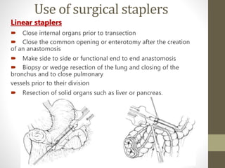
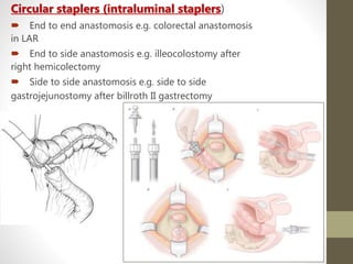
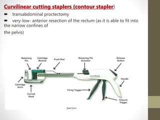
2. INTRODUCTION
• Suturing of any incision or wound needs to take into
consideration the site and tissues involved.
• A wound with proper blood supply and uninfected, heals
well by primary intension or suturing, thus requiring
accurate apposition of wound edges.
• Why we need Suturing of a Wound or Incision Site?
To Prevent Infection.
To Stop Bleeding from the site.
To enhance the process of Healing.
• Suturing is an art that requires Practise and proper
observation.
3. • “ Sutures are best made of soft thread, not too hard twisted
that it may sit easier on the tissue, nor are too few nor too
many of either of them to be put in.”
-Aurelius Cornelius Celsus.
• Early surgery abound suturing was done
by ligating blood vessels with Tendon strips
and closing of the wound with acacia thorns pushed
through the wound with vegetable matter- East African
Tribes.
History
4. Cont…
• A south American method: using Black/ Army ants to bite
the wound together with pincers or jaws acting like skin
clips and then the ant’s body was twisted off leaving
behind the head.
• By 1000 BC Surgeons were using horsehair, cotton and
leather sutures. Romans were using linen, silk and metal
clips called Fibulae.
• With the advancement of textile industries
both silk and catgut became popular as suture
materials.
Lord Moynihan
5. Desired Characteristics of
Suture material:
• Easy to Handle
• Predictable behaviour in tissues.
• Predictable tensile strength
• Sterile
• Glides through tissue easily
• Secure knotting ability
• Inexpensive.
• Minimal tissue reaction
• Non capillary
• Non allergenic
• Non carcinogenic
• Non Electrolytic
• Non Shrinkage.
6. SUTURE CHARACTERISTICS:
5 Characteristics are present that need to be considered:-
A. PHYSICAL STRUCTURE:
Can be of two Types:
1. MonoFilament:
They are smooth.
No sawing action.
Difficult to knot effectively
2. MultiFilament:
They are Braided.
Better Grip.
Much larger surface area.
Thus have interstices where bacteria
can lodge and lead to infections.
7. B. STRENGTH:
• It depends upon the thickness and the constituent of the suture
material.
• Strength can be Tensile strength or the material’s own In Vivo
strength.
• However Absorbable suture material show decay in strength.
• Non absorbable suture materials have indefinite Tensile Strength.
• However biological Non absorbable materials such as silk will
definitely fragment with time and lose their strength.
8. C. TENSILE BEHAVIOUR:
• Based on their Deformability and Flexibility.
• They can be:
Elastic: material will return to its original length once tension is
released.
Plastic: Stiff and above mentioned phenomenon does not occur.
• Memory: many synthetic materials show Memory ie.
They keep curling up in the shape they adopted . The
more memory the suture material has the less is the knot
Security.
Best Example of Memory is Prolene.
9. D. ABSORBABILITY:
• Suture materials may be
1. ABSORBABLE:
Required in cases for use in Biliary or urinary tract where it
needs to be absorbed in order to minimize the risk of stone
production.
1. NON-ABSORBABLE
In Vascular anastomosis, artificial graft or Prosthesis.
10. E. BIOLOGICAL BEHAVIOUR
• It depends upon the constituent raw material.
• Some of the biological sutures, such as catgut, are
proteolysed, but this involves a process that is not
entirely predictable and may even cause local
irritation.
• Synthetic materials are hydrolysed and their
disappearance in the tissue is predictable.
11.
12.
13. General Guidelines
• Careful evaluation of the wound is essential to assess for wound tension
and whether deep layer closure will be required
• Wound tension is the amount of perpendicular force that must be applied to
a wound to approximate the edges
• Higher tension = more force
• Wounds under higher tension require a larger gauge suture that will tolerate
a greater amount of force
• Deep layer closure and other advanced repair techniques can be used to
reduce wound tension
• Lower tension repairs are ideal as they result in a better cosmetic outcome
• Anatomic region tends to determine amount of tension on a wound
• Facial wounds are often lower tension than wounds on trunk or extremities
• Within these areas, there are regions of higher tension, like the chin or
flexor surfaces which may require different material
• Nonabsorbable . monofilament commonly used for skin closure
• Nylon (Ethilon) tends to be favored over polypropylene (Prolene) for its
lower cost and its slightly easier handling.
• Polypropylene (Prolene)
Favored in Tissues which requires Prologed Support.
Prolene is often colored blue, so may be chosen in areas with hair to help
identify sutures for removal.
Also used in Cardiovascular, Ophthalmic and Plastic Surgeries.
14. Absorbable Sutures
•VICRYL*
•MONOCRYL*
•Coated
VICRYL*
•Coated
VICRYL* Plus
Antibacterial Suture
•PDS* II
•Traumatology
• Ligaments
• Fascia
• Vessel
anastomosis
•Mass Absorption
•Wound Support
•10 days •By 42 days
•Typical Uses
•Skin
•Perineum
•Oral
•Lacerations
•30 days
•60 days
•20 days
•30 days • 56 - 70
days
•90 - 120 days
•56 - 70 days
•180 - 210
days
•Ligature
•General
•Bowel
•Orthopaedics
•Ligature
•General
•Bowel
•Ophthalmic
•Mucosa
•Obstetrics
•Bowel
•Skin& Ligature
15. Cont…
• Absorbable materials
• Reasons to select
• Suture will not be accessible for later removal
• Oral injuries
• Suture removal may be difficult (i.e. pediatric patients, patients with poor
follow-up)
• Polygalactin 910 (Vicryl)
• Useful in deep layer closures
• Provides long-term tensile strength and mid range absorption time (50%
at 21d), minimizing tissue reaction.
• Chromic gut
• Plain gut treated with chromic salts to slow absorption (50% at 21-28d)
and minimize tissue reaction
• Useful in oral repairs, nailbed laceration repair, Superficial Vessels,
Subcutaneous tissues and others that heals rapidly.
• Gamma irradiated Polygalactin 910 (Vicryl Rapide)
• Preferred by some oral surgeons for mucosal and dental injuries as may
lead to more rapid healing over chromic gut .Increasingly popular in
repair of lacerations on the torso and extremities in patients whom
suture removal may be difficult
• Available data suggests noninferiority to polypropylene (Prolene) in
cosmetic outcome with a nonsignificant greater incidence of infection
with absorbable material.
20. Cont…
Types
Cutting and reverse cutting most common for skin closure
Cutting: sharp edge of needle on inside of curve
Reverse cutting
Sharp edge of needle on outside of curve
Theoretically reduces chances of suture pulling through tissue
Taper point: used for surgical applications in soft tissue
21. STAPLES
Goals of stapler
• Close abdominal wounds
• Join internal organs to restore to normal function
• Maintain hemostasis
• Reduce tissue trauma
• Reduce contamination
• Prevent postoperative morbidity and infections
22. HISTORY OF SURGICAL
STAPLERS
1880s
• Reports of first stapler by Dr Henroz- everted bowel
anastomosis in dogs.
1908
• Professor Humer Hultl with Victor Fischer created a stapler
with emphasis of following
• principles-
• Tissue compression
• B-shaped configuration of closed staples
• placement of staples in double staggered rows
• use of fine wire as the staple material.
• But it was heavy and its assembly was difficult and
time-consuming
23. Cont…
1976
• Ethicon, Inc. introduces the first completely disposable, single
patient use mechanical stapler
1989
• Titanium replaces stainless steel as the key component for
staples
24. Advantages of stapling
Stapling anastomosis is faster than traditional
suturing techniques, hence reduced operating time.
Reduces tissue trauma by minimizing tissue
handling.
Prevents contamination
The availability of staplers has fostered the
development of procedures that were
difficult with traditional techniques because of limited
access.
Stapled tissue and anastomoses heal as reliably
and rapidly as sutured anastomoses
Not user dependent
26. • Living tissue before compression
• Living tissue compressed to adequate
thickness for stapling.
• Living tissue after stapling. The material
composition of the staple should avoid
spring back to keep the tissue compressed.
27. STAPLING BIOMECHANICS
Under-sizing staple cartridge
lead to excessive tissue compression, which
exceeds the tissue’s tensile strength
increases the risk for inadequate staple formation
Over sizing staple cartridges
Poor haemostasis
Inadequate opposition of tissue edges
Poor staple line formation predisposed to
anastomotic leakage
28. Use of surgical staplers
Linear staplers
Close internal organs prior to transection
Close the common opening or enterotomy after the creation
of an anastomosis
Make side to side or functional end to end anastomosis
Biopsy or wedge resection of the lung and closing of the
bronchus and to close pulmonary
vessels prior to their division
Resection of solid organs such as liver or pancreas.
29. Circular staplers (intraluminal staplers)
End to end anastomosis e.g. colorectal anastomosis
in LAR
End to side anastomosis e.g. illeocolostomy after
right hemicolectomy
Side to side anastomosis e.g. side to side
gastrojejunostomy after billroth II gastrectomy
30. Curvilinear cutting staplers (contour stapler)
transabdominal proctectomy
very-low- anterior resection of the rectum (as it is able to fit into
the narrow confines of
the pelvis)
31. Functional end to end anastomosis:
CRITICAL CONCEPTS
• Non-tension
• GIA stapler
• Align anti-mesenteric
sides of bowel
together
• Staggered staple lines
33. INTRODUCTION
• The word anastomosis comes from the Greek ‘ana’,
without, and ‘stoma’, a mouth, i.e. when a tubular
viscus (bowel) or vessel is joined after resection or
bypass without exteriorisation with a stoma.
• Intestinal anastomosis is the surgical
connection of separate or severed bowel to
form a continuous channel.
34. Cont…
• Early phase (0–4days): There is an acute
inflammatory response, but no intrinsic cohesion.
• Fibroplasia (3–14days): Fibroblast proliferation
occurs with collagen formation.
• Maturation stage (>10 days): This is the
period of collagen remodeling, when the
stability and strength of the anastomosis
increase
35. 3
5
TYPES
• Orientation of bowel
• Side-to-side
• End-to-end
• End-to-side
• Technique
• Hand sewed
• Stapling technique
• Part of the bowel involved
• Gastro-jejunostomy
• Jejuno-jejunostomy
• Ileo-colic anastomosis
• Base on the number of layers
• Single
• Double layer
37. (A) BOWEL ANASTOMOSES
• Ensure Good Blood supply to bowel ends before and after
formation of anastomosis.
• Ensure the anastomosis is under no tension.
• Avoid risk to mesenteric vessels by clamps or sutures.
• Use atraumatic bowel clamps to minimise contamination.
• Interrupted and continuous single layer suture techniques
are and safe.
• Stapling devices are an alternative when speed is required
or access is major factor.
38. SINGLE LAYER
ANASTOMOSIS
• An interrrupted seromuscular suture, with
absorbable thread. The submucosal layer is
strong and the blood supply is only minimally
damaged
• Lembert stitch
39. DOUBLE LAYER ANASTOMOSIS
• An inner continuous
absorbable suture, with
stitching of all layers
• An outer,
seromuscular,
interrupted
nonabsorbable
suture
• Serosa apposition and
mucosa inversion; the
inner layer has a
hemostatic effect, but
the mucosa is
strangulated
• Connell stitch- continuous
42. (B) VASCULAR ANASTOMOSIS
• Non Absorbable monofilament suture material should be
used. Eg. Prolene.
• A smooth intimal suture line is essential.
• Knots require multiple throws in order to ensure security.
• The suture must pass from within outwards on the down
flow aspect of the anastomosis.