Downloaded 636 times

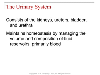
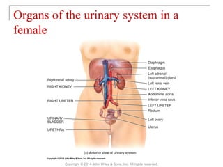
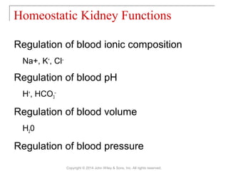
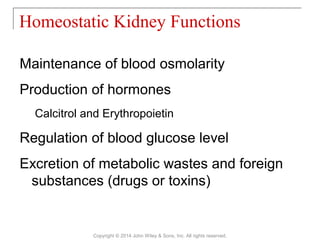

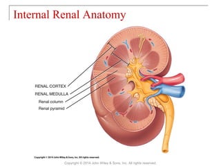
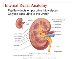
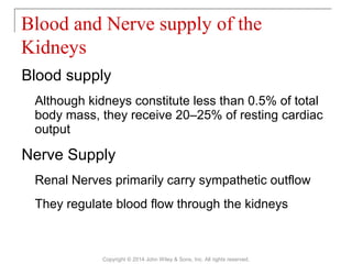
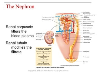
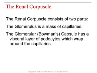
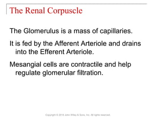
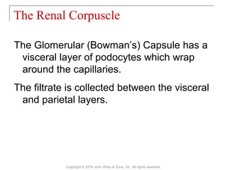
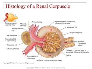
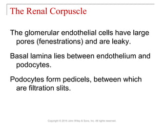
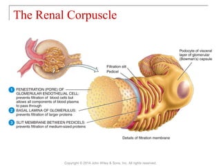
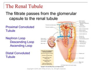

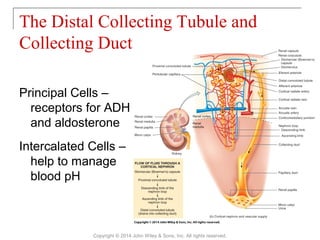
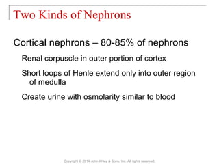
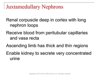
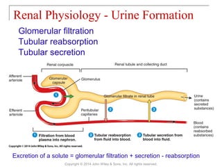
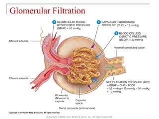
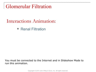
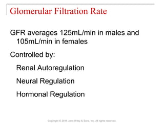

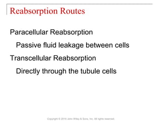
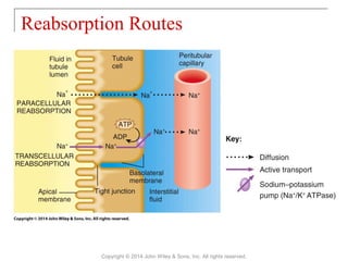
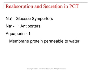
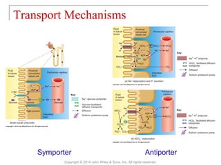
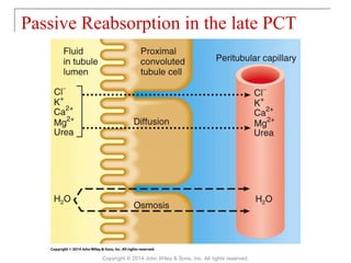

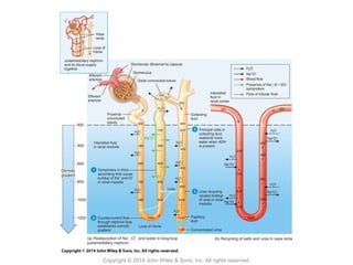


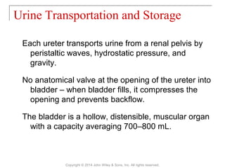
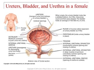


The document summarizes key aspects of kidney and urinary system anatomy and physiology. It describes how the kidneys, along with the ureters, bladder, and urethra, work together to maintain fluid homeostasis and remove wastes from the blood. Key functions of the kidneys include regulating blood volume, pressure, pH, and ion concentrations through glomerular filtration, tubular reabsorption and secretion. The nephron is the functional unit of the kidney, consisting of the renal corpuscle and renal tubule.
![28 [chapter 28 the reproductive system]](https://cdn.slidesharecdn.com/ss_thumbnails/28chapter28thereproductivesystem-170828134050-thumbnail.jpg?width=640&height=640&fit=bounds)
![29 [chapter 29 development and inheritance]](https://cdn.slidesharecdn.com/ss_thumbnails/29chapter29developmentandinheritance-170828044352-thumbnail.jpg?width=640&height=640&fit=bounds)
![14 [chapter 14 the brain and cranial nerves]](https://cdn.slidesharecdn.com/ss_thumbnails/14chapter14thebrainandcranialnerves-170828133437-thumbnail.jpg?width=640&height=640&fit=bounds)
![23 [chapter 23 the respiratory system]](https://cdn.slidesharecdn.com/ss_thumbnails/23chapter23therespiratorysystem-170828043650-thumbnail.jpg?width=640&height=640&fit=bounds)
![22 [chapter 22 the lymphatic system and immunity]](https://cdn.slidesharecdn.com/ss_thumbnails/22chapter22thelymphaticsystemandimmunity-170828153258-thumbnail.jpg?width=640&height=640&fit=bounds)
![24 [chapter 24 the digestive system][11e]](https://cdn.slidesharecdn.com/ss_thumbnails/24chapter24thedigestivesystem11e-170828043714-thumbnail.jpg?width=640&height=640&fit=bounds)
![08 [chapter 8 the skeletal system appendicular skeleton]](https://cdn.slidesharecdn.com/ss_thumbnails/08chapter8theskeletalsystem-appendicularskeleton-170828041008-thumbnail.jpg?width=640&height=640&fit=bounds)
![15 [chapter 15 the autonomic nervous system]](https://cdn.slidesharecdn.com/ss_thumbnails/15chapter15theautonomicnervoussystem-170828041929-thumbnail.jpg?width=640&height=640&fit=bounds)
![18 [chapter 18 the endocrine system]](https://cdn.slidesharecdn.com/ss_thumbnails/18chapter18theendocrinesystem-170828042016-thumbnail.jpg?width=640&height=640&fit=bounds)
![19 [chapter 19 the cardiovascular system the blood]](https://cdn.slidesharecdn.com/ss_thumbnails/19chapter19thecardiovascularsystem-theblood-170828042033-thumbnail.jpg?width=640&height=640&fit=bounds)
![17 [chapter 17 the special senses]](https://cdn.slidesharecdn.com/ss_thumbnails/17chapter17thespecialsenses-170828041636-thumbnail.jpg?width=640&height=640&fit=bounds)
![03 [chapter 3 the cellular level of organization]](https://cdn.slidesharecdn.com/ss_thumbnails/03chapter3thecellularleveloforganization-170828035521-thumbnail.jpg?width=640&height=640&fit=bounds)
![25 [chapter 25 metabolism and nutrition]](https://cdn.slidesharecdn.com/ss_thumbnails/25chapter25metabolismandnutrition-170828145139-thumbnail.jpg?width=640&height=640&fit=bounds)
![20 [chapter 20 the cardiovascular system the heart]](https://cdn.slidesharecdn.com/ss_thumbnails/20chapter20thecardiovascularsystem-theheart-170828133506-thumbnail.jpg?width=640&height=640&fit=bounds)
![13 [chapter 13 the spinal cord and spinal nerves]](https://cdn.slidesharecdn.com/ss_thumbnails/13chapter13thespinalcordandspinalnerves-170828040950-thumbnail.jpg?width=640&height=640&fit=bounds)
![27 [chapter 27 fluid, electrolyte and acid base homeostasis]](https://cdn.slidesharecdn.com/ss_thumbnails/27chapter27fluidelectrolyteandacid-basehomeostasis-170828044023-thumbnail.jpg?width=640&height=640&fit=bounds)
![10 [chapter 10 muscular tissue]](https://cdn.slidesharecdn.com/ss_thumbnails/10chapter10musculartissue-170828040153-thumbnail.jpg?width=640&height=640&fit=bounds)
![04 [chapter 4 the tissue level of organization][11e]](https://cdn.slidesharecdn.com/ss_thumbnails/04chapter4thetissueleveloforganization11e-170828035609-thumbnail.jpg?width=640&height=640&fit=bounds)
![12 [chapter 12 nervous tissue]](https://cdn.slidesharecdn.com/ss_thumbnails/12chapter12nervoustissue-170828041102-thumbnail.jpg?width=640&height=640&fit=bounds)
![11 [chapter 11 the muscular system]](https://cdn.slidesharecdn.com/ss_thumbnails/11chapter11themuscularsystem-170828041038-thumbnail.jpg?width=640&height=640&fit=bounds)
![05 [chapter 5 the integumentary system]](https://cdn.slidesharecdn.com/ss_thumbnails/05chapter5theintegumentarysystem-170828035624-thumbnail.jpg?width=640&height=640&fit=bounds)
![07 [chapter 7 the skeletal system the axial skeleton]](https://cdn.slidesharecdn.com/ss_thumbnails/07chapter7theskeletalsystem-theaxialskeleton-170828035650-thumbnail.jpg?width=640&height=640&fit=bounds)

![01 [chapter 1 an introduction to the human body]](https://cdn.slidesharecdn.com/ss_thumbnails/01chapter1anintroductiontothehumanbody-170828035545-thumbnail.jpg?width=640&height=640&fit=bounds)


![16 [chapter 16 sensory, motor, and integrative systems]](https://cdn.slidesharecdn.com/ss_thumbnails/16chapter16sensorymotorandintegrativesystems-170828041940-thumbnail.jpg?width=640&height=640&fit=bounds)


![PERI-PROSTHETIC FRACTURE NAIL-PLATE CONSTRUCT [NPC].pptx](https://cdn.slidesharecdn.com/ss_thumbnails/drarunkumardrmohamedashrafperiprostheticfrasturenail-plateconstructnpc-260209164459-7e9d15a1-thumbnail.jpg?width=640&height=640&fit=bounds)


