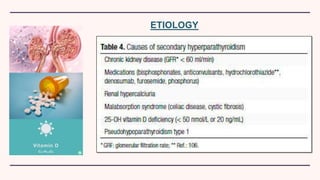
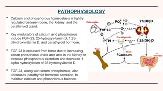
SECONDARY
HYPERPARATHYROIDISM
By Dr. Sarah D’souza
BDS
©
INTRODUCTION
• Secondary hyperparathyroidism is the medical condition of
excessive secretion of parathyroid hormone (PTH) by the parathyroid
glands in response to hypocalcemia (low blood calcium levels), with
resultant hyperplasia of these glands.
• This disorder is primarily seen in patients with chronic kidney failure.
ETIOLOGY
PATHOPHYSIOLOGY
• Calcium and phosphorous homeostasis is tightly
regulated between bone, the kidney, and the
parathyroid gland.
• Key modulators of calcium and phosphorous
include FGF-23, 25-hydroxyvitamin D, 1,25-
dihydroxyvitamin D, and parathyroid hormone.
• FGF-23 is released from bone due to increasing
serum phosphorus levels and acts in the kidney to
increase phosphorous excretion and decrease 1
alpha hydroxylation of 25-hydroxyvitamin D.
• FGF-23, along with serum phosphorous, also
decreases parathyroid hormone secretion, to
maintain calcium and phosphorous balance.
PATHOPHYSIOLOGY
• In CKD, stages 3-5, FGF-23 levels increase, initially leading to
phosphaturia and decreased parathyroid hormone excretion.
• As the CKD progresses, there is a resistance in the kidney and
parathyroid gland to FGF-23 and a deficiency in the kidney of 1 alpha
hydroxylation of vitamin D, both of which result in reduced phosphorous
excretion.
• The deficiency of 1,25-dihydroxyvitamin D, along with the decreased
phosphorus excretion, results in hypocalcemia and hyperphosphatemia,
thereby maintaining stimulation of parathyroid hormone synthesis and
parathyroid gland hyperplasia.
PATHOPHYSIOLOGY
• Pallor
• Skin pigmentation, peripheral oedema
• Features of rickets: Widening of wrist and ankle joint,
bowing of the legs, rickety rossery, curvature of the
spine, softening of skull bones.
CLINICAL EXAMINATION
• Arrhythmia, features of heart failure: elevated jugular
venous pressure. hepatomegaly, peripheral oedema,
fine end inspiratory bibasal crepitations
• Development of vascular calcification
• Scratch marks (due to pruritus)
CLINICAL EXAMINATION
INVESTIGATIONS
• Serum PTH: 35-70 pg/ml in stage 3, 70-110 pg/ml in stage 4 and
150-300 pg/ml in stage 5.
• Serum calcium level: Reduced to normal levels in stage 3 and 4,
and 2.1-2.4 mmol/l in stage 5.
• Serum phosphate level: 2.7 to 4.6 mg/dl in patients with CKD
stage 3 to 4 and 3.5 to 5.5 mg/dl in CKD stage 5.
• Arterial blood gas analysis: Hyperchloremic metabolic acidosis
• Screen for diabetes mellitus: HbA1c, fasting blood sugar
INVESTIGATIONS
• Electrocardiogram: Myocardial fibrosis secondary cardiac
calcification can leads to left ventricular hypertrophy.
Prominent R wave in V5 or V6 and prominent S wave in V1 or V2
is seen on ECG.
• Echocardiogram: There can be left ventricular hypertrophy and
myocardial calcific deposits.
• DEXA scan (Dual-emission X-ray absorptiometry): To detect
osteoporosis and Z -score <-2.5 is associated with high risk of
fractures.
• Renal function tests
MANAGEMENT
• Management of causative factors and complications.
• Dietary phosphate restriction.
• Vitamin D supplementation.
• Phosphate binders.
• Calcimimetics eg:- Cinacalcet.
• If the PTH levels remains >1000 pg/ml with hypercalcemia even after medical
therapy or when the volume of gland is >500 mm3, parathyroidectomy is
recomended.
PROGNOSIS
If left untreated, the disease will progress to tertiary
hyperparathyroidism, where correction of the underlying cause will
not stop excess PTH secretion, i.e. parathyroid gland hypertrophy
becomes irreversible.

Secondary Hyperparathyroidism

  • 1.
  • 2.
    INTRODUCTION • Secondary hyperparathyroidismis the medical condition of excessive secretion of parathyroid hormone (PTH) by the parathyroid glands in response to hypocalcemia (low blood calcium levels), with resultant hyperplasia of these glands. • This disorder is primarily seen in patients with chronic kidney failure.
  • 3.
  • 4.
    PATHOPHYSIOLOGY • Calcium andphosphorous homeostasis is tightly regulated between bone, the kidney, and the parathyroid gland. • Key modulators of calcium and phosphorous include FGF-23, 25-hydroxyvitamin D, 1,25- dihydroxyvitamin D, and parathyroid hormone. • FGF-23 is released from bone due to increasing serum phosphorus levels and acts in the kidney to increase phosphorous excretion and decrease 1 alpha hydroxylation of 25-hydroxyvitamin D. • FGF-23, along with serum phosphorous, also decreases parathyroid hormone secretion, to maintain calcium and phosphorous balance.
  • 5.
    PATHOPHYSIOLOGY • In CKD,stages 3-5, FGF-23 levels increase, initially leading to phosphaturia and decreased parathyroid hormone excretion. • As the CKD progresses, there is a resistance in the kidney and parathyroid gland to FGF-23 and a deficiency in the kidney of 1 alpha hydroxylation of vitamin D, both of which result in reduced phosphorous excretion. • The deficiency of 1,25-dihydroxyvitamin D, along with the decreased phosphorus excretion, results in hypocalcemia and hyperphosphatemia, thereby maintaining stimulation of parathyroid hormone synthesis and parathyroid gland hyperplasia.
  • 6.
  • 7.
    • Pallor • Skinpigmentation, peripheral oedema • Features of rickets: Widening of wrist and ankle joint, bowing of the legs, rickety rossery, curvature of the spine, softening of skull bones. CLINICAL EXAMINATION
  • 8.
    • Arrhythmia, featuresof heart failure: elevated jugular venous pressure. hepatomegaly, peripheral oedema, fine end inspiratory bibasal crepitations • Development of vascular calcification • Scratch marks (due to pruritus) CLINICAL EXAMINATION
  • 9.
    INVESTIGATIONS • Serum PTH:35-70 pg/ml in stage 3, 70-110 pg/ml in stage 4 and 150-300 pg/ml in stage 5. • Serum calcium level: Reduced to normal levels in stage 3 and 4, and 2.1-2.4 mmol/l in stage 5. • Serum phosphate level: 2.7 to 4.6 mg/dl in patients with CKD stage 3 to 4 and 3.5 to 5.5 mg/dl in CKD stage 5. • Arterial blood gas analysis: Hyperchloremic metabolic acidosis • Screen for diabetes mellitus: HbA1c, fasting blood sugar
  • 10.
    INVESTIGATIONS • Electrocardiogram: Myocardialfibrosis secondary cardiac calcification can leads to left ventricular hypertrophy. Prominent R wave in V5 or V6 and prominent S wave in V1 or V2 is seen on ECG. • Echocardiogram: There can be left ventricular hypertrophy and myocardial calcific deposits. • DEXA scan (Dual-emission X-ray absorptiometry): To detect osteoporosis and Z -score <-2.5 is associated with high risk of fractures. • Renal function tests
  • 11.
    MANAGEMENT • Management ofcausative factors and complications. • Dietary phosphate restriction. • Vitamin D supplementation. • Phosphate binders. • Calcimimetics eg:- Cinacalcet. • If the PTH levels remains >1000 pg/ml with hypercalcemia even after medical therapy or when the volume of gland is >500 mm3, parathyroidectomy is recomended.
  • 12.
    PROGNOSIS If left untreated,the disease will progress to tertiary hyperparathyroidism, where correction of the underlying cause will not stop excess PTH secretion, i.e. parathyroid gland hypertrophy becomes irreversible.