INTRODUCTION
• The fourparathyroid glands lie behind the lobes of thyroid
and weigh between 25 - 40 mg.
• Parathyroid hormone plays an important role in calcium
and phosphate homeostasis and vitamin D metabolism.
HYPERCALCAEMIA
• One ofthe most common biochemical abnormalities. And is
often detected during routine biochemical analysis in
asymptomatic patients.
• It can also present with chronic symptoms and acute
emergency with severe hypercalcaemia and dehydration .
• Affects 0.5 - 1 % of general population.
• Hypercalcaemia is corrected total serum calcium value
above the upper limit of normal range or an elevated
ionized calcium value.
7.
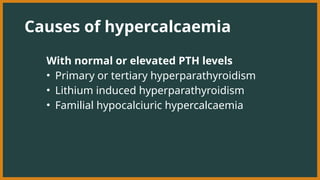
Causes of hypercalcaemia
Withnormal or elevated PTH levels
• Primary or tertiary hyperparathyroidism
• Lithium induced hyperparathyroidism
• Familial hypocalciuric hypercalcaemia
8.
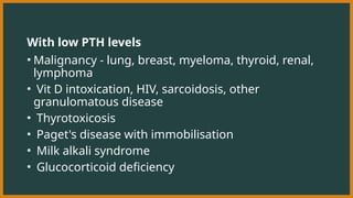
With low PTHlevels
• Malignancy - lung, breast, myeloma, thyroid, renal,
lymphoma
• Vit D intoxication, HIV, sarcoidosis, other
granulomatous disease
• Thyrotoxicosis
• Paget's disease with immobilisation
• Milk alkali syndrome
• Glucocorticoid deficiency
9.
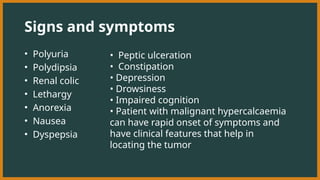
Signs and symptoms
•Polyuria
• Polydipsia
• Renal colic
• Lethargy
• Anorexia
• Nausea
• Dyspepsia
• Peptic ulceration
• Constipation
• Depression
• Drowsiness
• Impaired cognition
• Patient with malignant hypercalcaemia
can have rapid onset of symptoms and
have clinical features that help in
locating the tumor
10.
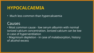
HYPOCALCAEMIA
• Much lesscommon than hypercalcaemia
Causes
• Most common cause - low serum albumin with normal
ionized calcium concentration. Ionized calcium can be low
in case of hyperventilation
• Magnisium depletion - in case of malabsorption, history
of alcohol excess
11.
Signs and symptoms
•Mild - asymptomatic
• Severe - tetany - characterized by muscle spasms due to
increased excitability of peripheral nerves
• Carpopedal spasm - hands adopt a characteristic position
with flexion of metacarpophalangeal joints of fingers and
adduction of thumb
• Stridor - caused by spasm of glottis
12.
• Latent tetanyis detcted by Trousseau's sign - inflation of
sphygmomanometer cuff on upper arm to more than systolic
pressure is followed by carpal spasm within 3 mins.
• Chvostek sign - Tapping over the branches of facial nerve as
they emerge from parotid gland produces twitching of facial
muscles
• papilloedema and prolongation of the ECG QT interval, which
may predispose to ventricular arrhythmias.
• Prolonged hypocalcaemia and hyperphosphataemia (as in
hypoparathyroidism) may cause calcification of the basal
ganglia, grand mal epilepsy, psychosis and cataracts.
• Hypocalcaemia associated with hypophosphataemia, as in
vitamin D deficiency, causes rickets in children and
osteomalacia in adults
13.
Management
Immediate management -10-20mL 10% calcium gluconate
IV over 10-20 mins
• Continuous IV infusion may be required for several hours
(equivalent of 10 mL 10% calcium gluconate/hr)
• Cardiac monitoring is recommended
• If associated with hypomagnesaemia - magnesium
chloride IV over 24 hrs . Most parenteral magnesium will be
excreted in the urine, so further doses may be required to
replenish body stores
14.
PRIMARY HYPERPARATHYROIDISM
• Primaryhyperparathyroidism is caused by autonomous
secretion of PTH, usually by a single parathyroid adenoma,
which can vary in diameter from a few millimetres to several
centimetres.
• It should be distinguished from secondary
hyperparathyroidism, in which there is a physiological
increase in PTH secretion to compensate for prolonged
hypocalcaemia (such as in vitamin D deficiency) and from
tertiary hyperparathyroidism, in which continuous stimulation
of the parathyroids over a prolonged period of time results in
adenoma formation and autonomous PTH secretion
15.
• The prevalenceof primary hyperparathyroidism is about 1 in
800
• it is 2-3 times more common in women than men;
• 90% of patients are over 50 years of age.
• It also occurs in the familial MEN syndromes
16.
Clinical features
• Osteitisfibrosa - bone pain, tenderness, fracture, deformity
• Chondrocalcinosis - resulting in secondary degenerative
arthritis or predispose to attacks of acute pseudogout
• Skeletal X-rays are usually normal in mild primary
hyperparathyroidism, but in patients with advanced disease
characteristic changes are observed.
• A 'pepper-pot' appearance may be seen on lateral X-rays of the
skull.
• Reduced bone mineral density
• osteopenia, osteoporosis
17.
Investigations
• The diagnosiscan be confirmed by finding a raised PTH the
presence of hypercalcaemia, provided that FHH is excluded
• Parathyroid scanning
• USG
18.
Management
• surgery, withexcision of a solitary parathyroid adenoma or
hyperplastic glands
• Patients who are treated conservatively without surgery should
have calcium biochemistry and renal function checked annually
and bone density monitored periodically. They should be
encouraged to maintain a high oral fluid intake to avoid renal
stones.
• Occasionally, primary hyperparathyroidism presents with
severe life-threatening hypercalcaemia. This is often due to
dehydration and should be managed medically with
intravenous fluids and bisphosphonates
19.
• If thisis not effective, then urgent parathyroidectomy
should be considered
• Surgery is usually indicated for individuals aged less than
50 years, with clear-cut symptoms or documented
complications (such as renal stones, renal impairment or
osteoporosis), and In asymptomatic patients) significant
hypercalcaemia (corrected serum calcium >2.85 mmol/L
(>11.4 mg/dL)).
20.
FAMILIAL HYPOCALCIURIC
HYPERCALCAEMIA
• Thisautosomal dominant disorder is caused by an
inactivating mutation in one of the alleles of the calcium-
sensing receptor gene, which reduces the ability of the
parathyroid gland to 'sense' ionised calcium concentrations.
• As a result, higher than normal calcium levels are required
to suppress PTH secretion.
21.
• The typicalpresentation is with mild hypercalcaemia with
PTH concentrations that are 'inappropriately' at the upper
end of the reference range or are slightly elevated.
• Calcium-sensing receptors in the renal tubules are also
affected and this leads to increased renal tubular
reabsorption of calcium and hypocalciuria (as measured in
the vitamin D-replete individual by a fractional calcium
excretion or 24-hour calcium excretion).
• The hypercalcaemia of FHH is always asymptomatic and
complications do not occur.
22.
• The mainrisk of FHH is that of the patient being subjected
to an unnecessary (and ineffective) parathyroidectomy if
misdiagnoser as having primary hyperparathyroidism.
• Testing of family members for hypercalcaernia is helpful in
confirming the diagnosis and it is also possible to perform
genetic testing. No treatment is necessary.
23.
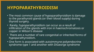
HYPOPARATHYROIDISM
• The mostcommon cause of hypoparathyroidism is damage
to the parathyroid glands (or their blood supply) during
thyroid surgery.
• Rarely, hypoparathyroidism can occur as a result of
infiltration of the glands with iron in haemochromatosis or
copper in Wilson's disease
• There are a number of rare congenital or inherited forms of
hypoparathyroidism.
• One form is associated with autoimmune polyendocrine
syndrome type 1 and another with DiGeorge syndrome
24.
Pseudohypoparathyroidism
• In thisdisorder, the individual is functionally
hypoparathyroid but,
• instead of PTH deficiency, there is tissue
resistance to the effects of PTH, such that PTH
concentrations are markedly elevated.
25.
Characteristics
There are severalsubtypes but the most common
(pseudohypoparathyroidism type 1a) is ;
• characterised by hypocalcaemia and hyperphosphataemia,
• in association with short stature,
• short fourth metacarpals and metatarsals,
• rounded face,
• obesity and subcutaneous calcification
• These features are collectively referred to as Albright's
hereditary osteodystrophy (AHO).
26.
Management
• Persistent hypoparathyroidismand
pseudohypoparathyroidism are treated with
• oral calcium salts and vitamin D analogues
• This therapy needs careful monitoring because of the risks
of iatrogenic hypercalcaemia. hypercalciuria and
nephrocalcinosis.
• Recombinant PTH is available as subcutaneous injection
therapy for osteoporosis and, although not currently
licensed, has been used in hypoparathyroidism