Cardiopulmonarybypass
•
5 likes•318 views

Sandeep Mewada Perfusionist Cardiopulmonarybypass

Recommended
More Related Content
What's hot
What's hot (20)
Similar to Cardiopulmonarybypass
Similar to Cardiopulmonarybypass (20)
More from SANDEEP MEWADA
More from SANDEEP MEWADA (20)
Recently uploaded
Recently uploaded (20)
Cardiopulmonarybypass
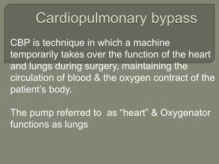
- 1. CBP is technique in which a machine temporarily takes over the function of the heart and lungs during surgery, maintaining the circulation of blood & the oxygen contract of the patient’s body. The pump referred to as “heart” & Oxygenator functions as lungs
- 2. Circuit :- Chart review and selection of equipment . Before assembling the perfusion circuit information from the patient’s chart is obtained regarding the proposal surgical procedure and relevant history.
- 3. 1 Oxygenator 2 Custom tubing pack 3 ALF 4 Cardioplegia delivery set 5 hamofilter 6 Cannulae 1 Aortic 2 Venous 3 CPG 4 Vent 5 Cardiotomy sucker
- 4. Conduction :- The perfusionist is responsible for the setup of CPB circuit & priming of the HL –Machine & Oxygenator the conduct of CPB & anticoagulation activity drugs & write the records.
- 5. 1. Check all machines, HL –machine, all pumps are in well working condition, battery backup, spot light, oxygen supply & cranks (handle). 2. TCM - check Water Levels (h20) both cold & warm tubing are proper function is not & ice making properly. 3. Also check JABP Machine according to case.
- 6. 4. Holder – according to the type of oxygenator, CPG system, arterial filter, cardioplegia device holder & hemofliter holder. 5. Check oxygen source supply & blender also. 6. Clamps & available. 7. ACT tubes & machine. 8. All disposable items should be available.
- 7. 9. All drugs & fluid for priming. 10. Check the pressure transducers are working properly. 11. Perfusion record chart is prepared for the pts and Hct (hemotocrit) level predict.
- 8. 1. Assembly of HL machine & circuit:- Disposable items available for adult & pediatric preparations. Assembly of circuit is done by done by all sterile procedures, while pts is being prepared by anesthetic & nurses. Oxygenator assembled with the circuit. Gas lines are connected. Water lines are connected to heat exchanger, CPG System & check for any leakage.
- 9. CPB Circuit is primed with crystalloid, Colloid & with heparin.
- 10. All outlets & venting parts & ancillary lines from the oxygenator closed. From above pump head 30 inch tubing keep according to rate of falling 1 am/ min is observed according to which is adjust the compression of roller pump.
- 11. Lines are taken from surgeon side & connected to venous inlet part & arterial filter outlet, suckers & vent. Sucker & vent are checked CPG lines is flushed for de-airing.
- 12. AV loop is circulated fast to de-airing the circuit properly Lines are tapped from the surgical sides as well as arterial filter is de-aired by tapping the lines
- 14. 3-4 mg/kg, heparin is given , a period of 3 min is allowed to elapse after heparin administration before cannulation ,is begun while at others, ACT>300 is necessary for cannulation. Cardiotomy suckers, On when ACT is reached at >250,400-480 sec ACT is required for going bypass.
- 15. Aortic cannulation is always performed first. This help in managing blood loss during cannulation & in case of hypotension allow commencement of CPB with a single venous cannula or “suker bypass”. During aortic cannulation, Perfusionist run the aortic pump on surgeon’s calling to make air free connection. After cannulation, check swing & resistance.
- 16. Line pressure checked, which is nearly equal to perfusion pressure indicate position of cannula is proper.
- 17. Retrograde autologous, priming is done after aortic cannulation to reduce the prime volume by maintain adequate reservoir volume. To decrease hemodiludtion.
- 18. This is depending on the surgical procedure. Which types of cannulation is to be done. 1. Bicaval cannulation 2. Two stage cannulation 3. Single stage cannulation SVC Cannulation is always done first as this involves less retraction of heart & hence less chance of hypotension during cannualtion.
- 19. IVC Cannulation :- On the posterior inferior RA wall. CPG Cannulation :- Depends on the surgical procedure, CPG Cannulation is done. 1. Autograde CPG Cannulation :- Placed to aortic root. P – 80- 120 mmHg. Flow – 200 -300 mmHg. 2. Ostial CPG :- Directly into the ostial by hand handling into the coronary ostia. 2. P – It ostia RT coronary ostia flow
- 20. Retrograde CPG – Cannula is inserted through lower part of RA into the coronary sinuous. P – Flow- Vent placement – in some special cases, like AVR if AR is present then surgeons uses LV vent cannulation by RSPV to prevent cardiac distension. ACT- when ACT>400-480 Sec.
- 21. CPB is initiated, after instructions from the surgeon, by the perfusionist slowly transfusing the pts with the CPB prime. Arterial flow should be unobstructed & an initial line pressure <100 mmHg. Confirm oxygenator gases & CPB safely alarms are switched on prior to the CPB. Venous clamp is gradually released after confirmation the arterial line is unobstructed, the pts is blood is diverted into the reservior.
- 22. P – informs the surgeon, On full bypass, Surgeon checks following 1. RA empty totally, with CVP<0 mmHg. 1. MPA should be soft. Then surgeon ready for aortic cross clamp cooling of the pt,if required by the surgeon, monitors, ECG at this stage, so that VF may be noted & actions taken to prevent cardiac distension.
- 23. Once, aortic has been clamped(check L.P) the required temperature has been achieved. Cardioplegia has been administered if required & steady state of perfusion has been attained the first sample for blood gases & ACT is checked. Perfusionist maintain perfusion pressure & all ABG parameter, ACT with in normal limit. CPG is repeated every 25-30
- 24. Pt’s flow & Hypothermic management:- pt’s blood flow depends on the C.I & Temp. As a general rule flow should be reduce with temp. (as metabolic required diminish) & vice versa.
- 25. Hyothermia Temp. Flow index (L/min) Fi o2 Gas : Blood flow Normothermi a 37 2.4 80% 1:1 Tepid (mild) 34 2.2 70% .8:1 Mod. 30 2.0 65% .7:1 Mod. 28 1.8 60% .6:1 Deep hypothermia 22 1.6 50% .5:1
- 26. Sys O2 consumption, V O2 is reduced by approx .50% for every 7*C reduction in core temp. below normothermia . At 30 *C - VO2 -50 % At 23 *C – VO2 -25% At 16 *C – VO2 -12.5% As relative, small decreases in temp.reduce requirement for sys.O2 delivery, making reduce in pump flow, DO2 sufficient to meet VO2.
- 27. 0 5 10 15 20 25 30 35 pO2(mmHG) 10 20 30 oxyhemoglobin(% Saturation)
- 28. Ct & Rt shifts in O2- Hb dissociation curve also occures with temp.changes during CPB. At lower temp. Hb has greater affinity for binding O2, Consquently, O2 is also released less readily & the dissociation curve shift to the Ct. At high temp. converse is true & curve shift to Rt. Pump flow rate must be adjusted with due consideration to temp. if metabolic demands for O2 are to be matched by delivery.
- 29. During CPB, Monitoring this parameters Moitoring continously intermittently
- 30. • Continously • Intermittently 1) ECG 1) ABG 2) Perfusion pressure/ line pressure (MP 40-80mmHg) 2) pH/Base deficit 3) CVP 3) Hb/Hct 4) Arterial pump flow rates 4) K+ conc. 5) Suction pump flow rates 5) Urine output ( 0.5 -1.0 ml/kg/hr) 6) Core temp & Hypothermia 6) LA/PA Pressure 7) Venous reservoir level 7) Glucose/ACT
- 31. During CPB, good renal function is an indication of the adequacy of perfusion . pH, lactate conc. SvO2 > 65% on bypass. Mixed venous satuartion is an indicator of matching of DO2 & VO2.
- 32. As margin between sys. O2 delivery & demand narrow, O2 extraction increase & SVO2 decrease. O2 Delivery equals pump flow time CaO2 & should be above 250 ml/min/m2 during normothermic perfusion. Metabolic acidosis ( base deficit) or eleveted lactic acid level also indicate inadequate perfusion.
- 33. The pt’s should be rewarmed using the arterial blood temp.& pt’s core temp. By maintaining the temp.gradient 4 -6 *C Rewarming the pt’s to 37*C more than 37*C should not be exceeded. On rewarming , appropriate adjustments gas flows & to the blood flow must be made Blood : Gas (1:1.5)
- 34. After intracardiac procedure, all cardiotomies are closed & the heart is de-aired these achieved by Start HOTSPOT ( if requires depending on surgical procedure,& surgeon.) Patient in head down position. (tender burg position) Anesthetic starts ventiation (active filling )
- 35. Perfusionist by partially clamping the venous line fills the heart .(passive filling) Surgeon massages LV & de-airs the heart through aortic root.
- 36. After complete de-airing, aortic is declamped in head down position. Aortic root vent is on slowly 1 ABh repeated,according to ABG,correct blood gas, metabolic abnormalities & Ca2+ & Mg2+.
- 37. After aortic declamping, heart regains its activity & contractility. Perfusionist to restore the blood volume to the heart by gradual occlusion of the venous return. Perfusionist now increasingly fill the heart guided by the CVP & PA distolic pressure ( CVP 8 -12 mmHg) Termination of CPB is achieved by complete occlusion of venous line & STOP the arterial pump.
- 38. Once, hemodynamic stability is established after weaning off CPB. Venous cannula is removed process of reserving of heparin with protamine. Prior to protamine administration,cardiotomy suction is stopped to avoid clotting with in the circuit. When protamine started residual blood is transfused to the pt’s by main taining safe reservior volume by supporting B.P.