SlideShare a Scribd company logo
1 of 127
RETINAL
DETACHMENT
DR.p K
junIORRESIDENT
OPHTHALMOLOGY
ANATOMY OF THE PERIPHERAL RETINA
1.Pars Plana
 The ciliary body starts 1mm from the limbus and extends posteriorly for about
6mm.
 The first 2mm = pars plicata and the remaining 4mm = flattened pars plana.
 In order not to endanger the lens or retina, the optimal location for a pars plana
surgical incision is 4 mm from the limbus in phakic eyes
and
3.5 mm from the limbus in pseudophakic eyes.
2. Ora Serrata
Forms the junction between the retina and ciliary body and is
characterized by the following :
1.Dentate processes
are teeth-like extensions of retina onto the pars plana; more
marked nasally than temporally.
2. Oral bays
are the scalloped edges of the pars plana epithelium in between
the dentate processes.
3. A meridional fold
a small radial fold of thickened retinal tissue in line with a
dentate process, usually located in the superonasal quadrant .
A fold may occasionally exhibit a small retinal hole at its apex
4. An enclosed oral bay
a small island of pars plana surrounded by retina as a result of
meeting of two adjacent dentate processes.
*It should not be mistaken for a retinal hole because it is
located anterior to the ora serrata.
5 Granular tissue
multiple white opacities within the vitreous base can
sometimes be mistaken for small peripheral opercula
DESCRIPTION LENGTH
Width of ora serrata 2.1 mm temporally
0.7-0.8 mm nasally
Location from limbus 6mm nasally
7mm temporally
From equator 6-8 mm
From optic disc 25 mm
At the ora, fusion of the sensory retina with the RPE and choroid
limits forward extension of SRF.
However, there being no equivalent adhesion between the choroid
and sclera, choroidal detachments may progress anteriorly to
involve the ciliary body (CILIOCHOROIDAL DETACHMENT).
**IMP** Choroidal detachments do not extend to posterior pole
bcoz they are limited by the VORTEX VEINS entering their
scleral channels.
VITREOUS BASE
 3–4 mm wide zone straddling the ora serrata.
 The cortical vitreous is strongly attached at the vitreous base,
so that following acute PVD, the posterior
hyaloid face remains attached to the posterior border of the
vitreous base.
 Pre-existing retinal holes within the vitreous base do not lead
to RD.
 Severe blunt trauma avulsion of the vitreous
base with tearing of the NPE of the pars plana along its
anterior border and of the retina along its posterior border.
VITREOUS ADHESIONS
1 Normal.
The peripheral cortical vitreous is
loosely attached to ILM of the
sensory retina. Stronger adhesions :
• Vitreous base, strongest.
• Around the ONH fairly strong.
• Around the fovea, fairly weak,
except in eyes with VMT and
macular hole formation.
• Along peripheral blood vessels,
usually weak.
2 Abnormal adhesions at the
following :
• Posterior border of islands of
lattice degeneration.
• Retinal pigment clumps.
• Peripheral paravascular
condensations.
• Vitreous base anomalies eg. tongue
like extensions & posterior islands.
• ‘WWP’ and ‘WWOP’.
VITREORETINAL TRACTION
 Is a force exerted on the retina by structures originating in
the vitreous, and may be dynamic or static.
1 Dynamic traction
is induced by eye movements
and
exerts a centripetal force towards the
vitreous cavity.
It plays an important role in the
pathogenesis of retinal tears and
Rhegmatogenous RD.
2 Static traction
is independent of ocular
movements.
It plays a key role in the
pathogenesis of tractional RD and
proliferative vitreoretinopathy
PERIPHERAL LESIONS PREDISPOSING
TO RETINAL DETACHMENT
LATTICE DEGENERATION
1. Prevalence - 8% of the population.
 Develops early in life, with a peak incidence in the 2nd and 3rd decades.
 More commonly in moderate myopes
 and is the most important degeneration directly related to RD.
 Bilateral
 Temporal and superiorly
 Lattice is present in about 40% of eyes with RD.
2. Pathology - There is discontinuity of the ILM with variable atrophy of
the underlying NSR.
 The vitreous overlying an area of lattice is synchytic but the vitreous
attachments around the margins are exaggerated.
3.Signs-
 Spindle-shaped areas of retinal thinning, commonly located
between the equator and the posterior border of the vitreous
base.
 Arborizing network of white lines within the islands. **
 Some lattice lesions may be associated with
‘snowflakes’ (remnants of degenerate Müller
cells.)
 Associated hyperplasia of the RPE is common.
 Small holes within lattice lesions are common and usually
innocuous.
4.Complications –
 No complications are encountered in most patients.
 Tears may occasionally develop in eyes with acute PVD.
 Atrophic holes may rarely lead to RD, particularly in young
myopes. The fellow eye often has a ‘mirror-image’ distribution
of holes.
SNAILTRACK DEGENERATION
 Characterized by sharply
demarcated bands of tightly
packed ‘snowflakes’which give
the peripheral retina a white frost-
like appearance.
 The islands are usually longer
than in lattice degeneration and
may be associated with overlying
vitreous liquefaction.
o However, marked vitreous traction at the posterior border of the
lesions is seldom present so that tractional U-tears rarely occur,
although round holes within the snailtracks may be present.
RETINOSCHISIS
Symptoms.
 Common in HYPERMETROPES.
 SPLIT IN RETINAL LAYERS.
 Cystoid degeneration.
 Photopsia and floaters are absent. ( because there is no
vitreoretinal traction. )
 Occasionally symptoms occur as a result of either VH or the
development of progressive RD.
Typical RS /DEGENERATIVE
More common
Low lying
Split in OPL
Less complications
Located Ant to
equator
Less common
Absent
Reticular
/JUVENILE RS
Less common
BULLOUS
Split in NFL
more
Can be Posterior to
equator
Microaneursyms &
small telengeictasias
Outer layer holes
 Signs :
Early retinoschisis - extreme inferotemporal periphery of both
fundi.
** appearing as an exaggeration of microcystoid degeneration with a
smooth immobile elevation of the retina.
**The surface of the inner layer may show snowflakes as well as
sheathing or ‘silver-wiring’ of blood vessels and the schisis
cavity may be bridged by rows of torn grey-white tissue.
 Breaks may be +nt in one or both layers.
 The elevation is convex, smooth, thin and relatively immobile,
unlike the opaque and corrugated appearance of a rhegmatogenous
RD.
RETINOSCHISIS RD
TYPICAL PT AGE Middle to elderly Middle age
Refractive association Hyperopia Myopia
Symptoms Almost always absent Acute : present
Chronic : absent
Scotoma Absolute Relative
VH or pigment Absent Present
Location Inferotemporal> ST Acute : usually superior
Chronic:usually inferior
Texture Smooth Ac: corrugated
Ch: smooth
Muller footplates Common Absent
Mobility Relatively immobile Ac: very often mobile
Ch: relatively immobile
Movement with scleral depression Moves as a single unit Height decreases
RETINOSCHISIS RD
Color with scleral
depression
WWP may be seen in
outer layer
NOT
Breaks May be present Present
RPE Normal Ac: normal
Ch: atrophy & demarcation lines
OCT Splitting of retinal layers SRF
Effect of LASER
application through
retinal break
Through inner layer
break: Uptake
Through full thickness break :
no uptake
DIFFUSE CHORIORETINAL ATROPHY
 Characterized by choroidal
depigmentation and thinning
of the overlying retina in the
equatorial area of highly myopic
eyes.
 Retinal holes developing in the atrophic retina may lead to RD.
 Because of lack of contrast between the depigmented choroid
and sensory retina, small holes may be very difficult to
visualize without the help of slit-lamp biomicroscopy.
WHITE WITH PRESSURE
 Retinal areas in which a translucent
white-grey appearance can be induced
by scleral indentation.
 Each area has a fixed configuration
that does not change when
indentation is moved to an
adjacent area.
 Along posterior border of lattice deg,
snail-track deg and the outer layers
of acquired retinoschisis..
 Can be seen in normal eyes === abnormally strong attachment of
vitreous gel.
WHITE WITHOUT PRESSURE
 Present without scleral
indentation.
 Area with fairly strong
adhesion of condensed
vitreous.
 Can be mistakes for flat retinal
hole.
 Retinal breaks, GRTs can
develop along the posterior
border of WWOP.
 Prohylactic therapy thus
considered in WWOP in
fellow eye of a patient with
spontaneous GRT in other eye.
POSTERIOR VITREOUS DETACHMENT
 Separation of the cortical vitreous from the ILM of the NSR posterior to the
vitreous base.
 Classification :
1 Onset
 Acute : most common Develops suddenly and usually becomes
complete soon after onset.
 Chronic : gradually weeks or months to become complete.
2 Extent
 Complete PVD entire vitreous cortex detaches up to the posterior
margin of the vitreous base.
 Incomplete PVD residual vitreoretinal attachments remain
posterior to the vitreous base.
Symptoms :
1. Photopsia -- inc in dim light, common in temporal periphery.
2.Myodesopsia (floaters) : spots/ cobwebs/ flies.
3. Blurred vision : d/t vitreous haemorrhage or d/t PHM or floaters in
visual axis.
 Weiss ring :-
Detached former attachment of the margin of optic disc.
Can be seen by patient as a circle or other solitary lesion.
Signs :
 Detached PHM seen on S/L as CRUMPLED TRANSLUCENT
membrane in mid vitreous cavity which is optically clear.
 Haemorrhage---rbcs in ant vitreous.
 Pigment granules in ant. vitreous in abscence
of inflammation, trauma, surgery on S/L -
k/a
SHAFER SIGN OR TOBACCO DUST
Presence of pigment granules in ant vitreous 95% sensitivity for
possibility of a retinal break
PHM as Crumpled translucent
membrane
Tobacco dust
Tobacco dust
COMPLETE PVD
INCOMPLETE PVD
 Rhegmatogenous RD is usually associated with acute
PVD;
 tractional RD is associated with chronic, incomplete PVD;
 exudative RD is unrelated to the presence of PVD.
MANAGEMENT
FACTORS PREVENTING RD
 Metabolic pump of RPE.
 Osmotic pressure of choroid.
 Minor mechanical processes of the interphotoreceptor
matrix.
compromised
RETINAL DETACHMENT
RD
Traction
component
present
Rhegmatogenous RD
Tractional RD
Combined Tractional-
rhegmatogenous RD
Central RD in myopic eye
Traction component
absent
Exudative RD
Haemorrhagic
RD
RHEGMATOGENOUSRETINAL
DETACHMENT
RHEGMA= RENT/ RUPTURE
 A Condition in which the fluid from the vitreous cavity
passes through a retinal defect into the subretinal space
to cause seperation of neural retina from the underlying
RPE
 Retinal break and vitreous liquefaction is must.**
 Even though a retinal break is present, a RD will not
occur if the vitreous is not at least partially liquefied and
if the necessary traction is not present.
PVD V. .liquefaction Retinal tears at site of significant
VR adhesions
CONDITIONS THAT PREDISPOSE AN EYE TO RD
 High myopia.------3 fold in RD
 PP or Aphakia.
 Blunt trauma and penetrating ocular trauma.
( 10-15%)
 CMV retinits ass. AIDS.
 YAG capsulotomy
FACTORS CAUSING RD
A. RETINAL BREAKS
B. VITREOUS LIQUEFACTION AND DETACHMENT
C. VR TRACTION
D. INTRAOCULAR FLUID CURRENTS ASSOCIATED WITH MOVEMENT OF
LIQUID VITREOUS AND SRF
RETINAL BREAKS
RETINAL BREAKS
 Full thickness defects in neurosensory retina.
 Typical location- near vitreous base.
 Holes/ tears/ dialysis.
 M=F
 R/F :
1. Myopia.
2. Lattice degeneration.
3. Ocular contusion and Penerating trauma.
Most common of retinal break after ocular contusion is :-
Retinal Dialysis
OCULAR MANIFESTATIONS:
1. RETINAL TEARS :
 Full thickness breaks that occur secondary to vitreous traction.
 (spontaneous posterior vitreous detachment).
horseshoe-shaped
 Types
flap shaped
 Most common site- vitreous base.
 Symptoms: Floaters, Flashes.
 In lattice deg, mc site of retinal tear is POSTERO-LATERAL
margin of lattice.
2. ROUND HOLES WITH OPERCULA
Persistent traction on HST/ flap tear
Avulsion at base
Complete relief of VRT in that area
Small round defect in neural retina with an overlying opercula
 3.Round holes without opercula – ATROPHIC HOLES
 Secondary to retinal thinning.
 No role of vitreous traction.
 Common in areas of lattice deg.
4. TRAUMATIC retinal breaks :
HST Retinal dialysis Macular holes
 MC site : INFEROTEMPORALLY &
SUPERONASALLLY
5. MACULAR BREAKS :
 Secondary to tangential traction from precortical vitreous
Pars plana cysts
Enclosed oral
bays
Meridional folds
Ora serrata pearls
Paving stone deg Chorioretinal scars
WWP
WWOP
D/D OF RETINAL BREAKS
INDICATIONS FOR T/T OF RETINAL TEAR/ HOLE IN
ASYMPTOMATIC PATIENT
Type of lesion Phakic Highly myopic Fellow eye Aphakic or
Pseudophakic
Retinal
dialysis
Almost
always
Almost always Almost always Almost always
HST sometimes sometimes sometimes sometimes
Operculated tears no rarely rarely rarely
Atrophic holes rarely rarely rarely rarely
Lattice deg’n
w/ or w/o
holes
no no sometimes rarely
Lincoff and Geiser reported 4 guidelines for locating RB causing
RRD *
Determined by
 Location of causative break
 Anatomic barriers (optic n.,ora serrata, existing chorioretinal
adhesions)
 Effect gravity on SRF in upright position
Note : onlyfor freshRDwith1 RB
1. A shallow inferior RD in which the SRF is slightly higher on
the temporal side points to a primary break located inferiorly on
that side.
2. A primary break located at 6 o’clock will cause an inferior
RD with equal fluid levels.
3. In a bullous inferior RD the primary break usually lies
above the horizontal meridian.
4. If the primary break is located in the upper nasal quadrant
the SRF will revolve around the optic disc and then rise on the
temporal side until it is level with the primary break.
5. A subtotal RD with a superior wedge of attached retina
points to a primary break located in the periphery nearest its
highest border.
6. When the SRF crosses the vertical midline above, the
primary break is near to 12 o’clock, the lower edge of the RD
corresponding to the side of the break .
B. VITREOUS LIQUEFACTION AND DETACHMENT
Ageing of human vitreous (SYNCHYSIS)
Liquefaction of vitreous gel with development of
optically emtpy lacunae in gel
Extensive liquefaction
Decrease in shock absorbing capacity and stability of gel
C. VR TRACTION
VR TRACTION
Gravitational
force
Rotational eye
movements
Fibrous tissue
contracture
Liquid
currents
Higher percentage
of SUPERIOR
retinal tears (80%)
Trauma/ retinal
vascular
proliferative
disorders
Continous flow of
liquid vitreous
through a retinal
break into subretinal
space is necessary to
maintain a RRD
OCULAR MANIFESTATIONS
SYMPTOMS
 Sudden onset of tiny dark floating objects
 Associated with Photopsia
**usually brief
**in the temporal visual field
SUBCLINICAL DETACHMENTS :
RD with small SRF (<2 DD) not acc by visual field loss
SIGNS
1. RAPD
2. IOP is usually lower by about 5 mmHg compared with the
eye.
If the intraocular pressure is extremely low, an associated
choroidal detachment may be present.
3. Iritis is very common but usually mild.
4. ‘Tobacco dust’ consisting of pigment cells is seen in the
anterior vitreous.
It may normal be raised, characteristically in SCHWARTZ-MATSUO syndrome in
which RRD is associated with an apparent mild anterior uveitis , often due to a
DIALYSIS due to prior blunt trauma in a young man.
5. Retinal breaks--- appear as discontinuities in the retinal
surface.
They are usually red because of the colour contrast between
the sensory retina and underlying choroid.
(However, in eyes with hypopigmented choroid (as in high myopia), the colour
contrast is decreased and small breaks may be overlooked unless careful slit-lamp
and indirect ophthalmoscopic examination is performed.)
6. Retinal signs depend on the
-- duration of RD and
-- the presence or absence of PVR.
Or tide marks. Take 3 months to develop
MODIFIED AMSLER –DUBOIS CHART
PROLIFERATIVE VITREORETINOPATHY
 Caused by epiretinal and subretinal membrane formation.
 Most common cause of ultimate failure after surgical tt for
RRD.
 Retinal break cells in vitreous cavity cells form
membranes on inner retinal surface or posterior vitreous
surface RRD.
 Usually, Occurs following surgery for RRD or penetrating
injury.
However, it may also occur in eyes with RRD that have not
had previous vitreoretinal surgery.
RISK FACTORS FOR PVR
 1. Giant retinal tear ( >3 clock hour).
 2. Number and size of retinal break.
 3. No.of previous operations.
 4. Presence of choroidal effusions.
 5. Use of cryotherapy.
 6. Intraocular haemorrhage.
 7.Aphakia.
 8. Vitreous protein levels.
1 Grade A (minimal) PVR : Characterized by diffuse vitreous haze and
tobacco dust. There may also be pigmented clumps on the inferior surface
of the retina.
2 Grade B (moderate) PVR : Characterized by wrinkling of the inner
retinal surface, tortuosity of blood vessels, retinal stiffness, decreased
mobility of vitreous gel and rolled edges of retinal breaks.
** The epiretinal membranes responsible for these findings cannot be
identified clinically.**
3 Grade C (marked) PVR Characterized by rigid full-thickness retinal
folds with heavy vitreous condensation and strands.
It can be either anterior (A) or posterior (P), the approximate dividing
line being the equator of the globe. The severity of proliferation in each
area is expressed by the number of clock hours of retina involved although
proliferations need not be contiguous.
4 B-scan ultrasonography in advanced disease shows gross reduction of
retinal mobility with retinal shortening and the characteristic triangular
sign. (FUNNEL LIKE)
T/T FOR PVR
1. No T/T if: retinal breaks absent unless macula is
involved.
localised TRD posterior to scleral buckle.
2. Macular pucker membrane peeling.
3. RRD location and severity of PVR
If surface membranes do not
prevent break closure……
SCLERAL BUCKLING
If surface membrane occur
in close association with
retinal break
Closed intraocular microsx with
membrane peeling
 RECENT ADVANCES :
1. PERIOPERATIVE DUANOMYCIN (on going trial) .
2.Adjuvant 5-HYDROXYURACIL
+
LOWER MOLECULE WEIGHT HEPARIN
reduce PVR in high-risk cases indergoing vitrectomy.
D/D FOR RRD
 Traction RD.
 Exudative RD.
 Retinoschisis.
 Elevated choroidal lesions.
 Intravitreal optical illusions (vitreous h’age).
SYSTEMIC ASSOCIATIONS
 Most important ocular disorder : STICKLER $
 Most important systemic association : DM, AIDS
TREATMENT OF RD
AIM : to counter the factors and forces that causes RD and to re-
establish physiological conditions that normally maintain contact
between neural retina and RPE.
T/T OPTIONS AVAILABLE :
 Primary vitrectomy
 Laser demarcation
 Permanent scleral buckle
## Encircling with/without drainage.
## Segmental with/without drainage
 Temporary scleral buckle
## Lincoff ballon.
## Absorbable buckling sutures.
 Pnematic retinopexy - routine
- with drainage of SRF.
VITRECTOMY
INDICATIONS :
1. RD with PVR
2. Giant retinal Tears.
3.RD with posterior retinal breaks
.
4.RD with vitreous hemorrhage.
5. RD with intraocular foreign body.
6. TRD threatening to or involving the macula.
Less likely to be closed with
scleral buckle
Retinal breaks can not be
visualized
VITRECTOMY
 VIDEO
CONTRAINDICATIONS :
1. Bleeding disorders.
2. Suspected tumors like retinoblastoma, melanoma.
COMPLICATIONS :
1.Iatrogenic breaks.
2.Lens trauma.
3.Redetachment.
4.Secondary glaucoma.
5. PVR
6.Cataract progression.
SCLERALBUCKLING SURGERY
 INDICATIONS :
1. RRD (PVR less than C1)
2. Inferior retinal dialysis.
3. Retinal dialysis.
4.Pediatric RD
SEGMENTAL BUCKLING
Usually reserved for
** RRD < 1 clock hour
** Anterior breaks
CONTRA-INDICATIONS :
1. Posterior breaks.
2. Opaque media.
3. Vaso-occlusive diseases.
4. PVR more than C2.
COMPLICATIONS :
1. Diplopia. (mc)
2.Perforation.
3. Rise in IOP.
4. Extrusion.
5. Infection.
6. AS ischemia.
7. ERM
8. Recurrent RD.
9. Surgical failure : ‘FISH-MOUTHING’
Phenomenon of a tear, typically a large superior equatorial U-
tear in a bullous RD,
to open widely following scleral
buckling requiring further operative tt
PNEUMATIC RETINOPEXY
 INDICATIONS :
1. A detachment caused by a single break, in superior 8 clock hours.
2. The break should not be more than 1clock hour.
3. Multiple breaks but in 1-2 clock hours of each other.
 CONTRA-INDICATIONS :
1. Break > 1 clock hr.
2. Break inferior to 4 clock hr.
3. PVR grade C,D.
4. Cloudy ocular media.
5. Uncontrolled or severe glaucoma.
6. Can’t maintain Head position.
GAS EXPANSION NON-EXPANSIBLE
CONC
AVERAGE
DURATION
VOLUME
USED FOR PR
SF6 2 times 20 % 10-14 days 0.5 ml
C3F8 4 times 12% 30-45 days 0.35 ml
AIR Non expansile - 5-7 days 0.8 ml
SILICONE OILS :
-Lighter than water,
-Commonly used for : intraop retinal manipulation
prolonged postop intraocular tamponade.
-Particularly useful in PVR.
DRAINAGE OF SUBRETINAL FLUID
 In deep and long standing viscous SRF.
 Complications : incarceration at drainage site ,
Retinal perforation,
Choroidal haemorrhage
SEAL THE BREAK(S)
SEAL
Diathermy
(obsolete)
Laser CRYO
LASER PHOTOCOAGULATION
 Usually cannot seal RB if presence SRF may be used to
create barrier to prevent progression of RD
 Esp. useful in
Chronic inferior RD
Systemic illness contraindicated to surgery
Laser
photocoagulation
Slit-lamp
biomicroscope with
contact lens
laser indirect
ophthalmoscope
(LIO)
Slit-lamp LIO
Indentation +
better magnified significant cataracts, PCO, mild VH more
easily treated with LIO
Safer in inexpertise
operator
Need more skill
Less need of corneal
care during laser
Less pain
not be readily
available
Any patient position
IMP**
Compared with diathermy and cryopexy
 less breakdown of blood–ocular barrier.
 thermal effect confined predominantly to retina and RPE
with little or no effect on choroid or sclera.
 Induces adhesive effect between neurosensory retina &
RPE within 24hr.
CRYORETINOPEXY
 RD with very shallow fluid can be cured by cryoretinopexy.
 Test cryoprobe prior to use to make sure probe is freezing.
 Freezing or whitening of RPE will noticed first,
Followed by delineation of edges of retinal tear
And whitening of retina
** Excessive freezing or ice crystal formation should be avoided
Histologic response depends on whether
 RPE alone or RPE and overlying detached retina together are
frozen
Disadvantage :
 Dispersion of pigment epithelial cells, which can result in
subretinal pigmentary changes after reattachment.
 Dispersion of viable pigment epithelial cells capable of
causing PVR following cryopexy.
 Induce choroidal congestion & hyperemia (transient)
 May complicate drainage of SRF through treated areas.
 Breakdown of BRB cause post-op CME and ERD
TRACTIONAL RETINAL
DETACHMENT
 Main causes of tractional RD
penetrating posterior segment
trauma
proliferative
retinopathy such as
diabetic and ROP
PATHOGENESIS OF DIABETIC TRACTIONAL RD
 Caused by progressive contraction of
fibrovascular membranes over large areas of vitreoretinal
adhesion.
 PVD in diabetic eyes is gradual and frequently incomplete.
 RD is Caused by leakage of plasma constituents into the
vitreous gel from a fibrovascular network adherent to the
posterior vitreous surface.
 In the very rare event of a subsequent complete PVD, the new
blood vessels are avulsed and RD does not develop.
Static vitreoretinal traction of the following three types is
recognized --
A Tangential traction
Caused by the contraction of
epiretinal fibrovascular membranes
with puckering of the retina and
distortion of retinal blood vessels.
 B Anteroposterior traction is
Caused by the contraction of
fibrovascular membranes
extending from the posterior retina,
usually in association with the
major arcades, to the vitreous base
anteriorly
C Bridging (trampoline) traction
is the result of contraction of fibrovascular membranes
which stretch from one part of the posterior retina to
another or between the vascular arcades,
tending to pull the two involved points together.
DIAGNOSIS
 Symptoms : Photopsia and floaters are usually absent
 Signs :
Concave configuration
breaks are absent.
Retinal mobility is severely reduced
shifting fluid is absent.
 The SRF is shallower than in a RRD and seldom extends to the ora serrata.
 Highest elevation of the retina at sites of vitreoretinal traction.
 If a tractional RD develops a break it assumes the characteristics of a RRD
and progresses more quickly (combined TRD-RRD).
TREATMENT
 GOAL : To release antero-posterior and/or circumferential
traction.
 Simple peeling------------------NOT POSSIBLE(vascularity and
friable retina)
 Methods of removing fibrovascular membranes in Diabetic TRDs.
DELAMINATION SEGMENTATION
(Vertical cutting) (horizontal cutting)
EXUDATIVE RETINAL
DETACHMENT
PATHOGENESIS
 Characterized by the accumulation of SRF in the absence of
retinal breaks or traction.
 As long as the RPE is able to compensate by pumping the
leaking fluid into the choroidal circulation,
no fluid accumulates in the subretinal space
RD does not occur.
 However, when the normal RPE pump is overwhelmed, or if
the RPE activity is decreased,
then fluid starts to accumulate in the subretinal space.
ETIOLOGY
1 .Choroidal tumours
such as melanomas, haemangiomas and metastases; it is therefore very
important to consider that exudative RD is caused by an intraocular tumour
until proved otherwise.
2 .Inflammation such as Harada disease and posterior scleritis.
3 .Bullous central serous chorioretinopathy : rare
4 .Iatrogenic causes - RD surgery & PRP.
5. Subretinal neovascularization which may leak and give rise to extensive
subretinal accumulation of fluid at the posterior pole.
6. Hypertensive choroidopathy, as may occur in toxaemia of pregnancy:rare.
7. Idiopathic such as the uveal effusion syndrome.
Posterior scleritis
Harada disease
Elshnig’s spots
SEIGERT’S
STREAK
DIAGNOSIS
Symptoms:-
**Photopsia is absent because there is no vitreoretinal traction,
although
floaters may be present if there is associated
vitritis.
Signs:-
Convex configuration, just like a RRD,
BUT
its surface is smooth and not corrugated.
 The detached retina is very mobile and exhibits the phenomenon of
‘shifting fluid’
in which SRF responds to the force of gravity and detaches the area
of retina under which it accumulates.
(For example, in the upright position the SRF collects under the
inferior retina, but on assuming the supine position for several
minutes, the inferior retina flattens and the SRF shifts posteriorly
detaching the superior retina.)
 The cause of the RD, such as a choroidal tumour, may be apparent
when the fundus is examined, or the patient may have an associated
systemic disease responsible for the RD (e.g. Harada disease,
toxaemia of pregnancy).
 ‘Leopard spots’ consisting of scattered areas of subretinal pigment
clumping may be seen after the detachment has flattened.
 Leopard sign pigmentation following resolution of
exudative RD
IS THIS RRD OR TRD OR ERD ??????
SYMPTOMS RRD TRD ERD
Floaters ++ +/- +/-
Flashes ++ - -
Progression of VA
loss
acute chronic Subacute/
chronic
Fluctuation of vision - +/- +
SIGNS RRD TRD ERD
Shaffer’s
sign
+ - -
PVD ++ - +/-
VH +/- +/- -
RD contour convex concave bullous
RD surface corrugate Ridge/ wavy smooth
Shifting +/- - ++
Associated Myopia/
trauma/ Sx
DM CTD
THANK YOU

More Related Content

What's hot

Retinal detachment
Retinal detachment Retinal detachment
Retinal detachment jyotigontia
 
Choroidal detachment
Choroidal detachmentChoroidal detachment
Choroidal detachmentSSSIHMS-PG
 
Refrective surgery ppt
Refrective surgery pptRefrective surgery ppt
Refrective surgery pptsubhadri manna
 
Dry eye diagnosis and management
Dry eye diagnosis and managementDry eye diagnosis and management
Dry eye diagnosis and managementNIKHIL GOTMARE
 
Goldman applanation tonometry
 Goldman applanation tonometry Goldman applanation tonometry
Goldman applanation tonometryHossein Mirzaie
 
Optical coherence tomography in glaucoma - Dr Shylesh Dabke
Optical coherence tomography in glaucoma - Dr Shylesh DabkeOptical coherence tomography in glaucoma - Dr Shylesh Dabke
Optical coherence tomography in glaucoma - Dr Shylesh DabkeShylesh Dabke
 
DME Management Guidelines
DME Management GuidelinesDME Management Guidelines
DME Management GuidelinesReyad Yossif
 
Vitreous substitutes
Vitreous substitutesVitreous substitutes
Vitreous substitutesSSSIHMS-PG
 
Glaucoma drainage devices
Glaucoma drainage devicesGlaucoma drainage devices
Glaucoma drainage devicesmeenank
 
Clinical examination of squint
Clinical examination of squintClinical examination of squint
Clinical examination of squintReshma Peter
 
Microtropia - Definition, Types and Shot Note
Microtropia - Definition, Types and Shot NoteMicrotropia - Definition, Types and Shot Note
Microtropia - Definition, Types and Shot NoteMero Eye
 
Anterior segment OCT & UBM
Anterior segment OCT & UBMAnterior segment OCT & UBM
Anterior segment OCT & UBMDinesh Madduri
 
Peripheral fundus & its disorders
Peripheral fundus & its disordersPeripheral fundus & its disorders
Peripheral fundus & its disordersRohit Rao
 
A scan biometry | How to Use A-scan? Types of A-Scan Biometry?
A scan biometry | How to Use A-scan? Types of A-Scan Biometry?A scan biometry | How to Use A-scan? Types of A-Scan Biometry?
A scan biometry | How to Use A-scan? Types of A-Scan Biometry?Naeem Ahmad
 

What's hot (20)

Retinal detachment
Retinal detachment Retinal detachment
Retinal detachment
 
Fundus in Glaucoma
Fundus in GlaucomaFundus in Glaucoma
Fundus in Glaucoma
 
Choroidal detachment
Choroidal detachmentChoroidal detachment
Choroidal detachment
 
Refrective surgery ppt
Refrective surgery pptRefrective surgery ppt
Refrective surgery ppt
 
Ac/a ratio
Ac/a ratio Ac/a ratio
Ac/a ratio
 
Dry eye diagnosis and management
Dry eye diagnosis and managementDry eye diagnosis and management
Dry eye diagnosis and management
 
Goldman applanation tonometry
 Goldman applanation tonometry Goldman applanation tonometry
Goldman applanation tonometry
 
Optical coherence tomography in glaucoma - Dr Shylesh Dabke
Optical coherence tomography in glaucoma - Dr Shylesh DabkeOptical coherence tomography in glaucoma - Dr Shylesh Dabke
Optical coherence tomography in glaucoma - Dr Shylesh Dabke
 
DME Management Guidelines
DME Management GuidelinesDME Management Guidelines
DME Management Guidelines
 
Vitreous substitutes
Vitreous substitutesVitreous substitutes
Vitreous substitutes
 
Glaucoma drainage devices
Glaucoma drainage devicesGlaucoma drainage devices
Glaucoma drainage devices
 
Clinical examination of squint
Clinical examination of squintClinical examination of squint
Clinical examination of squint
 
Microtropia - Definition, Types and Shot Note
Microtropia - Definition, Types and Shot NoteMicrotropia - Definition, Types and Shot Note
Microtropia - Definition, Types and Shot Note
 
Coats' Disease
Coats' DiseaseCoats' Disease
Coats' Disease
 
Anterior segment OCT & UBM
Anterior segment OCT & UBMAnterior segment OCT & UBM
Anterior segment OCT & UBM
 
Peripheral fundus & its disorders
Peripheral fundus & its disordersPeripheral fundus & its disorders
Peripheral fundus & its disorders
 
Eccentric Fixation
Eccentric FixationEccentric Fixation
Eccentric Fixation
 
A scan biometry | How to Use A-scan? Types of A-Scan Biometry?
A scan biometry | How to Use A-scan? Types of A-Scan Biometry?A scan biometry | How to Use A-scan? Types of A-Scan Biometry?
A scan biometry | How to Use A-scan? Types of A-Scan Biometry?
 
Disc edema
Disc edemaDisc edema
Disc edema
 
Test for stereopsis
Test for stereopsisTest for stereopsis
Test for stereopsis
 

Similar to RETINAL DETACHMENT

RETINAL DETACHMENT
RETINAL DETACHMENTRETINAL DETACHMENT
RETINAL DETACHMENTslidenka
 
Retinal detachment
Retinal detachment Retinal detachment
Retinal detachment Nikhil Rp
 
Retina Detachment.pptx
Retina Detachment.pptxRetina Detachment.pptx
Retina Detachment.pptxSHAYRI PILLAI
 
Rhegmatogenous retinal detachment
Rhegmatogenous retinal detachmentRhegmatogenous retinal detachment
Rhegmatogenous retinal detachmentSamuel Ponraj
 
CONGENITAL OPTIC DISC ANOMALIES
CONGENITAL OPTIC DISC ANOMALIESCONGENITAL OPTIC DISC ANOMALIES
CONGENITAL OPTIC DISC ANOMALIESsatabdi89dec
 
3 mirror, retinal break.pptx
3 mirror, retinal break.pptx3 mirror, retinal break.pptx
3 mirror, retinal break.pptxTimothyLiew3
 
Rhegmatogenous retinal detachment (rrd)
Rhegmatogenous retinal detachment (rrd)Rhegmatogenous retinal detachment (rrd)
Rhegmatogenous retinal detachment (rrd)Pavan Mahajan
 
Retinal detachment presentation
Retinal detachment presentationRetinal detachment presentation
Retinal detachment presentationNirav Modi
 
Tractional RD
Tractional RD Tractional RD
Tractional RD Nikhil Rp
 
Hereditary macular dystrophies
Hereditary macular  dystrophiesHereditary macular  dystrophies
Hereditary macular dystrophiesShruti Laddha
 
Retinal Detachment_Pradeep Bastola.pptx
Retinal Detachment_Pradeep Bastola.pptxRetinal Detachment_Pradeep Bastola.pptx
Retinal Detachment_Pradeep Bastola.pptxDr. Pradeep Bastola
 
retinal_detachment 4.pptx
retinal_detachment 4.pptxretinal_detachment 4.pptx
retinal_detachment 4.pptxHarshika Malik
 

Similar to RETINAL DETACHMENT (20)

RETINAL DETACHMENT
RETINAL DETACHMENTRETINAL DETACHMENT
RETINAL DETACHMENT
 
Retinal detachment
Retinal detachment Retinal detachment
Retinal detachment
 
Retina Detachment.pptx
Retina Detachment.pptxRetina Detachment.pptx
Retina Detachment.pptx
 
Retinal detachment
Retinal detachment Retinal detachment
Retinal detachment
 
Rhegmatogenous retinal detachment
Rhegmatogenous retinal detachmentRhegmatogenous retinal detachment
Rhegmatogenous retinal detachment
 
CONGENITAL OPTIC DISC ANOMALIES
CONGENITAL OPTIC DISC ANOMALIESCONGENITAL OPTIC DISC ANOMALIES
CONGENITAL OPTIC DISC ANOMALIES
 
3 mirror, retinal break.pptx
3 mirror, retinal break.pptx3 mirror, retinal break.pptx
3 mirror, retinal break.pptx
 
Rhegmatogenous retinal detachment (rrd)
Rhegmatogenous retinal detachment (rrd)Rhegmatogenous retinal detachment (rrd)
Rhegmatogenous retinal detachment (rrd)
 
Retinal detachment presentation
Retinal detachment presentationRetinal detachment presentation
Retinal detachment presentation
 
Nw2013 RetinalDetachment
Nw2013 RetinalDetachmentNw2013 RetinalDetachment
Nw2013 RetinalDetachment
 
Corneal Ectasias
Corneal Ectasias Corneal Ectasias
Corneal Ectasias
 
Coloboma
ColobomaColoboma
Coloboma
 
Tractional RD
Tractional RD Tractional RD
Tractional RD
 
Hereditary macular dystrophies
Hereditary macular  dystrophiesHereditary macular  dystrophies
Hereditary macular dystrophies
 
Keratoconus
KeratoconusKeratoconus
Keratoconus
 
Retinal Detachment_Pradeep Bastola.pptx
Retinal Detachment_Pradeep Bastola.pptxRetinal Detachment_Pradeep Bastola.pptx
Retinal Detachment_Pradeep Bastola.pptx
 
Retinal detachment
Retinal detachment  Retinal detachment
Retinal detachment
 
MACULAR DYSTROPHY
MACULAR DYSTROPHYMACULAR DYSTROPHY
MACULAR DYSTROPHY
 
Myopia
MyopiaMyopia
Myopia
 
retinal_detachment 4.pptx
retinal_detachment 4.pptxretinal_detachment 4.pptx
retinal_detachment 4.pptx
 

Recently uploaded

Call Girl Bangalore Nandini 7001305949 Independent Escort Service Bangalore
Call Girl Bangalore Nandini 7001305949 Independent Escort Service BangaloreCall Girl Bangalore Nandini 7001305949 Independent Escort Service Bangalore
Call Girl Bangalore Nandini 7001305949 Independent Escort Service Bangalorenarwatsonia7
 
College Call Girls Vyasarpadi Whatsapp 7001305949 Independent Escort Service
College Call Girls Vyasarpadi Whatsapp 7001305949 Independent Escort ServiceCollege Call Girls Vyasarpadi Whatsapp 7001305949 Independent Escort Service
College Call Girls Vyasarpadi Whatsapp 7001305949 Independent Escort ServiceNehru place Escorts
 
Asthma Review - GINA guidelines summary 2024
Asthma Review - GINA guidelines summary 2024Asthma Review - GINA guidelines summary 2024
Asthma Review - GINA guidelines summary 2024Gabriel Guevara MD
 
Call Girls Frazer Town Just Call 7001305949 Top Class Call Girl Service Avail...
Call Girls Frazer Town Just Call 7001305949 Top Class Call Girl Service Avail...Call Girls Frazer Town Just Call 7001305949 Top Class Call Girl Service Avail...
Call Girls Frazer Town Just Call 7001305949 Top Class Call Girl Service Avail...narwatsonia7
 
Call Girls Hosur Just Call 7001305949 Top Class Call Girl Service Available
Call Girls Hosur Just Call 7001305949 Top Class Call Girl Service AvailableCall Girls Hosur Just Call 7001305949 Top Class Call Girl Service Available
Call Girls Hosur Just Call 7001305949 Top Class Call Girl Service Availablenarwatsonia7
 
Call Girls Thane Just Call 9910780858 Get High Class Call Girls Service
Call Girls Thane Just Call 9910780858 Get High Class Call Girls ServiceCall Girls Thane Just Call 9910780858 Get High Class Call Girls Service
Call Girls Thane Just Call 9910780858 Get High Class Call Girls Servicesonalikaur4
 
Call Girls Jp Nagar Just Call 7001305949 Top Class Call Girl Service Available
Call Girls Jp Nagar Just Call 7001305949 Top Class Call Girl Service AvailableCall Girls Jp Nagar Just Call 7001305949 Top Class Call Girl Service Available
Call Girls Jp Nagar Just Call 7001305949 Top Class Call Girl Service Availablenarwatsonia7
 
Russian Call Girls in Pune Riya 9907093804 Short 1500 Night 6000 Best call gi...
Russian Call Girls in Pune Riya 9907093804 Short 1500 Night 6000 Best call gi...Russian Call Girls in Pune Riya 9907093804 Short 1500 Night 6000 Best call gi...
Russian Call Girls in Pune Riya 9907093804 Short 1500 Night 6000 Best call gi...Miss joya
 
Low Rate Call Girls Pune Esha 9907093804 Short 1500 Night 6000 Best call girl...
Low Rate Call Girls Pune Esha 9907093804 Short 1500 Night 6000 Best call girl...Low Rate Call Girls Pune Esha 9907093804 Short 1500 Night 6000 Best call girl...
Low Rate Call Girls Pune Esha 9907093804 Short 1500 Night 6000 Best call girl...Miss joya
 
Ahmedabad Call Girls CG Road 🔝9907093804 Short 1500 💋 Night 6000
Ahmedabad Call Girls CG Road 🔝9907093804  Short 1500  💋 Night 6000Ahmedabad Call Girls CG Road 🔝9907093804  Short 1500  💋 Night 6000
Ahmedabad Call Girls CG Road 🔝9907093804 Short 1500 💋 Night 6000aliya bhat
 
Call Girls Budhwar Peth 7001305949 All Area Service COD available Any Time
Call Girls Budhwar Peth 7001305949 All Area Service COD available Any TimeCall Girls Budhwar Peth 7001305949 All Area Service COD available Any Time
Call Girls Budhwar Peth 7001305949 All Area Service COD available Any Timevijaych2041
 
Call Girl Service Bidadi - For 7001305949 Cheap & Best with original Photos
Call Girl Service Bidadi - For 7001305949 Cheap & Best with original PhotosCall Girl Service Bidadi - For 7001305949 Cheap & Best with original Photos
Call Girl Service Bidadi - For 7001305949 Cheap & Best with original Photosnarwatsonia7
 
Russian Call Girl Brookfield - 7001305949 Escorts Service 50% Off with Cash O...
Russian Call Girl Brookfield - 7001305949 Escorts Service 50% Off with Cash O...Russian Call Girl Brookfield - 7001305949 Escorts Service 50% Off with Cash O...
Russian Call Girl Brookfield - 7001305949 Escorts Service 50% Off with Cash O...narwatsonia7
 
Bangalore Call Girls Majestic 📞 9907093804 High Profile Service 100% Safe
Bangalore Call Girls Majestic 📞 9907093804 High Profile Service 100% SafeBangalore Call Girls Majestic 📞 9907093804 High Profile Service 100% Safe
Bangalore Call Girls Majestic 📞 9907093804 High Profile Service 100% Safenarwatsonia7
 
Bangalore Call Girls Marathahalli 📞 9907093804 High Profile Service 100% Safe
Bangalore Call Girls Marathahalli 📞 9907093804 High Profile Service 100% SafeBangalore Call Girls Marathahalli 📞 9907093804 High Profile Service 100% Safe
Bangalore Call Girls Marathahalli 📞 9907093804 High Profile Service 100% Safenarwatsonia7
 
Call Girls Electronic City Just Call 7001305949 Top Class Call Girl Service A...
Call Girls Electronic City Just Call 7001305949 Top Class Call Girl Service A...Call Girls Electronic City Just Call 7001305949 Top Class Call Girl Service A...
Call Girls Electronic City Just Call 7001305949 Top Class Call Girl Service A...narwatsonia7
 
VIP Call Girls Lucknow Nandini 7001305949 Independent Escort Service Lucknow
VIP Call Girls Lucknow Nandini 7001305949 Independent Escort Service LucknowVIP Call Girls Lucknow Nandini 7001305949 Independent Escort Service Lucknow
VIP Call Girls Lucknow Nandini 7001305949 Independent Escort Service Lucknownarwatsonia7
 
VIP Call Girls Mumbai Arpita 9910780858 Independent Escort Service Mumbai
VIP Call Girls Mumbai Arpita 9910780858 Independent Escort Service MumbaiVIP Call Girls Mumbai Arpita 9910780858 Independent Escort Service Mumbai
VIP Call Girls Mumbai Arpita 9910780858 Independent Escort Service Mumbaisonalikaur4
 
Housewife Call Girls Bangalore - Call 7001305949 Rs-3500 with A/C Room Cash o...
Housewife Call Girls Bangalore - Call 7001305949 Rs-3500 with A/C Room Cash o...Housewife Call Girls Bangalore - Call 7001305949 Rs-3500 with A/C Room Cash o...
Housewife Call Girls Bangalore - Call 7001305949 Rs-3500 with A/C Room Cash o...narwatsonia7
 
Call Girls Whitefield Just Call 7001305949 Top Class Call Girl Service Available
Call Girls Whitefield Just Call 7001305949 Top Class Call Girl Service AvailableCall Girls Whitefield Just Call 7001305949 Top Class Call Girl Service Available
Call Girls Whitefield Just Call 7001305949 Top Class Call Girl Service Availablenarwatsonia7
 

Recently uploaded (20)
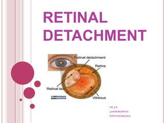
Call Girl Bangalore Nandini 7001305949 Independent Escort Service Bangalore
Call Girl Bangalore Nandini 7001305949 Independent Escort Service BangaloreCall Girl Bangalore Nandini 7001305949 Independent Escort Service Bangalore
Call Girl Bangalore Nandini 7001305949 Independent Escort Service Bangalore
 
College Call Girls Vyasarpadi Whatsapp 7001305949 Independent Escort Service
College Call Girls Vyasarpadi Whatsapp 7001305949 Independent Escort ServiceCollege Call Girls Vyasarpadi Whatsapp 7001305949 Independent Escort Service
College Call Girls Vyasarpadi Whatsapp 7001305949 Independent Escort Service
 
Asthma Review - GINA guidelines summary 2024
Asthma Review - GINA guidelines summary 2024Asthma Review - GINA guidelines summary 2024
Asthma Review - GINA guidelines summary 2024
 
Call Girls Frazer Town Just Call 7001305949 Top Class Call Girl Service Avail...
Call Girls Frazer Town Just Call 7001305949 Top Class Call Girl Service Avail...Call Girls Frazer Town Just Call 7001305949 Top Class Call Girl Service Avail...
Call Girls Frazer Town Just Call 7001305949 Top Class Call Girl Service Avail...
 
Call Girls Hosur Just Call 7001305949 Top Class Call Girl Service Available
Call Girls Hosur Just Call 7001305949 Top Class Call Girl Service AvailableCall Girls Hosur Just Call 7001305949 Top Class Call Girl Service Available
Call Girls Hosur Just Call 7001305949 Top Class Call Girl Service Available
 
Call Girls Thane Just Call 9910780858 Get High Class Call Girls Service
Call Girls Thane Just Call 9910780858 Get High Class Call Girls ServiceCall Girls Thane Just Call 9910780858 Get High Class Call Girls Service
Call Girls Thane Just Call 9910780858 Get High Class Call Girls Service
 
Call Girls Jp Nagar Just Call 7001305949 Top Class Call Girl Service Available
Call Girls Jp Nagar Just Call 7001305949 Top Class Call Girl Service AvailableCall Girls Jp Nagar Just Call 7001305949 Top Class Call Girl Service Available
Call Girls Jp Nagar Just Call 7001305949 Top Class Call Girl Service Available
 
Russian Call Girls in Pune Riya 9907093804 Short 1500 Night 6000 Best call gi...
Russian Call Girls in Pune Riya 9907093804 Short 1500 Night 6000 Best call gi...Russian Call Girls in Pune Riya 9907093804 Short 1500 Night 6000 Best call gi...
Russian Call Girls in Pune Riya 9907093804 Short 1500 Night 6000 Best call gi...
 
Low Rate Call Girls Pune Esha 9907093804 Short 1500 Night 6000 Best call girl...
Low Rate Call Girls Pune Esha 9907093804 Short 1500 Night 6000 Best call girl...Low Rate Call Girls Pune Esha 9907093804 Short 1500 Night 6000 Best call girl...
Low Rate Call Girls Pune Esha 9907093804 Short 1500 Night 6000 Best call girl...
 
Ahmedabad Call Girls CG Road 🔝9907093804 Short 1500 💋 Night 6000
Ahmedabad Call Girls CG Road 🔝9907093804  Short 1500  💋 Night 6000Ahmedabad Call Girls CG Road 🔝9907093804  Short 1500  💋 Night 6000
Ahmedabad Call Girls CG Road 🔝9907093804 Short 1500 💋 Night 6000
 
Call Girls Budhwar Peth 7001305949 All Area Service COD available Any Time
Call Girls Budhwar Peth 7001305949 All Area Service COD available Any TimeCall Girls Budhwar Peth 7001305949 All Area Service COD available Any Time
Call Girls Budhwar Peth 7001305949 All Area Service COD available Any Time
 
Call Girl Service Bidadi - For 7001305949 Cheap & Best with original Photos
Call Girl Service Bidadi - For 7001305949 Cheap & Best with original PhotosCall Girl Service Bidadi - For 7001305949 Cheap & Best with original Photos
Call Girl Service Bidadi - For 7001305949 Cheap & Best with original Photos
 
Russian Call Girl Brookfield - 7001305949 Escorts Service 50% Off with Cash O...
Russian Call Girl Brookfield - 7001305949 Escorts Service 50% Off with Cash O...Russian Call Girl Brookfield - 7001305949 Escorts Service 50% Off with Cash O...
Russian Call Girl Brookfield - 7001305949 Escorts Service 50% Off with Cash O...
 
Bangalore Call Girls Majestic 📞 9907093804 High Profile Service 100% Safe
Bangalore Call Girls Majestic 📞 9907093804 High Profile Service 100% SafeBangalore Call Girls Majestic 📞 9907093804 High Profile Service 100% Safe
Bangalore Call Girls Majestic 📞 9907093804 High Profile Service 100% Safe
 
Bangalore Call Girls Marathahalli 📞 9907093804 High Profile Service 100% Safe
Bangalore Call Girls Marathahalli 📞 9907093804 High Profile Service 100% SafeBangalore Call Girls Marathahalli 📞 9907093804 High Profile Service 100% Safe
Bangalore Call Girls Marathahalli 📞 9907093804 High Profile Service 100% Safe
 
Call Girls Electronic City Just Call 7001305949 Top Class Call Girl Service A...
Call Girls Electronic City Just Call 7001305949 Top Class Call Girl Service A...Call Girls Electronic City Just Call 7001305949 Top Class Call Girl Service A...
Call Girls Electronic City Just Call 7001305949 Top Class Call Girl Service A...
 
VIP Call Girls Lucknow Nandini 7001305949 Independent Escort Service Lucknow
VIP Call Girls Lucknow Nandini 7001305949 Independent Escort Service LucknowVIP Call Girls Lucknow Nandini 7001305949 Independent Escort Service Lucknow
VIP Call Girls Lucknow Nandini 7001305949 Independent Escort Service Lucknow
 
VIP Call Girls Mumbai Arpita 9910780858 Independent Escort Service Mumbai
VIP Call Girls Mumbai Arpita 9910780858 Independent Escort Service MumbaiVIP Call Girls Mumbai Arpita 9910780858 Independent Escort Service Mumbai
VIP Call Girls Mumbai Arpita 9910780858 Independent Escort Service Mumbai
 
Housewife Call Girls Bangalore - Call 7001305949 Rs-3500 with A/C Room Cash o...
Housewife Call Girls Bangalore - Call 7001305949 Rs-3500 with A/C Room Cash o...Housewife Call Girls Bangalore - Call 7001305949 Rs-3500 with A/C Room Cash o...
Housewife Call Girls Bangalore - Call 7001305949 Rs-3500 with A/C Room Cash o...
 
Call Girls Whitefield Just Call 7001305949 Top Class Call Girl Service Available
Call Girls Whitefield Just Call 7001305949 Top Class Call Girl Service AvailableCall Girls Whitefield Just Call 7001305949 Top Class Call Girl Service Available
Call Girls Whitefield Just Call 7001305949 Top Class Call Girl Service Available
 

RETINAL DETACHMENT

  • 2. ANATOMY OF THE PERIPHERAL RETINA 1.Pars Plana  The ciliary body starts 1mm from the limbus and extends posteriorly for about 6mm.  The first 2mm = pars plicata and the remaining 4mm = flattened pars plana.  In order not to endanger the lens or retina, the optimal location for a pars plana surgical incision is 4 mm from the limbus in phakic eyes and 3.5 mm from the limbus in pseudophakic eyes.
  • 3.
  • 4. 2. Ora Serrata Forms the junction between the retina and ciliary body and is characterized by the following : 1.Dentate processes are teeth-like extensions of retina onto the pars plana; more marked nasally than temporally. 2. Oral bays are the scalloped edges of the pars plana epithelium in between the dentate processes.
  • 5. 3. A meridional fold a small radial fold of thickened retinal tissue in line with a dentate process, usually located in the superonasal quadrant . A fold may occasionally exhibit a small retinal hole at its apex 4. An enclosed oral bay a small island of pars plana surrounded by retina as a result of meeting of two adjacent dentate processes. *It should not be mistaken for a retinal hole because it is located anterior to the ora serrata. 5 Granular tissue multiple white opacities within the vitreous base can sometimes be mistaken for small peripheral opercula
  • 6.
  • 7. DESCRIPTION LENGTH Width of ora serrata 2.1 mm temporally 0.7-0.8 mm nasally Location from limbus 6mm nasally 7mm temporally From equator 6-8 mm From optic disc 25 mm
  • 8. At the ora, fusion of the sensory retina with the RPE and choroid limits forward extension of SRF. However, there being no equivalent adhesion between the choroid and sclera, choroidal detachments may progress anteriorly to involve the ciliary body (CILIOCHOROIDAL DETACHMENT). **IMP** Choroidal detachments do not extend to posterior pole bcoz they are limited by the VORTEX VEINS entering their scleral channels.
  • 9.
  • 10. VITREOUS BASE  3–4 mm wide zone straddling the ora serrata.  The cortical vitreous is strongly attached at the vitreous base, so that following acute PVD, the posterior hyaloid face remains attached to the posterior border of the vitreous base.  Pre-existing retinal holes within the vitreous base do not lead to RD.  Severe blunt trauma avulsion of the vitreous base with tearing of the NPE of the pars plana along its anterior border and of the retina along its posterior border.
  • 11.
  • 12. VITREOUS ADHESIONS 1 Normal. The peripheral cortical vitreous is loosely attached to ILM of the sensory retina. Stronger adhesions : • Vitreous base, strongest. • Around the ONH fairly strong. • Around the fovea, fairly weak, except in eyes with VMT and macular hole formation. • Along peripheral blood vessels, usually weak. 2 Abnormal adhesions at the following : • Posterior border of islands of lattice degeneration. • Retinal pigment clumps. • Peripheral paravascular condensations. • Vitreous base anomalies eg. tongue like extensions & posterior islands. • ‘WWP’ and ‘WWOP’.
  • 13. VITREORETINAL TRACTION  Is a force exerted on the retina by structures originating in the vitreous, and may be dynamic or static. 1 Dynamic traction is induced by eye movements and exerts a centripetal force towards the vitreous cavity. It plays an important role in the pathogenesis of retinal tears and Rhegmatogenous RD.
  • 14. 2 Static traction is independent of ocular movements. It plays a key role in the pathogenesis of tractional RD and proliferative vitreoretinopathy
  • 16. LATTICE DEGENERATION 1. Prevalence - 8% of the population.  Develops early in life, with a peak incidence in the 2nd and 3rd decades.  More commonly in moderate myopes  and is the most important degeneration directly related to RD.  Bilateral  Temporal and superiorly  Lattice is present in about 40% of eyes with RD. 2. Pathology - There is discontinuity of the ILM with variable atrophy of the underlying NSR.  The vitreous overlying an area of lattice is synchytic but the vitreous attachments around the margins are exaggerated.
  • 17.
  • 18. 3.Signs-  Spindle-shaped areas of retinal thinning, commonly located between the equator and the posterior border of the vitreous base.  Arborizing network of white lines within the islands. **  Some lattice lesions may be associated with ‘snowflakes’ (remnants of degenerate Müller cells.)  Associated hyperplasia of the RPE is common.  Small holes within lattice lesions are common and usually innocuous.
  • 19. 4.Complications –  No complications are encountered in most patients.  Tears may occasionally develop in eyes with acute PVD.  Atrophic holes may rarely lead to RD, particularly in young myopes. The fellow eye often has a ‘mirror-image’ distribution of holes.
  • 20.
  • 21. SNAILTRACK DEGENERATION  Characterized by sharply demarcated bands of tightly packed ‘snowflakes’which give the peripheral retina a white frost- like appearance.  The islands are usually longer than in lattice degeneration and may be associated with overlying vitreous liquefaction. o However, marked vitreous traction at the posterior border of the lesions is seldom present so that tractional U-tears rarely occur, although round holes within the snailtracks may be present.
  • 22. RETINOSCHISIS Symptoms.  Common in HYPERMETROPES.  SPLIT IN RETINAL LAYERS.  Cystoid degeneration.  Photopsia and floaters are absent. ( because there is no vitreoretinal traction. )  Occasionally symptoms occur as a result of either VH or the development of progressive RD.
  • 23. Typical RS /DEGENERATIVE More common Low lying Split in OPL Less complications Located Ant to equator Less common Absent Reticular /JUVENILE RS Less common BULLOUS Split in NFL more Can be Posterior to equator Microaneursyms & small telengeictasias Outer layer holes
  • 24.  Signs : Early retinoschisis - extreme inferotemporal periphery of both fundi. ** appearing as an exaggeration of microcystoid degeneration with a smooth immobile elevation of the retina. **The surface of the inner layer may show snowflakes as well as sheathing or ‘silver-wiring’ of blood vessels and the schisis cavity may be bridged by rows of torn grey-white tissue.  Breaks may be +nt in one or both layers.  The elevation is convex, smooth, thin and relatively immobile, unlike the opaque and corrugated appearance of a rhegmatogenous RD.
  • 25.
  • 26.
  • 27. RETINOSCHISIS RD TYPICAL PT AGE Middle to elderly Middle age Refractive association Hyperopia Myopia Symptoms Almost always absent Acute : present Chronic : absent Scotoma Absolute Relative VH or pigment Absent Present Location Inferotemporal> ST Acute : usually superior Chronic:usually inferior Texture Smooth Ac: corrugated Ch: smooth Muller footplates Common Absent Mobility Relatively immobile Ac: very often mobile Ch: relatively immobile Movement with scleral depression Moves as a single unit Height decreases
  • 28. RETINOSCHISIS RD Color with scleral depression WWP may be seen in outer layer NOT Breaks May be present Present RPE Normal Ac: normal Ch: atrophy & demarcation lines OCT Splitting of retinal layers SRF Effect of LASER application through retinal break Through inner layer break: Uptake Through full thickness break : no uptake
  • 29. DIFFUSE CHORIORETINAL ATROPHY  Characterized by choroidal depigmentation and thinning of the overlying retina in the equatorial area of highly myopic eyes.  Retinal holes developing in the atrophic retina may lead to RD.  Because of lack of contrast between the depigmented choroid and sensory retina, small holes may be very difficult to visualize without the help of slit-lamp biomicroscopy.
  • 30. WHITE WITH PRESSURE  Retinal areas in which a translucent white-grey appearance can be induced by scleral indentation.  Each area has a fixed configuration that does not change when indentation is moved to an adjacent area.  Along posterior border of lattice deg, snail-track deg and the outer layers of acquired retinoschisis..  Can be seen in normal eyes === abnormally strong attachment of vitreous gel.
  • 31. WHITE WITHOUT PRESSURE  Present without scleral indentation.  Area with fairly strong adhesion of condensed vitreous.  Can be mistakes for flat retinal hole.  Retinal breaks, GRTs can develop along the posterior border of WWOP.  Prohylactic therapy thus considered in WWOP in fellow eye of a patient with spontaneous GRT in other eye.
  • 32. POSTERIOR VITREOUS DETACHMENT  Separation of the cortical vitreous from the ILM of the NSR posterior to the vitreous base.  Classification : 1 Onset  Acute : most common Develops suddenly and usually becomes complete soon after onset.  Chronic : gradually weeks or months to become complete. 2 Extent  Complete PVD entire vitreous cortex detaches up to the posterior margin of the vitreous base.  Incomplete PVD residual vitreoretinal attachments remain posterior to the vitreous base.
  • 33. Symptoms : 1. Photopsia -- inc in dim light, common in temporal periphery. 2.Myodesopsia (floaters) : spots/ cobwebs/ flies. 3. Blurred vision : d/t vitreous haemorrhage or d/t PHM or floaters in visual axis.
  • 34.  Weiss ring :- Detached former attachment of the margin of optic disc. Can be seen by patient as a circle or other solitary lesion.
  • 35. Signs :  Detached PHM seen on S/L as CRUMPLED TRANSLUCENT membrane in mid vitreous cavity which is optically clear.  Haemorrhage---rbcs in ant vitreous.  Pigment granules in ant. vitreous in abscence of inflammation, trauma, surgery on S/L - k/a SHAFER SIGN OR TOBACCO DUST Presence of pigment granules in ant vitreous 95% sensitivity for possibility of a retinal break
  • 36. PHM as Crumpled translucent membrane Tobacco dust Tobacco dust
  • 39.  Rhegmatogenous RD is usually associated with acute PVD;  tractional RD is associated with chronic, incomplete PVD;  exudative RD is unrelated to the presence of PVD.
  • 41.
  • 42. FACTORS PREVENTING RD  Metabolic pump of RPE.  Osmotic pressure of choroid.  Minor mechanical processes of the interphotoreceptor matrix. compromised RETINAL DETACHMENT
  • 43. RD Traction component present Rhegmatogenous RD Tractional RD Combined Tractional- rhegmatogenous RD Central RD in myopic eye Traction component absent Exudative RD Haemorrhagic RD
  • 45.  A Condition in which the fluid from the vitreous cavity passes through a retinal defect into the subretinal space to cause seperation of neural retina from the underlying RPE  Retinal break and vitreous liquefaction is must.**  Even though a retinal break is present, a RD will not occur if the vitreous is not at least partially liquefied and if the necessary traction is not present. PVD V. .liquefaction Retinal tears at site of significant VR adhesions
  • 46. CONDITIONS THAT PREDISPOSE AN EYE TO RD  High myopia.------3 fold in RD  PP or Aphakia.  Blunt trauma and penetrating ocular trauma. ( 10-15%)  CMV retinits ass. AIDS.  YAG capsulotomy
  • 47. FACTORS CAUSING RD A. RETINAL BREAKS B. VITREOUS LIQUEFACTION AND DETACHMENT C. VR TRACTION D. INTRAOCULAR FLUID CURRENTS ASSOCIATED WITH MOVEMENT OF LIQUID VITREOUS AND SRF
  • 49. RETINAL BREAKS  Full thickness defects in neurosensory retina.  Typical location- near vitreous base.  Holes/ tears/ dialysis.  M=F  R/F : 1. Myopia. 2. Lattice degeneration. 3. Ocular contusion and Penerating trauma. Most common of retinal break after ocular contusion is :- Retinal Dialysis
  • 50. OCULAR MANIFESTATIONS: 1. RETINAL TEARS :  Full thickness breaks that occur secondary to vitreous traction.  (spontaneous posterior vitreous detachment). horseshoe-shaped  Types flap shaped  Most common site- vitreous base.  Symptoms: Floaters, Flashes.  In lattice deg, mc site of retinal tear is POSTERO-LATERAL margin of lattice.
  • 51. 2. ROUND HOLES WITH OPERCULA Persistent traction on HST/ flap tear Avulsion at base Complete relief of VRT in that area Small round defect in neural retina with an overlying opercula
  • 52.  3.Round holes without opercula – ATROPHIC HOLES  Secondary to retinal thinning.  No role of vitreous traction.  Common in areas of lattice deg.
  • 53. 4. TRAUMATIC retinal breaks : HST Retinal dialysis Macular holes  MC site : INFEROTEMPORALLY & SUPERONASALLLY 5. MACULAR BREAKS :  Secondary to tangential traction from precortical vitreous
  • 54.
  • 55. Pars plana cysts Enclosed oral bays Meridional folds Ora serrata pearls Paving stone deg Chorioretinal scars WWP WWOP D/D OF RETINAL BREAKS
  • 56.
  • 57. INDICATIONS FOR T/T OF RETINAL TEAR/ HOLE IN ASYMPTOMATIC PATIENT Type of lesion Phakic Highly myopic Fellow eye Aphakic or Pseudophakic Retinal dialysis Almost always Almost always Almost always Almost always HST sometimes sometimes sometimes sometimes Operculated tears no rarely rarely rarely Atrophic holes rarely rarely rarely rarely Lattice deg’n w/ or w/o holes no no sometimes rarely
  • 58.
  • 59. Lincoff and Geiser reported 4 guidelines for locating RB causing RRD * Determined by  Location of causative break  Anatomic barriers (optic n.,ora serrata, existing chorioretinal adhesions)  Effect gravity on SRF in upright position Note : onlyfor freshRDwith1 RB
  • 60. 1. A shallow inferior RD in which the SRF is slightly higher on the temporal side points to a primary break located inferiorly on that side.
  • 61. 2. A primary break located at 6 o’clock will cause an inferior RD with equal fluid levels.
  • 62. 3. In a bullous inferior RD the primary break usually lies above the horizontal meridian. 4. If the primary break is located in the upper nasal quadrant the SRF will revolve around the optic disc and then rise on the temporal side until it is level with the primary break.
  • 63. 5. A subtotal RD with a superior wedge of attached retina points to a primary break located in the periphery nearest its highest border. 6. When the SRF crosses the vertical midline above, the primary break is near to 12 o’clock, the lower edge of the RD corresponding to the side of the break .
  • 64. B. VITREOUS LIQUEFACTION AND DETACHMENT Ageing of human vitreous (SYNCHYSIS) Liquefaction of vitreous gel with development of optically emtpy lacunae in gel Extensive liquefaction Decrease in shock absorbing capacity and stability of gel
  • 65. C. VR TRACTION VR TRACTION Gravitational force Rotational eye movements Fibrous tissue contracture Liquid currents Higher percentage of SUPERIOR retinal tears (80%) Trauma/ retinal vascular proliferative disorders Continous flow of liquid vitreous through a retinal break into subretinal space is necessary to maintain a RRD
  • 66. OCULAR MANIFESTATIONS SYMPTOMS  Sudden onset of tiny dark floating objects  Associated with Photopsia **usually brief **in the temporal visual field SUBCLINICAL DETACHMENTS : RD with small SRF (<2 DD) not acc by visual field loss
  • 67.
  • 68. SIGNS 1. RAPD 2. IOP is usually lower by about 5 mmHg compared with the eye. If the intraocular pressure is extremely low, an associated choroidal detachment may be present. 3. Iritis is very common but usually mild. 4. ‘Tobacco dust’ consisting of pigment cells is seen in the anterior vitreous. It may normal be raised, characteristically in SCHWARTZ-MATSUO syndrome in which RRD is associated with an apparent mild anterior uveitis , often due to a DIALYSIS due to prior blunt trauma in a young man.
  • 69. 5. Retinal breaks--- appear as discontinuities in the retinal surface. They are usually red because of the colour contrast between the sensory retina and underlying choroid. (However, in eyes with hypopigmented choroid (as in high myopia), the colour contrast is decreased and small breaks may be overlooked unless careful slit-lamp and indirect ophthalmoscopic examination is performed.) 6. Retinal signs depend on the -- duration of RD and -- the presence or absence of PVR.
  • 70.
  • 71. Or tide marks. Take 3 months to develop
  • 73. PROLIFERATIVE VITREORETINOPATHY  Caused by epiretinal and subretinal membrane formation.  Most common cause of ultimate failure after surgical tt for RRD.  Retinal break cells in vitreous cavity cells form membranes on inner retinal surface or posterior vitreous surface RRD.  Usually, Occurs following surgery for RRD or penetrating injury. However, it may also occur in eyes with RRD that have not had previous vitreoretinal surgery.
  • 74. RISK FACTORS FOR PVR  1. Giant retinal tear ( >3 clock hour).  2. Number and size of retinal break.  3. No.of previous operations.  4. Presence of choroidal effusions.  5. Use of cryotherapy.  6. Intraocular haemorrhage.  7.Aphakia.  8. Vitreous protein levels.
  • 75. 1 Grade A (minimal) PVR : Characterized by diffuse vitreous haze and tobacco dust. There may also be pigmented clumps on the inferior surface of the retina. 2 Grade B (moderate) PVR : Characterized by wrinkling of the inner retinal surface, tortuosity of blood vessels, retinal stiffness, decreased mobility of vitreous gel and rolled edges of retinal breaks. ** The epiretinal membranes responsible for these findings cannot be identified clinically.** 3 Grade C (marked) PVR Characterized by rigid full-thickness retinal folds with heavy vitreous condensation and strands. It can be either anterior (A) or posterior (P), the approximate dividing line being the equator of the globe. The severity of proliferation in each area is expressed by the number of clock hours of retina involved although proliferations need not be contiguous. 4 B-scan ultrasonography in advanced disease shows gross reduction of retinal mobility with retinal shortening and the characteristic triangular sign. (FUNNEL LIKE)
  • 76.
  • 77.
  • 78. T/T FOR PVR 1. No T/T if: retinal breaks absent unless macula is involved. localised TRD posterior to scleral buckle. 2. Macular pucker membrane peeling. 3. RRD location and severity of PVR If surface membranes do not prevent break closure…… SCLERAL BUCKLING If surface membrane occur in close association with retinal break Closed intraocular microsx with membrane peeling
  • 79.  RECENT ADVANCES : 1. PERIOPERATIVE DUANOMYCIN (on going trial) . 2.Adjuvant 5-HYDROXYURACIL + LOWER MOLECULE WEIGHT HEPARIN reduce PVR in high-risk cases indergoing vitrectomy.
  • 80. D/D FOR RRD  Traction RD.  Exudative RD.  Retinoschisis.  Elevated choroidal lesions.  Intravitreal optical illusions (vitreous h’age). SYSTEMIC ASSOCIATIONS  Most important ocular disorder : STICKLER $  Most important systemic association : DM, AIDS
  • 81. TREATMENT OF RD AIM : to counter the factors and forces that causes RD and to re- establish physiological conditions that normally maintain contact between neural retina and RPE.
  • 82. T/T OPTIONS AVAILABLE :  Primary vitrectomy  Laser demarcation  Permanent scleral buckle ## Encircling with/without drainage. ## Segmental with/without drainage  Temporary scleral buckle ## Lincoff ballon. ## Absorbable buckling sutures.  Pnematic retinopexy - routine - with drainage of SRF.
  • 83. VITRECTOMY INDICATIONS : 1. RD with PVR 2. Giant retinal Tears. 3.RD with posterior retinal breaks . 4.RD with vitreous hemorrhage. 5. RD with intraocular foreign body. 6. TRD threatening to or involving the macula. Less likely to be closed with scleral buckle Retinal breaks can not be visualized
  • 85. CONTRAINDICATIONS : 1. Bleeding disorders. 2. Suspected tumors like retinoblastoma, melanoma. COMPLICATIONS : 1.Iatrogenic breaks. 2.Lens trauma. 3.Redetachment. 4.Secondary glaucoma. 5. PVR 6.Cataract progression.
  • 86. SCLERALBUCKLING SURGERY  INDICATIONS : 1. RRD (PVR less than C1) 2. Inferior retinal dialysis. 3. Retinal dialysis. 4.Pediatric RD SEGMENTAL BUCKLING Usually reserved for ** RRD < 1 clock hour ** Anterior breaks
  • 87.
  • 88. CONTRA-INDICATIONS : 1. Posterior breaks. 2. Opaque media. 3. Vaso-occlusive diseases. 4. PVR more than C2. COMPLICATIONS : 1. Diplopia. (mc) 2.Perforation. 3. Rise in IOP. 4. Extrusion. 5. Infection. 6. AS ischemia.
  • 89. 7. ERM 8. Recurrent RD. 9. Surgical failure : ‘FISH-MOUTHING’ Phenomenon of a tear, typically a large superior equatorial U- tear in a bullous RD, to open widely following scleral buckling requiring further operative tt
  • 90. PNEUMATIC RETINOPEXY  INDICATIONS : 1. A detachment caused by a single break, in superior 8 clock hours. 2. The break should not be more than 1clock hour. 3. Multiple breaks but in 1-2 clock hours of each other.  CONTRA-INDICATIONS : 1. Break > 1 clock hr. 2. Break inferior to 4 clock hr. 3. PVR grade C,D. 4. Cloudy ocular media. 5. Uncontrolled or severe glaucoma. 6. Can’t maintain Head position.
  • 91. GAS EXPANSION NON-EXPANSIBLE CONC AVERAGE DURATION VOLUME USED FOR PR SF6 2 times 20 % 10-14 days 0.5 ml C3F8 4 times 12% 30-45 days 0.35 ml AIR Non expansile - 5-7 days 0.8 ml SILICONE OILS : -Lighter than water, -Commonly used for : intraop retinal manipulation prolonged postop intraocular tamponade. -Particularly useful in PVR.
  • 92.
  • 93. DRAINAGE OF SUBRETINAL FLUID  In deep and long standing viscous SRF.  Complications : incarceration at drainage site , Retinal perforation, Choroidal haemorrhage
  • 95. LASER PHOTOCOAGULATION  Usually cannot seal RB if presence SRF may be used to create barrier to prevent progression of RD  Esp. useful in Chronic inferior RD Systemic illness contraindicated to surgery Laser photocoagulation Slit-lamp biomicroscope with contact lens laser indirect ophthalmoscope (LIO)
  • 96. Slit-lamp LIO Indentation + better magnified significant cataracts, PCO, mild VH more easily treated with LIO Safer in inexpertise operator Need more skill Less need of corneal care during laser Less pain not be readily available Any patient position
  • 97. IMP** Compared with diathermy and cryopexy  less breakdown of blood–ocular barrier.  thermal effect confined predominantly to retina and RPE with little or no effect on choroid or sclera.  Induces adhesive effect between neurosensory retina & RPE within 24hr.
  • 98. CRYORETINOPEXY  RD with very shallow fluid can be cured by cryoretinopexy.  Test cryoprobe prior to use to make sure probe is freezing.  Freezing or whitening of RPE will noticed first, Followed by delineation of edges of retinal tear And whitening of retina
  • 99. ** Excessive freezing or ice crystal formation should be avoided Histologic response depends on whether  RPE alone or RPE and overlying detached retina together are frozen
  • 100. Disadvantage :  Dispersion of pigment epithelial cells, which can result in subretinal pigmentary changes after reattachment.  Dispersion of viable pigment epithelial cells capable of causing PVR following cryopexy.  Induce choroidal congestion & hyperemia (transient)  May complicate drainage of SRF through treated areas.  Breakdown of BRB cause post-op CME and ERD
  • 101.
  • 103.  Main causes of tractional RD penetrating posterior segment trauma proliferative retinopathy such as diabetic and ROP
  • 104. PATHOGENESIS OF DIABETIC TRACTIONAL RD  Caused by progressive contraction of fibrovascular membranes over large areas of vitreoretinal adhesion.  PVD in diabetic eyes is gradual and frequently incomplete.  RD is Caused by leakage of plasma constituents into the vitreous gel from a fibrovascular network adherent to the posterior vitreous surface.  In the very rare event of a subsequent complete PVD, the new blood vessels are avulsed and RD does not develop.
  • 105. Static vitreoretinal traction of the following three types is recognized -- A Tangential traction Caused by the contraction of epiretinal fibrovascular membranes with puckering of the retina and distortion of retinal blood vessels.
  • 106.  B Anteroposterior traction is Caused by the contraction of fibrovascular membranes extending from the posterior retina, usually in association with the major arcades, to the vitreous base anteriorly
  • 107. C Bridging (trampoline) traction is the result of contraction of fibrovascular membranes which stretch from one part of the posterior retina to another or between the vascular arcades, tending to pull the two involved points together.
  • 108.
  • 109. DIAGNOSIS  Symptoms : Photopsia and floaters are usually absent  Signs : Concave configuration breaks are absent. Retinal mobility is severely reduced shifting fluid is absent.  The SRF is shallower than in a RRD and seldom extends to the ora serrata.  Highest elevation of the retina at sites of vitreoretinal traction.  If a tractional RD develops a break it assumes the characteristics of a RRD and progresses more quickly (combined TRD-RRD).
  • 110.
  • 111.
  • 112. TREATMENT  GOAL : To release antero-posterior and/or circumferential traction.  Simple peeling------------------NOT POSSIBLE(vascularity and friable retina)  Methods of removing fibrovascular membranes in Diabetic TRDs. DELAMINATION SEGMENTATION (Vertical cutting) (horizontal cutting)
  • 114. PATHOGENESIS  Characterized by the accumulation of SRF in the absence of retinal breaks or traction.  As long as the RPE is able to compensate by pumping the leaking fluid into the choroidal circulation, no fluid accumulates in the subretinal space RD does not occur.  However, when the normal RPE pump is overwhelmed, or if the RPE activity is decreased, then fluid starts to accumulate in the subretinal space.
  • 115. ETIOLOGY 1 .Choroidal tumours such as melanomas, haemangiomas and metastases; it is therefore very important to consider that exudative RD is caused by an intraocular tumour until proved otherwise. 2 .Inflammation such as Harada disease and posterior scleritis. 3 .Bullous central serous chorioretinopathy : rare 4 .Iatrogenic causes - RD surgery & PRP. 5. Subretinal neovascularization which may leak and give rise to extensive subretinal accumulation of fluid at the posterior pole. 6. Hypertensive choroidopathy, as may occur in toxaemia of pregnancy:rare. 7. Idiopathic such as the uveal effusion syndrome.
  • 118. DIAGNOSIS Symptoms:- **Photopsia is absent because there is no vitreoretinal traction, although floaters may be present if there is associated vitritis. Signs:- Convex configuration, just like a RRD, BUT its surface is smooth and not corrugated.
  • 119.  The detached retina is very mobile and exhibits the phenomenon of ‘shifting fluid’ in which SRF responds to the force of gravity and detaches the area of retina under which it accumulates. (For example, in the upright position the SRF collects under the inferior retina, but on assuming the supine position for several minutes, the inferior retina flattens and the SRF shifts posteriorly detaching the superior retina.)  The cause of the RD, such as a choroidal tumour, may be apparent when the fundus is examined, or the patient may have an associated systemic disease responsible for the RD (e.g. Harada disease, toxaemia of pregnancy).  ‘Leopard spots’ consisting of scattered areas of subretinal pigment clumping may be seen after the detachment has flattened.
  • 120.
  • 121.
  • 122.  Leopard sign pigmentation following resolution of exudative RD
  • 123.
  • 124. IS THIS RRD OR TRD OR ERD ??????
  • 125. SYMPTOMS RRD TRD ERD Floaters ++ +/- +/- Flashes ++ - - Progression of VA loss acute chronic Subacute/ chronic Fluctuation of vision - +/- +
  • 126. SIGNS RRD TRD ERD Shaffer’s sign + - - PVD ++ - +/- VH +/- +/- - RD contour convex concave bullous RD surface corrugate Ridge/ wavy smooth Shifting +/- - ++ Associated Myopia/ trauma/ Sx DM CTD

Editor's Notes

  1. TREATMENT :
  2. ( BECAUSE vitreoretinal traction develops insidiously and is not associated with acute PVD. )