SlideShare a Scribd company logo
1 of 15
Download to read offline
Esophageal carcinoma
Esophageal cancer - a malignant tumor emanating from the
mucous membrane of the esophagus makes up a significant part
of all diseases of this organ. The main symptoms of this disease
are: progressive impairment of swallowing (first solid food, then
liquid) and unintentional weight loss. As of 2012, esophageal
cancer was the eighth most common cancer in the world, with
456,000 new cases during the year. In men, esophageal cancer
occurs about 3 times more often than in women.
Etiology
The main risk factors that can cause esophageal cancer include:
Consuming hot, rough, and poorly chewed foods
Drinking alcohol
Frequent drinking of hot drinks (tea, coffee)
Smoking
Barrett's esophagus
Esophagitis
Diverticulitis
Thermal and chemical burns of the esophagus, accompanied by
the formation of scars
Leukoplakia
Tumor growth types
There are 3 types of esophageal cancer:
Exophytic (nodular, mushroom, papillomatous -
growth into the lumen of the organ)
Endophytic (ulcerative)
Infiltrative sclerosing (circular form)
Histological forms
Most often (97-99% of cases), squamous cell
carcinoma with keratinization and without
keratinization is found. There are also glandular
forms of esophageal cancer and tumors originating
from dystopic epithelium. The international
classification also includes small cell carcinoma,
adenoacanthoma, and carcinosarcoma. [7]
Undifferentiated cancers are also rarely found.
Metastasis
Metastasis of esophageal cancer is due to the developed lymphatic network
of the esophagus. Metastases spread to the adjacent lymphatic vessels, and
then to the lymph nodes. Cancer of the cervical esophagus metastasizes to
the deep cervical lymph nodes, cancer of the upper thoracic and middle
thoracic esophagus affects paraesophageal, tracheobronchial and posterior
mediastinal lymph nodes with metastases; cancer of the lower thoracic and
abdominal regions spreads to the subphrenic, paraesophageal, paracardial
lymph nodes, as well as to the lymph nodes along the lesser curvature of
the stomach and the left gastric artery. In addition, metastases are found in
the lymph nodes of the lesser omentum, along the left gastric artery, and in
the cervical and supraclavicular lymph nodes.
Distant metastases affect the liver, lungs, and skeletal system.
Clinical classification
According to TNM classification:
T - primary tumor
Tх - insufficient information to assess the primary tumor
T0 - no primary tumor found
Tis - carcinoma in situ.
T1 - the tumor invades the wall of the esophagus up to the submucosa
T2 - the tumor invades the wall of the esophagus to the muscle layer
T3 - the tumor invades the wall of the esophagus to the adventitia.
T4 - the tumor process spreads to neighboring organs
N - regional lymph nodes
Nx - insufficient data to assess regional lymph nodes
N0 - no metastases to regional lymph nodes were found
N1 - metastases to regional lymph nodes are detected
M - distant metastases
Mx - insufficient information to determine distant metastases
M0 - distant metastases not found
M1 - distant metastases are detected
In accordance with the Russian classification, esophageal cancer is
divided into 4 stages:
Stage 1 - a small neoplasm affects the mucous membrane and the
submucosa of the esophageal wall, but does not narrow the lumen of the
esophagus and its muscle layer does not grow. No metastases (T1N0M0)
Stage 2 - tumor masses penetrate into the muscular membrane of the
esophagus and narrow the lumen, but do not leave the organ. Single
metastases are found in regional lymph nodes (stage 2A: T2N0M0,
T3N0M0; stage 2B: T1N1M0, T2N1M0)
Stage III - all layers of the esophageal wall are involved in the tumor
process, as well as the peri-esophageal tissue or serous membrane, but
neighboring organs are not affected. Numerous metastases are found in
regional lymph nodes (T3N1M0, T4 any N M0)
Stage IV - cancer affects all layers of the esophagus wall and spreads
to neighboring organs. Metastases are found in regional and distant lymph
nodes (any T, any N, M1)
Clinical picture
The clinical symptoms of esophageal cancer can be divided into three
groups: primary or local symptoms caused by damage to the walls of the
esophagus; secondary symptoms resulting from the spread of the tumor
process to neighboring organs and tissues; general symptoms caused by
intoxication and malnutrition.
Primary symptoms include dysphagia, chest pain, chest fullness,
regurgitation, and increased salivation. Almost all of these symptoms
indicate a fairly large spread of the pathological process along the
esophagus.
Typical symptoms of esophageal cancer are caused by obstruction. The
most striking of them is dysphagia - the difficulty in passing food
through the esophagus. Dysphagia is caused by a narrowing of the organ
lumen by a growing tumor (mechanical dysphagia), but sometimes it
depends on spasm in the overlying parts of the esophagus (reflex
dysphagia).
In most cases, dysphagia increases gradually. At first, there are subtle
delays in the passage of solid food through the esophagus. The patient, as
it were, feels a solid lump of food moving along the esophagus. The
narrowing progresses, and soon the patient is forced to drink solid food
with a sip of water or refuse to take second courses. In the future, after a
few weeks or months, semi-liquid food ceases to pass, and then liquid.
This sequential development of dysphagia is not always observed.
Sometimes, as a result of the disintegration of the tumor or drug
treatment, the patency of the esophagus is partially or completely restored.
The improvement in the condition does not last long, and soon the
dysphagia begins to progress again.
DIAGNOSTICS
At LISOD, effective diagnosis is based on a comprehensive examination. The first in
this series is the endoscopic examination of the esophagus: a flexible endoscope is
inserted into the esophagus, with the help of which a full examination of the entire
mucosa is performed; if necessary, a biopsy is taken - a small piece of tissue for
histological examination.
To find out the extent of the tumor (the degree of involvement of other tissues and
organs in it), i.e. determining the stage of the disease, use additional research methods:
computed tomography of the chest and abdomen;
ultrasound examination of the abdominal cavity;
chest x-ray;
a unique for Ukraine transesophageal ultrasound examination of the walls of the
esophagus and structures of the mediastinum with a possible biopsy of the formations
located in the immediate vicinity of the esophagus;
bronchoscopy;
video laparoscopy and video thoracoscopy;
laboratory research.
LISOD uses a modern research method - PET-CT. This
study is prescribed for patients who are indicated for
radical treatment, as well as for patients who have
undergone neoadjuvant chemotherapy. PET-CT is also
used to accurately plan radiation therapy, to assess the
outcome of treatment and to identify possible recurrence
of the disease.
The use of a number of diagnostic measures allows
specialists to identify the extent of the spread of the
disease and begin complex treatment.
TREATMENT
Treatment for esophageal cancer depends on the extent of the tumor and the underlying
pathology. Often, esophageal cancer is detected too late for radical treatment.
In such cases, we offer procedures aimed at improving the quality of life of patients:
placement with an endoscope of a special tube (stent) in the esophagus in order to allow
the passage of food and liquid;
radiation therapy aimed at shrinking the tumor;
expansion (bougienage) of the esophagus or argon plasma destruction of the esophageal
tumor.
These methods are aimed at eliminating esophageal stenosis - closure of it by a tumor -
and associated severe symptoms such as dysphagia, vomiting, and weight loss.
Patients without pronounced concomitant diseases, with a localized tumor, are offered
surgical intervention, which gives a good chance of recovery.
The operation involves removing most of the swollen esophagus and connecting the
remainder to the stomach. Surgery is often accompanied by pre- or postoperative
chemotherapy and radiation therapy. In LISOD, the most complex operations on the
esophagus are performed using a low-traumatic laparoscopic method.
SYMPTOMS
Signs of esophageal cancer usually do not appear until the growth is of sufficient
size. The most common symptom is difficulty swallowing solid food. But after a
while, even swallowing liquid becomes difficult. A tumor of the esophagus is
also characterized by other signs.
Any of the following symptoms should be considered as a serious reason for
seeking medical attention and a comprehensive examination:
pain in the center of the chest;
vomiting;
pain when swallowing;
hoarseness and persistent cough (it happens if the tumor spreads to the trachea
and larynx);
weight loss;
signs of gastrointestinal bleeding: vomiting of blood or a mass that resembles
coffee grounds;
black feces (melena).
PREVENTION
Quitting smoking and drinking alcohol can reduce the incidence of
squamous cell carcinoma of the esophagus. At least 90% of this type of
cancer is associated with alcohol and tobacco abuse.
Esophageal adenocarcinoma is a common complication of Barrett's
esophagus, affecting more than 20% of people with reflux symptoms.
People with frequent reflux symptoms (heartburn or belching) should
have regular endoscopic examinations.
Patients with Barrett's esophagus should undergo endoscopy in order to
identify precancerous changes in the esophageal mucosa and receive
quality adequate treatment. They also need treatment for
gastroesophageal reflux, which includes diet and lifestyle changes.

More Related Content

Similar to Esophageal carcinoma.pdf

Similar to Esophageal carcinoma.pdf (20)

Colorectal cancer
Colorectal cancerColorectal cancer
Colorectal cancer
 
Diagnosis of tumor.ppt
Diagnosis of tumor.pptDiagnosis of tumor.ppt
Diagnosis of tumor.ppt
 
11 esophageal cancer
11 esophageal cancer11 esophageal cancer
11 esophageal cancer
 
Gastric Carcinoma
Gastric CarcinomaGastric Carcinoma
Gastric Carcinoma
 
1GASTRIC_CARCINOMA.ppt
1GASTRIC_CARCINOMA.ppt1GASTRIC_CARCINOMA.ppt
1GASTRIC_CARCINOMA.ppt
 
• Esophageal cancer tumor.pptx
• Esophageal cancer tumor.pptx• Esophageal cancer tumor.pptx
• Esophageal cancer tumor.pptx
 
Bladder cancer
Bladder cancerBladder cancer
Bladder cancer
 
Ca bladder (2)
Ca bladder (2)Ca bladder (2)
Ca bladder (2)
 
ORAL CANCER- OM VERMA 2022.pdf
ORAL CANCER- OM VERMA 2022.pdfORAL CANCER- OM VERMA 2022.pdf
ORAL CANCER- OM VERMA 2022.pdf
 
Esophageal carcinoma
Esophageal carcinomaEsophageal carcinoma
Esophageal carcinoma
 
colon ca.pptx
colon ca.pptxcolon ca.pptx
colon ca.pptx
 
Esophageal cancer
Esophageal cancerEsophageal cancer
Esophageal cancer
 
Gastric cancer
Gastric cancerGastric cancer
Gastric cancer
 
Gastric cancer
Gastric cancerGastric cancer
Gastric cancer
 
Colorectal cancer
Colorectal cancerColorectal cancer
Colorectal cancer
 
Malignant Neoplasms of Stomach.pptx
Malignant Neoplasms of Stomach.pptxMalignant Neoplasms of Stomach.pptx
Malignant Neoplasms of Stomach.pptx
 
Carcinoma stomach
Carcinoma stomachCarcinoma stomach
Carcinoma stomach
 
Gastric Carcinoma
Gastric CarcinomaGastric Carcinoma
Gastric Carcinoma
 
Cancer (Diet therapy, Nutritional care)
Cancer (Diet therapy, Nutritional care)Cancer (Diet therapy, Nutritional care)
Cancer (Diet therapy, Nutritional care)
 
Carcinoma of Stomach
 Carcinoma of Stomach Carcinoma of Stomach
Carcinoma of Stomach
 

Recently uploaded

MuleSoft Integration with AWS Textract | Calling AWS Textract API |AWS - Clou...
MuleSoft Integration with AWS Textract | Calling AWS Textract API |AWS - Clou...MuleSoft Integration with AWS Textract | Calling AWS Textract API |AWS - Clou...
MuleSoft Integration with AWS Textract | Calling AWS Textract API |AWS - Clou...
MysoreMuleSoftMeetup
 
Contoh Aksi Nyata Refleksi Diri ( NUR ).pdf
Contoh Aksi Nyata Refleksi Diri ( NUR ).pdfContoh Aksi Nyata Refleksi Diri ( NUR ).pdf
Contoh Aksi Nyata Refleksi Diri ( NUR ).pdf
cupulin
 
Spellings Wk 4 and Wk 5 for Grade 4 at CAPS
Spellings Wk 4 and Wk 5 for Grade 4 at CAPSSpellings Wk 4 and Wk 5 for Grade 4 at CAPS
Spellings Wk 4 and Wk 5 for Grade 4 at CAPS
AnaAcapella
 
Transparency, Recognition and the role of eSealing - Ildiko Mazar and Koen No...
Transparency, Recognition and the role of eSealing - Ildiko Mazar and Koen No...Transparency, Recognition and the role of eSealing - Ildiko Mazar and Koen No...
Transparency, Recognition and the role of eSealing - Ildiko Mazar and Koen No...
EADTU
 

Recently uploaded (20)

FICTIONAL SALESMAN/SALESMAN SNSW 2024.pdf
FICTIONAL SALESMAN/SALESMAN SNSW 2024.pdfFICTIONAL SALESMAN/SALESMAN SNSW 2024.pdf
FICTIONAL SALESMAN/SALESMAN SNSW 2024.pdf
 
MuleSoft Integration with AWS Textract | Calling AWS Textract API |AWS - Clou...
MuleSoft Integration with AWS Textract | Calling AWS Textract API |AWS - Clou...MuleSoft Integration with AWS Textract | Calling AWS Textract API |AWS - Clou...
MuleSoft Integration with AWS Textract | Calling AWS Textract API |AWS - Clou...
 
OSCM Unit 2_Operations Processes & Systems
OSCM Unit 2_Operations Processes & SystemsOSCM Unit 2_Operations Processes & Systems
OSCM Unit 2_Operations Processes & Systems
 
TỔNG HỢP HƠN 100 ĐỀ THI THỬ TỐT NGHIỆP THPT TOÁN 2024 - TỪ CÁC TRƯỜNG, TRƯỜNG...
TỔNG HỢP HƠN 100 ĐỀ THI THỬ TỐT NGHIỆP THPT TOÁN 2024 - TỪ CÁC TRƯỜNG, TRƯỜNG...TỔNG HỢP HƠN 100 ĐỀ THI THỬ TỐT NGHIỆP THPT TOÁN 2024 - TỪ CÁC TRƯỜNG, TRƯỜNG...
TỔNG HỢP HƠN 100 ĐỀ THI THỬ TỐT NGHIỆP THPT TOÁN 2024 - TỪ CÁC TRƯỜNG, TRƯỜNG...
 
male presentation...pdf.................
male presentation...pdf.................male presentation...pdf.................
male presentation...pdf.................
 
Observing-Correct-Grammar-in-Making-Definitions.pptx
Observing-Correct-Grammar-in-Making-Definitions.pptxObserving-Correct-Grammar-in-Making-Definitions.pptx
Observing-Correct-Grammar-in-Making-Definitions.pptx
 
How to Manage Website in Odoo 17 Studio App.pptx
How to Manage Website in Odoo 17 Studio App.pptxHow to Manage Website in Odoo 17 Studio App.pptx
How to Manage Website in Odoo 17 Studio App.pptx
 
Mattingly "AI and Prompt Design: LLMs with NER"
Mattingly "AI and Prompt Design: LLMs with NER"Mattingly "AI and Prompt Design: LLMs with NER"
Mattingly "AI and Prompt Design: LLMs with NER"
 
Andreas Schleicher presents at the launch of What does child empowerment mean...
Andreas Schleicher presents at the launch of What does child empowerment mean...Andreas Schleicher presents at the launch of What does child empowerment mean...
Andreas Schleicher presents at the launch of What does child empowerment mean...
 
When Quality Assurance Meets Innovation in Higher Education - Report launch w...
When Quality Assurance Meets Innovation in Higher Education - Report launch w...When Quality Assurance Meets Innovation in Higher Education - Report launch w...
When Quality Assurance Meets Innovation in Higher Education - Report launch w...
 
Contoh Aksi Nyata Refleksi Diri ( NUR ).pdf
Contoh Aksi Nyata Refleksi Diri ( NUR ).pdfContoh Aksi Nyata Refleksi Diri ( NUR ).pdf
Contoh Aksi Nyata Refleksi Diri ( NUR ).pdf
 
Spellings Wk 4 and Wk 5 for Grade 4 at CAPS
Spellings Wk 4 and Wk 5 for Grade 4 at CAPSSpellings Wk 4 and Wk 5 for Grade 4 at CAPS
Spellings Wk 4 and Wk 5 for Grade 4 at CAPS
 
Supporting Newcomer Multilingual Learners
Supporting Newcomer  Multilingual LearnersSupporting Newcomer  Multilingual Learners
Supporting Newcomer Multilingual Learners
 
Improved Approval Flow in Odoo 17 Studio App
Improved Approval Flow in Odoo 17 Studio AppImproved Approval Flow in Odoo 17 Studio App
Improved Approval Flow in Odoo 17 Studio App
 
e-Sealing at EADTU by Kamakshi Rajagopal
e-Sealing at EADTU by Kamakshi Rajagopale-Sealing at EADTU by Kamakshi Rajagopal
e-Sealing at EADTU by Kamakshi Rajagopal
 
Transparency, Recognition and the role of eSealing - Ildiko Mazar and Koen No...
Transparency, Recognition and the role of eSealing - Ildiko Mazar and Koen No...Transparency, Recognition and the role of eSealing - Ildiko Mazar and Koen No...
Transparency, Recognition and the role of eSealing - Ildiko Mazar and Koen No...
 
AIM of Education-Teachers Training-2024.ppt
AIM of Education-Teachers Training-2024.pptAIM of Education-Teachers Training-2024.ppt
AIM of Education-Teachers Training-2024.ppt
 
How to Send Pro Forma Invoice to Your Customers in Odoo 17
How to Send Pro Forma Invoice to Your Customers in Odoo 17How to Send Pro Forma Invoice to Your Customers in Odoo 17
How to Send Pro Forma Invoice to Your Customers in Odoo 17
 
The Liver & Gallbladder (Anatomy & Physiology).pptx
The Liver &  Gallbladder (Anatomy & Physiology).pptxThe Liver &  Gallbladder (Anatomy & Physiology).pptx
The Liver & Gallbladder (Anatomy & Physiology).pptx
 
PSYPACT- Practicing Over State Lines May 2024.pptx
PSYPACT- Practicing Over State Lines May 2024.pptxPSYPACT- Practicing Over State Lines May 2024.pptx
PSYPACT- Practicing Over State Lines May 2024.pptx
 

Esophageal carcinoma.pdf

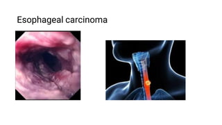
  • 2. Esophageal cancer - a malignant tumor emanating from the mucous membrane of the esophagus makes up a significant part of all diseases of this organ. The main symptoms of this disease are: progressive impairment of swallowing (first solid food, then liquid) and unintentional weight loss. As of 2012, esophageal cancer was the eighth most common cancer in the world, with 456,000 new cases during the year. In men, esophageal cancer occurs about 3 times more often than in women.
  • 3. Etiology The main risk factors that can cause esophageal cancer include: Consuming hot, rough, and poorly chewed foods Drinking alcohol Frequent drinking of hot drinks (tea, coffee) Smoking Barrett's esophagus Esophagitis Diverticulitis Thermal and chemical burns of the esophagus, accompanied by the formation of scars Leukoplakia
  • 4. Tumor growth types There are 3 types of esophageal cancer: Exophytic (nodular, mushroom, papillomatous - growth into the lumen of the organ) Endophytic (ulcerative) Infiltrative sclerosing (circular form)
  • 5. Histological forms Most often (97-99% of cases), squamous cell carcinoma with keratinization and without keratinization is found. There are also glandular forms of esophageal cancer and tumors originating from dystopic epithelium. The international classification also includes small cell carcinoma, adenoacanthoma, and carcinosarcoma. [7] Undifferentiated cancers are also rarely found.
  • 6. Metastasis Metastasis of esophageal cancer is due to the developed lymphatic network of the esophagus. Metastases spread to the adjacent lymphatic vessels, and then to the lymph nodes. Cancer of the cervical esophagus metastasizes to the deep cervical lymph nodes, cancer of the upper thoracic and middle thoracic esophagus affects paraesophageal, tracheobronchial and posterior mediastinal lymph nodes with metastases; cancer of the lower thoracic and abdominal regions spreads to the subphrenic, paraesophageal, paracardial lymph nodes, as well as to the lymph nodes along the lesser curvature of the stomach and the left gastric artery. In addition, metastases are found in the lymph nodes of the lesser omentum, along the left gastric artery, and in the cervical and supraclavicular lymph nodes. Distant metastases affect the liver, lungs, and skeletal system.
  • 7. Clinical classification According to TNM classification: T - primary tumor Tх - insufficient information to assess the primary tumor T0 - no primary tumor found Tis - carcinoma in situ. T1 - the tumor invades the wall of the esophagus up to the submucosa T2 - the tumor invades the wall of the esophagus to the muscle layer T3 - the tumor invades the wall of the esophagus to the adventitia. T4 - the tumor process spreads to neighboring organs N - regional lymph nodes Nx - insufficient data to assess regional lymph nodes N0 - no metastases to regional lymph nodes were found N1 - metastases to regional lymph nodes are detected M - distant metastases Mx - insufficient information to determine distant metastases M0 - distant metastases not found M1 - distant metastases are detected
  • 8. In accordance with the Russian classification, esophageal cancer is divided into 4 stages: Stage 1 - a small neoplasm affects the mucous membrane and the submucosa of the esophageal wall, but does not narrow the lumen of the esophagus and its muscle layer does not grow. No metastases (T1N0M0) Stage 2 - tumor masses penetrate into the muscular membrane of the esophagus and narrow the lumen, but do not leave the organ. Single metastases are found in regional lymph nodes (stage 2A: T2N0M0, T3N0M0; stage 2B: T1N1M0, T2N1M0) Stage III - all layers of the esophageal wall are involved in the tumor process, as well as the peri-esophageal tissue or serous membrane, but neighboring organs are not affected. Numerous metastases are found in regional lymph nodes (T3N1M0, T4 any N M0) Stage IV - cancer affects all layers of the esophagus wall and spreads to neighboring organs. Metastases are found in regional and distant lymph nodes (any T, any N, M1)
  • 9. Clinical picture The clinical symptoms of esophageal cancer can be divided into three groups: primary or local symptoms caused by damage to the walls of the esophagus; secondary symptoms resulting from the spread of the tumor process to neighboring organs and tissues; general symptoms caused by intoxication and malnutrition. Primary symptoms include dysphagia, chest pain, chest fullness, regurgitation, and increased salivation. Almost all of these symptoms indicate a fairly large spread of the pathological process along the esophagus. Typical symptoms of esophageal cancer are caused by obstruction. The most striking of them is dysphagia - the difficulty in passing food through the esophagus. Dysphagia is caused by a narrowing of the organ lumen by a growing tumor (mechanical dysphagia), but sometimes it depends on spasm in the overlying parts of the esophagus (reflex dysphagia).
  • 10. In most cases, dysphagia increases gradually. At first, there are subtle delays in the passage of solid food through the esophagus. The patient, as it were, feels a solid lump of food moving along the esophagus. The narrowing progresses, and soon the patient is forced to drink solid food with a sip of water or refuse to take second courses. In the future, after a few weeks or months, semi-liquid food ceases to pass, and then liquid. This sequential development of dysphagia is not always observed. Sometimes, as a result of the disintegration of the tumor or drug treatment, the patency of the esophagus is partially or completely restored. The improvement in the condition does not last long, and soon the dysphagia begins to progress again.
  • 11. DIAGNOSTICS At LISOD, effective diagnosis is based on a comprehensive examination. The first in this series is the endoscopic examination of the esophagus: a flexible endoscope is inserted into the esophagus, with the help of which a full examination of the entire mucosa is performed; if necessary, a biopsy is taken - a small piece of tissue for histological examination. To find out the extent of the tumor (the degree of involvement of other tissues and organs in it), i.e. determining the stage of the disease, use additional research methods: computed tomography of the chest and abdomen; ultrasound examination of the abdominal cavity; chest x-ray; a unique for Ukraine transesophageal ultrasound examination of the walls of the esophagus and structures of the mediastinum with a possible biopsy of the formations located in the immediate vicinity of the esophagus; bronchoscopy; video laparoscopy and video thoracoscopy; laboratory research.
  • 12. LISOD uses a modern research method - PET-CT. This study is prescribed for patients who are indicated for radical treatment, as well as for patients who have undergone neoadjuvant chemotherapy. PET-CT is also used to accurately plan radiation therapy, to assess the outcome of treatment and to identify possible recurrence of the disease. The use of a number of diagnostic measures allows specialists to identify the extent of the spread of the disease and begin complex treatment.
  • 13. TREATMENT Treatment for esophageal cancer depends on the extent of the tumor and the underlying pathology. Often, esophageal cancer is detected too late for radical treatment. In such cases, we offer procedures aimed at improving the quality of life of patients: placement with an endoscope of a special tube (stent) in the esophagus in order to allow the passage of food and liquid; radiation therapy aimed at shrinking the tumor; expansion (bougienage) of the esophagus or argon plasma destruction of the esophageal tumor. These methods are aimed at eliminating esophageal stenosis - closure of it by a tumor - and associated severe symptoms such as dysphagia, vomiting, and weight loss. Patients without pronounced concomitant diseases, with a localized tumor, are offered surgical intervention, which gives a good chance of recovery. The operation involves removing most of the swollen esophagus and connecting the remainder to the stomach. Surgery is often accompanied by pre- or postoperative chemotherapy and radiation therapy. In LISOD, the most complex operations on the esophagus are performed using a low-traumatic laparoscopic method.
  • 14. SYMPTOMS Signs of esophageal cancer usually do not appear until the growth is of sufficient size. The most common symptom is difficulty swallowing solid food. But after a while, even swallowing liquid becomes difficult. A tumor of the esophagus is also characterized by other signs. Any of the following symptoms should be considered as a serious reason for seeking medical attention and a comprehensive examination: pain in the center of the chest; vomiting; pain when swallowing; hoarseness and persistent cough (it happens if the tumor spreads to the trachea and larynx); weight loss; signs of gastrointestinal bleeding: vomiting of blood or a mass that resembles coffee grounds; black feces (melena).
  • 15. PREVENTION Quitting smoking and drinking alcohol can reduce the incidence of squamous cell carcinoma of the esophagus. At least 90% of this type of cancer is associated with alcohol and tobacco abuse. Esophageal adenocarcinoma is a common complication of Barrett's esophagus, affecting more than 20% of people with reflux symptoms. People with frequent reflux symptoms (heartburn or belching) should have regular endoscopic examinations. Patients with Barrett's esophagus should undergo endoscopy in order to identify precancerous changes in the esophageal mucosa and receive quality adequate treatment. They also need treatment for gastroesophageal reflux, which includes diet and lifestyle changes.