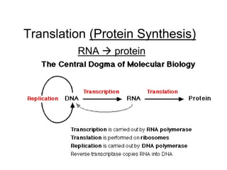
2. Translation
A. Translation-forming of a polypeptide
-uses mRNA as a template for a.a.
sequence
-4 steps (initiation, elongation,
translocation and termination)
-begins after mRNA enters cytoplasm
-uses tRNA (the interpreter of mRNA)
3. B. Ribosomes
-made of proteins and rRNA
-each has a large and small subunit
-each has three binding sites for tRNA on
its surface
-each has one binding site for mRNA
-facilitates codon and anticodon bonding
Translation
4. -the three tRNA binding sites are:
1. A site=holds tRNA that is carrying
the next amino acid to be added
2. P site= holds tRNA that is carrying
the growing polypeptide chain
3. E site= where discharged tRNAs
leave the ribosome
#8. Translation
5. Ribosomal structure
E
P A
Large subunit
Peptidyl-tRNA binding site
Aminoacyl-tRNA binding site
mRNA
5’
Exit
site
Small subunit
3’
6. P site (Peptidyl-tRNA
binding site)
E site
(Exit site)
mRNA
binding site
A site (Aminoacyl-
tRNA binding site)
Large
subunit
Small
subunit
Schematic model showing binding sites on ribosome
E P A
7. C. The genetic code
– Four RNA nucleotides are arranged 20
different ways to make 20 different amino
acids
– Nucleotide bases exist in triplets
– Triplets of bases are the smallest units that
can code for an a.a.
– 3 bases = 1 codon = 1 a.a.
– There are 64 possible codes (64=43)
Translation
8. C. The genetic code
– Most of the 20 a.a. have between 2 and 4
possible codes
– The mRNA base triplets are codons
– In translation the codons are decoded into
amino acids that make a polypeptide chain
Translation
9. C. The genetic code (continued)
– 61 of 64 codons code for a.a.
– Codon AUG has two functions
-codes for amino acid methionine (Met)
-functions as a start codon
– mRNA codon AUG starts translation
– The three ‘unaccounted for’ codons act as
stop codons (end translation)
Translation
10. D. How it works
DNA (antisense)
ACC AAA CCG
mRNA (transcription)
UGG UUU GGC
polypeptide (translation)
Trp-Phe-Gly-
Translation
11. E. More on tRNA
– tRNA is transcribed in the nucleus and must enter the
cytoplasm
– Each tRNA molecule links to a particular mRNA
codon with a particular amino acid
– When tRNA arrives at the ribosome it has a specific
amino acid on one end and an anticodon on the other
– Anticodons (tRNA) bond to codons (mRNA)
Translation
12. Where the a.a. attaches
Hydrogen bonds
Anticodon
=
tRNA diagrams
Although we
draw tRNA in
a clover
shape it’s
true 3-D
conformation
is L-shaped.
13. Amino acid
attachment site
Hydrogen
bonds
3
5
Two-dimensional structure
Anticodon
Amino acid
attachment site
3
5
Hydrogen
bonds
Anticodon Anticodon
Symbol used in this book
Three-dimensional structure
3 5
tRNA structure (Three different
schematics)
~ 80 nt long
14. Translation (Initiation)
A. Initiation
1. Brings together mRNA, tRNA (w/ 1st
a.a.) and ribosomal subunits
2. Small ribosomal subunit binds to mRNA
and an initiator tRNA
-start codon= AUG
-start anticodon-UAC
-small ribosomal subunit attaches to 5’
end of mRNA
15. -downstream from the 5’ end is the
start codon AUG (mRNA)
-the anticodon UAC carries the a.a.
Methionine
3.After the union of mRNA, tRNA and
small subunit, the large ribosomal subunit
attaches
4. Initiation is complete
#9. Translation (Initiation)
16. 5. The intitiator tRNA and a.a. will sit in the
P site of the large ribosomal subunit
6. The A site will remain vacant and ready
for the aminoacyl-tRNA
Translation (Initiation)
18. Translation (Elongation)
A. Amino acids are added one by one to the first
amino acid (remember, the goal is to make a
polypeptide)
B. Step 1- Codon recognition
a. mRNA codon in the A site forms hydrogen
bonds with the tRNA anitcodon
C. Step 2- Peptide bond formation
a. The ribosome catalyzes the formation of the
peptide bonds between the amino acids (the
one already in place and the one being
added)
b. The polypeptide extending from the P site
moves to the A site to attach to the new a.a.
19. A. The tRNA with the polypeptide chain in
the A site is translocated to the P site
B. tRNA at the P site moves to the E site
and leaves the ribosome
C. The ribosome moves down the mRNA in
the 5’→3’ direction
Translation (Translocation)
20. A. Happens at the stop codon
B. Stop codons are UAA, UAG and UGA
-they do not code for a.a.
C. The polypeptide is freed from the ribosome and
the rest of the translation assembly comes
apart
Translation (Termination)
21. Making a protein
• tRNA with the complementary anticodon
carries amino acid (a.a.) to bind to the codon
23. Ribosome ready for
next aminoacyl tRNA
mRNA
5
Amino end
of polypeptide
E
P
site
A
site
3
2
2 GDP
E
P A
GTP
GTP
GDP
E
P A
E
P A
1. Recognition
2. Peptide bond
formation
3. Translocation
24. 3
The release factor hydrolyzes the
bond between the tRNA in the
P site and the last amino acid of the
polypeptide chain. The polypeptide
is thus freed from the ribosome.
The two ribosomal subunits
and the other components
of the assembly dissociate.
Release
factor
Stop codon
(UAG, UAA, or UGA)
5
3
5
3
5
Free
polypeptide
When a ribosome reaches a stop
codon on mRNA, the A site of the
ribosome accepts a protein called
a release factor instead of tRNA.