Adrian Gadano - Argentina - Tuesday 29 - Liver Transplantation Towards New H...
Carley_SURF-2016-poster
1. Introduction
• Cholestatic liver diseases are characterized by inflammation,
mainly by liver kupffer cells (macrophages), which release pro-
inflammatory cytokines and chemokines, i.e. IL-1, TNF, IL-6, IL-8;
IL-8 is significantly elevated in chronic liver diseases, including
patients with cholestasis (1).
• Complications of unresolved cholestasis include prolonged
hepatic inflammation which progresses to fibrosis, cirrhosis and
end-stage liver disease or malignancies, i.e. liver, bile duct,
gallbladder, or colon cancer
• Currently, the only therapy available is ursodeoxycholic acid
(UDCA), yet, many patients have a sub-therapeutic response and
UDCA does not improve survival for some types of cholestasis.
Novel therapeutic strategies to target and reduce hepatic
inflammation are needed.
• Fenofibrate is FDA-approved for treatment of
hypercholesterolemia and a peroxisome proliferator-activated
receptor-alpha (PPARα) agonist.
• Fenofibrate reduced symptoms and liver function abnormalities in
patients with cholestasis who do not respond to UDCA (2), yet the
mechanism(s) remains unknown.
• Nuclear transcription factor NF-κB regulates the expression of
pro-inflammatory cytokines and chemokines and PPARα has been
shown to inhibit inflammation by negatively interfering in NF-κB
signaling (3).
Fenofibrate reduces cytokine and chemokine secretion in human
macrophages: implications for the treatment of cholestatic liver diseases
Rachel Carley and Nisanne Ghonem
Department of Biomedical and Pharmaceutical Sciences, University of Rhode Island, Kingston, RI
Cell Culture: Human THP-1 cells (ATCC) were cultured in RPMI-1640
medium containing 10% FBS and maintained in 5% CO2 and 95% air at
37 C. Cell suspensions were seeded at a density of 2 x 10⁵ cells/ml and
phorbol 12-myristate 13-acetate (PMA, 5 ng/ml) was applied to cells x
24- 48 hours to induce macrophage differentiation, illustrated below.
Cell treatment: Post-differentiation, cells were treated with DMSO (0.1%,
vehicle control), lipopolysaccharide (LPS, 0111:B4), or fenofibrate (FF, 5
– 125 µM) for 2 hours before LPS stimulation.
NF-κB translocation: Nuclear extracts of THP-1 cells treated with the
drugs listed above were analyzed for the nuclear translocation
(activation) of NF-κB by ELISA assay.
Statistical analysis: Data shown are mean ± SD and differences
between groups were analyzed by ANOVA and post-hoc analyses
(GraphPad). Significance will be determined (p < 0.05).
To characterize the inhibitory role of fenofibrate, a PPARα agonist,
against human pro-inflammatory cytokine and chemokine secretion.
• A human macrophage cell culture system was
successfully established with THP-1 cells, a human
leukemia monocyte cell line that differentiates into a
macrophage-like phenotype when treated with PMA.
• LPS stimulated a significant pro-inflammatory response in
human THP-1 differentiated macrophages, which lasted for
24 hours post-treatment.
• Fenofibrate pre-treatment reduced LPS-mediated pro-
inflammatory cytokine secretion of IL-1β, TNFα, IL-6, IL-8.
• Fenofibrate also reduced LPS-stimulated NF-κB
activation.
• Fenofibrate is currently under clinical investigation for its
therapeutic role in cholestatic liver diseases.
• Additional studies are underway to determine the
inhibitory role of fenofibrate on NF-κB-mediated activity.
Research reported in this poster was supported in part by RI-INBRE
SURF program Grant # P20 GM103430 from NIGMS, NIH. Printing
services provided by the RI-INBRE Centralized Research Core Facility
Conclusions
Methods and Methods
Objective
Results
Acknowledgements
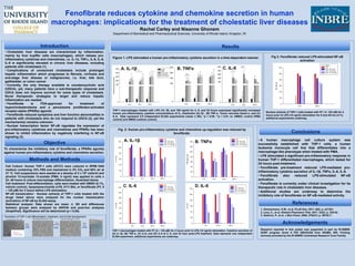
THP-1 macrophages treated with LPS (10, 50, and 100 ng/ml) for 4, 8, and 24 hours expressed significantly increased
human pro-inflammatory cytokine concentrations of IL, Interleukin (A)-1β , (B) TNF-α, Tumor necrosis factor-α, and (C)
IL-8. Data represent 3-5 independent ELISA experiments (mean ± SD), *p < 0.05, **p < 0.01 vs. DMSO; control (PMA
control) and DMSO (vehicle control).
A. IL-1β *
THP-1 macrophages treated with FF (5 – 125 µM) for 2 hours prior to LPS (10 ng/ml) stimulation. Cytokine secretion of
(A) IL-1β, (B) TNF-α, (C) IL-6, and (D) IL-8 at 2, 8, and 24 hour post-LPS treatment. Data represent one independent
ELISA experiment, additional experiments are underway.
1. Zimmermann, H.W., et al. PLoS One, 2011. 6(6): p. e21381.
2. Levy, C., et al. Aliment Pharmacol Ther, 2011. 33(2): p. 235-42.
3. Delerive, P., et al. J Biol Chem, 2000. 275(47): p. 36703-7.
References
A. IL-1β B. TNFα
Figure 1. LPS stimulated a human pro-inflammatory cytokine secretion in a time-dependent manner.
Fig. 2: Human pro-inflammatory cytokine and chemokine up-regulation was reduced by
fenofibrate.
C. IL-6 D. IL-8
B. TNFα
**
C. IL-8 **
*
Fig 3. Fenofibrate reduced LPS-stimulated NF-κB
activation
Nuclear extracts of THP-1 cells treated with FF ( 5- 125 uM) for 2
hours prior to LPS (10 ng/ml) stimulation for 8 and 48 hrs (n=1),
additional experiments underway.
Illustration of THP-1 cell differentiation, treatment, and ELISA development.
PMA x
24 hrs
Undifferentiated THP-1 cells Macrophage-induced THP-1 cells
Drug
treatments
ELISA
assay
Treated macrophages x 2-48 hrs