2. Nader et al
J Cardiovasc Thorac Res, 2015, 7(3), 81-8682
cus on Toll-like receptors superfamily (TLR-4) signaling
in which TNFα plays a major role. Additionally, it is diffi-
cult to assess the dose-response relationship between the
level of sepsis and myocardial injury in traditional models
of sepsis (ie, cecal puncture ligation model).
Materials and Methods
All experimental procedures and protocols used in this
study were reviewed and approved by the Animal Use
and Care Committee of the VA Medical Center at Buffa-
lo and the care and handling of the animals were in ac-
cord with National Institutes of Health Guidelines. New
Zealand White male rabbits, weighing 3.6 to 4.5 kg, were
used for these experiments. The rationale for using rabbit
model was due to its anatomical similarity to human cor-
onary vessels and its larger size comparing to other rodent
models.
Experimental Design and Lipopolysaccharide Injection
Rabbits were housed for a period of one week before the
experimentation. A total of 120 rabbits were used in these
experiments; 11 rabbits deceased in the process without
completing all the required steps and therefore were ex-
cluded from the final results. Deceased animals were then
replaced until the desired number of animals in each
group was achieved. On the day of the experiment, each
rabbit was injected with a pre-calculated dose of LPS. The
dose of LPS for each group was calculated on logarithmic
increments and injected intravenously, prior to ischemia
to stimulate TNFα production by the animal. Serum
TNFα was measured to confirm the response to LPS. E.
Coli-derived LPS was purchased from (Sigma Aldrich, St
Louis, MO) and suspended in sterile normal saline solu-
tion 1 mg/mL and stored in the refrigerator until it was
diluted and used. Sham-operated group was used as a con-
trol for surgical procedures. LPS administration was done
60 minutes prior to occlusion of left anterior descending
artery.
In a separate group, in order to examine the role of TNFα
in mediating the effects of LPS, soluble TNFα receptor
(sTNFαR) etanercept (Enbrel®
, Amgen Inc., Thousand
Oaks, CA) 5 mcg in saline solution was injected subcu-
taneously 48 hours prior to surgery to inhibit TNFα. This
dose (1.6 mcg/kg) was considered a “high dose regimen”
and it was previously used in a rabbit model of arthritis
to block the effects of TNFα.11
This soluble receptor was
a fusion protein engineered to link the human gene for
soluble TNF receptor 2 to the gene for the Fc component
of human immunoglobulin G1. Soluble TNFαR was only
used to in animals which received 100 µg of LPS since this
dose was associated with peak TNFα response. Controls
were injected with NS instead of LPS to examine the effect
of sTNFαR on ischemic myocardium.
Surgical Procedures, Anesthetic Techniques and Invasive
Monitoring
Each rabbit was anesthetized with 25 mg/kg of ketamine
intraperitoneally. Intubation of the trachea was done us-
ing a polyethylene tube No. 3.0 (Hudson RCI, Research
Triangle Park, NC) following anesthesia, and mechanical
ventilation was started using a tidal volume of 10 ml/kg at
the rate of 25 breaths per minute. Anesthesia was main-
tained throughout the study by administering additional
intra-peritoneal doses of ketamine 5 mg/kg, as needed and
buprenorphine 0.05 mg/kg every 2 hours. Body tempera-
ture was monitored using a rectal probe and was kept be-
tween 36.5°C-37.5°C by placing a controlled heating plate
under the animal during the experiment. Femoral arte-
rial catheter was used for blood pressure measurements.
Arterial blood pressures, electrocardiogram, and oxygen
saturations were monitored throughout the experiment.
Blood gas tensions and pH were monitored and were kept
within the normal physiologic range (pH of 7.35-7.45 and
PaCO2 between 30-40 torr). Experimental medications
and maintenance IV fluid PlasmaLyte (Life Technology,
Grand Island, NY) solutions at 25 ml/hour were adminis-
tered via a venous line placed in the femoral vein.
Exposition of the heart through a median sternotomy and
baseline readings were done at this time. Left ventricular
pressure was measured by surgical advancement of a PE-
15 (Med-Vet International, Libertyville, IL) catheter into
the left ventricle and its global function parameters were
calculated from pressure changes in one second (dp/dt)
and isovolumetric relaxation time (IVRT). The segmental
contractility of the myocardium was measured by sonomi-
crometer technology (Sonometrics®
, London, Ontario)
by attaching three crystal electrodes to the apex, anterior
wall and posterior wall of the left ventricular base. Sen-
sors were secured using a 6-0 prolene suture and segment
shortening was measured using ultrasonic recordings.
Hemodynamic parameters and cardiac function were
measured continuously and recorded at baseline, upon
induction of ischemia, immediately at the time of reperfu-
sion and at the conclusion of 4 hours of reperfusion. Blood
samples were collected from femoral arterial lines into
tubes containing heparin powder 300 mg as anticoagulant
(green top) and centrifuged at 2000 g to separate plasma
from blood cells. Plasma samples were stored at -80°C
until they were analyzed for TNFα and cardiac troponin
I (cTnI) concentrations upon conclusion of experiments.
Induction of Ischemia
After collection of baseline information, hearts from each
ischemia group were treated with a 9-0 prolene suture
loop around the left anterior descending artery, distal to
the first diagonal branch. This level of occlusion produced
a sublethal ischemia of the myocardium with ≤10% (11
out of 120 rabbits) mortality before completion of the ex-
perimental protocol. In the sham-operated group, the loop
was not tightened allowing normal blood flow, whereas
in the ischemia groups the loop was tightened to create
an occlusion and induce myocardial ischemia. The occlu-
sion of the coronary artery was maintained for 50 minutes
(Figure 1). The ischemic region was reperfused then by
loosening the suture loop for 4 additional hours. Ischemia
was confirmed by epicardial cyanosis of the ischemic re-
3. Myocardial Ischemia and Endotoxemia
J Cardiovasc Thorac Res, 2015, 7(3), 81-86 83
gion, and reperfusion by a hyperemic response.
CellInjuryandLocalInflammatoryResponseAssessment
Serum concentrations of cardiac troponin I (cTnI) were
measured as a surrogate to the extent of myocardial inju-
ry. A commercially available ELISA kit was used to mea-
sure cTnI on polyvinyl microtiter 96-well plates (Life Di-
agnostics Inc., West Chester, PA). Duplicate assays were
performed for each assay, and final colored plates were
analyzed at 450 nm using an ELISA plate reader (Bench-
mark, Bio-RAD). Numeric readings were expressed in pg/
ml of serum. TNFα levels in the serum were measured
prior to the induction of ischemia to examine the cyto-
kine response to LPS injection. For determination of se-
rum TNFα concentrations, we similarly used an ELISA
assay (Quantikine®
, R&D Systems, Minneapolis, MN).
The assays were performed on 96-well plates as described
in the product guide material. The sensitivity of the test
was 5 pg/mL and the range of measurement was 12.5-800
pg/mL.
The hearts were removed after 4 hours of reperfusion and
the anterior wall of the left ventricle was minced and ho-
mogenized using a tissue homogenizer. Oxidative stress in
ischemic tissue homogenates was analyzed by measuring
tissue malondialdehyde (MDA) levels using a commer-
cially available kit (Bioytech LPO-586TM Portland, OR).
The base of this assay was based on the reaction of a chro-
mogenic reagent, N-methyl-2-phenylindole, with MDA
and 4-hydroxyalkenals at 45°C secondary to the reaction
between one molecule of either MDA or 4-hydroxyalkenal
with 2 molecules of the chromogenic reagent.
Statistical Analysis
All data were maintained in a Microsoft Excel datasheet
and were exported into a NCSS 2007 (Kaysville, Utah)
database for statistical analyses. Differences of parametri-
cal variables within and among the groups were assessed
using one-way analysis of variance with repeated mea-
surements. Bonferroni correction was used for post hoc
analysis. Expressed data are presented as mean ± standard
deviation. Null hypotheses were rejected at alpha error set
at 0.05 or less.
Results
Ventricular Function
Baseline derivative of pressure over time (dP/dT) of the
left ventricle was 2531 ± 912 mm Hg/s similar among all
groups. After induction of ischemia, dP/dT decreased to
1422 ± 844 mm Hg/s from its respective values of 2445 ±
1018 mm Hg/s in SHAM-operated controls (P = .04) and
remained suppressed throughout reperfusion period. In-
travenous administration of low doses of LPS (<0.3 µg)
prior to the induction of ischemia partially restored dP/
dT. Additional increases in LPS dose were associated with
further decreases in dP/dT (Table 1). The left ventricle
was significantly improved when the ischemia plus 100 µg
LPS group was treated with sTNFαR (781 ± 355 mm Hg/s
vs. 1532 ± 854 mm Hg/s, P = .002) (Table 1).
Segment shortening was measured as a surrogate to re-
gional contractile function of the myocardium and was
examined in both ischemic and non-ischemic regions of
the heart. In comparison to SHAM control with a shorten-
ing percentage of 12 ± 2.6%, the anterior ventricular wall
(ischemic myocardium) segment shortening disappeared
during systole and in fact there was a systolic lengthening
of 5.3 ± 1.2% after ischemia was induced (Wall dyskine-
sia). Ventricular wall segment shortening in the ischemic
region (ie, dysfunction) was improved with low doses of
LPS treatment. The dyskinesia of the anterior ventricular
wall (-5.3 ± 1.2%) due to ischemia improved to akinesia or
hypokinesia of the ischemic wall at 0.1 ± 1.5% (P < .001)
when ischemic hearts were treated with low doses of LPS
(<0.3 µg) and reappeared when the dose of LPS exceeded
1 µg (Table 1).
Non-ischemic posterior segment shortening was mea-
sured to compare with the ischemic anterior wall segment
shortening values. Posterior segment shortening increases
with anterior wall ischemia in the control ischemia group
(compensatory hyperkinesia). Lower doses of LPS (0.1 µg
and 0.3 µg) increased the posterior wall segment short-
ening while higher LPS doses decrease the shortening
percentage to below the levels achieved in the SHAM-op-
erated control group. Soluble TNFαR treatment did not
improve the posterior wall segment shortening in the
ischemia plus 100 µg LPS group.
Relaxation of Ventricular Muscle
Negative dP/dT and isovolumetric relaxation time (IVRT)
were measured to assess ventricular relaxation. In the in
vivo experiment, the baseline IVRT increased from 50 ±
18 milliseconds in the control sham to 102 ± 64 millisec-
onds in the ischemia control group. Negative dP/dT, did
not change significantly with lower doses of LPS. LPS dos-
es over 30 µg, impaired ventricular relaxation significant-
ly, as shown by decreased dP/dT. Although adding sTN-
FαR alleviated ventricular pressure changes over the time,
but in overall sTNFαR did not help ischemic myocardial
function. Additionally, low doses of LPS (<30 µg) had no
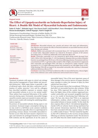
Figure 1. Timetable of the Treatments for the Experimental
Groups.
4. Nader et al
J Cardiovasc Thorac Res, 2015, 7(3), 81-8684
effect on the relaxation time of the ischemic hearts, while
further increases of LPS dose prolonged the isovolumet-
ric relaxation time (Table 1). IVRT was similar when the
ischemia plus LPS 100 µg group was treated with sTNFαR
(205 ± 101 milliseconds vs. 131 ± 96 milliseconds).
Cytokine Response and Cellular Injury
Serum TNFα increased from 23 ± 1.5 pg/mL to 180 ± 56
pg/mL following myocardial ischemia (P < .01) when ana-
lyzed after reperfusion. The serum concentration of TNFα
increased linearly in response to intravenous administra-
tion of LPS up to 30 µg and remained steady as the dose
of LPS increased (R2
= 0.65, P < .01). sTNFαR adminis-
tration had no effect on LPS-induced increases of serum
TNFα following myocardial ischemia (Table 1). Serum
cTnI concentrations, which were undetectable in SHAM
injury group, increased following ischemia (10.1 ± 3.2 ng/
dL) indicating structural myocardial damage. Serum cTnI
levels continued to rise as increasing doses of LPS were
administered, peaking at 37.5 ± 5.1 ng/dL following 100
µg of LPS. Further increases of the LPS dose did not in-
crease the serum concentrations of cTnI (Table 1). Serum
concentrations of cTnI and TNFα were directly correlat-
ed (R2
= 0.68, P < .001). Treating the rabbits with sTNFαR
prevented the increases in cTnI in the ischemia plus 100
µg LPS group, thus the cTnI levels were close to that on the
control ischemia group (Table 1).
Similarly, MDA concentrations increased from 23 ± 4.8
in SHAM controls to 180 ± 25 nmol/mL in ischemia con-
trols. Administering 100 µg LPS to the ischemic animals
further increased myocardial MDA levels to 650 ± 253
nmol/ml. Pretreating the animals with sTNFαR complete-
ly abolished LPS-induced increases of MDA levels (285 ±
115 nmol/ml) compared to controls (822 ± 119 nmol/ml).
Following administration of 300 µg of LPS, the myocardial
concentrations of MDA peaked at 2770 ± 1240 nmol/mL
(Table 1).
Discussion
This study made 2 new observations that might have im-
portant clinical implications. These findings were based
on the experimental model of intravenous administration
of LPS to animals with myocardial ischemia-reperfusion.
Firstly, lower doses of LPS partially reversed the isch-
emia-induced impairment of myocardial contractility, as
demonstrated by one grade improvement in fiber short-
ening of the myocardium from a dyskinetic to an akinetic
state. However, with higher doses of LPS the left ventricu-
lar wall dyskinesia reappeared.
In parallel to a linear pattern of cellular injury as evident
by constant rises of cTnI, myocardial contractility of the
ischemic region remained significantly suppressed in LPS
treated animals. This finding suggested that a significant
myocardial stunning was still present although the resul-
tant cell death was more moderated. These findings were
in a weak agreement with previous studies that had ob-
served beneficial effects of LPS pretreatment and its asso-
ciated increases in TNFα levels on ischemic myocardium.
Yao et al12
demonstrated that LPS pre-treatment reduced
apoptosis of myocardial cells following an ischemia reper-
fusion injury and decreased MDA levels in the myocardi-
um. Several other studies reported that LPS pre-treatment
Table 1. Physiological and Biochemical Variables for Various Treatment Groups
Variables 50 min Ischemia by applying LAD tourniquet sTNFαR
LPS (µg)
None
(n = 15)
0.1 µg
(n = 8)
0.3 µg
(n = 8)
1.0 µg
(n = 8)
10 µg
(n = 8)
30 µg
(n = 8)
100 µg
(n = 15)
300 µg
(n = 8)
100 µg
(n = 8)
Left Ventricle Contractile Function
+dP/dT (mm Hg/s) 1422±844 1752±967 1733±1021 1499±1211 1321 ± 1201 1354±1380 1002±488 762±348 989±1004
SS (ISW) % -5.3 ± 1.2 0.1 ± 1.5* 0.2 ± 1.6* -2.5 ± 1.3 -3.4 ± 1.5 -4.8 ± 1.8 -5.9 ± 2.4 -6.8 ± 2.1 -5.1 ± 2.3
SS (NIW) % 15.1 ± 4.2 18.5 ± 5.1 16.8 ± 5.3 14.1 ± 5.2 11.1 ± 3.5 12.3 ± 5.3 6.4 ± 3.8 * 5.2 ± 2.0 * 5.9 ± 3.1
Left Ventricle Relaxation
-dP/dT (mm Hg/s) 1022±944 1688±912 1622±895 1411±962 1123±745 1098±591 781±355 512±452 1032 ±854
IVRT (msec) 102 ± 64 86 ± 91 101 ± 84 109 ± 91 126 ± 95 116 ± 86 205 ± 101 311 ± 124 131 ± 96
Inflammatory Cytokines and myocardial Injury Biomarkers
TNFα (pg/mL) 180 ± 56 345 ±79 434 ± 102 604 ± 362 893 ± 330 988±287 2350±1220 2403±1340 2421±1620
cTnI (ng/dL) 10.1 ± 3.2 10.2 ± 4.1 11.2 ± 7.6 18.9 ± 10.2 25.6 ± 13.1 31.2 ± 6.9* 37.5± 5.1†* 37.4 ± 8.1* 9.5 ± 3.6†
MDA (pg/mL) 180 ± 25 NM NM NM NM NM
650 ± 253
†*
2770±1240
*
285 ± 115†
MDA (pg/mL) 180 ± 25 NM NM NM NM NM NM NM NM
All cardiac variables measured after 4 hours of reperfusion to examine the contractile and relaxation functions of the heart, as well as the marker of myocardial
injury. Markers of myocardial contraction included derivatives of the left ventricle (LV) pressure over the derivatives of time during systole (+dP/dT) and the
percent segment shortening (SS) of the anterior ischemic wall (ISW) and the posterior non-ischemic wall (NIW). In order to assess the relaxation of the LV,
negative values of dP/dT were measured during diastole along with isovolumetric relaxation time in millisecond. Biomarkers of cellular injury and inflammation
included serum concentrations of tumor necrosis factor alpha (TNFα, which are expressed in pg/mL, cardiac troponin I (cTnI) and malondialdehyde (MDA), a
marker of lipid peroxidation, which is also expressed in pg/mL of tissue homogenate. Asterisks indicate a significant difference (P < .05), when a comparison
was made to the ischemia controls (IS) and † indicates significant difference (P < .05), when a comparison was made between the groups treated with soluble
receptor of TNFα to their normal saline-treated control (IS + LPS 100 µg).
5. Myocardial Ischemia and Endotoxemia
J Cardiovasc Thorac Res, 2015, 7(3), 81-86 85
was associated with reduction in myocardial infarct size.13-
16
Since different models of ischemia-reperfusion injury
were used in these studies, no direct correlation could
be concluded with our findings. We further showed that
higher doses of LPS were not associated with improve-
ment in cardiac function, and in addition, excessive doses
of endotoxin had other deleterious effects on contractility
and relaxation of myocardium.
Since soluble TNFαR also blocked the LPS-induced
changes, these new findings appeared to result from
LPS-induced generation of TNFα, which rose linearly
until a plateau was reached. Effects of TNFα are induced
by its binding to 2 different receptors: TNFα receptor
type 1 and type 2. Both receptor types are found in the
myocardium and activated via trimerization upon TNFα
binding. In addition, it could potentially prevent NF-kB
linked anti-apoptotic pathways, which may protect the
heart during reperfusion.17-20
Our findings confirmed
that following administration of soluble TNFαR, ischemic
cellular injury was decreased, but ischemic impairment
of myocardial contractility remained unchanged. These
findings clearly demonstrated that detrimental effects of
LPS on ischemia-reperfusion induced insult were mediat-
ed through the action of TNFα on the myocardium. One
unique characteristic of this study was its graded action of
LPS-induced increases in TNFα. Additionally, Brown et
al demonstrated a decrease in myocardial injury of isch-
emia-reperfusion by TNFα pre-treatment.21
Even though
the direct administration of TNFα was not used in these
experiments, other studies clearly demonstrated a strong
association of this cytokine in mediating the actions of
LPS.22,23
Evaluation of LPS-induced TNFα levels showed
a dose-dependent changes in cardiac function (myocar-
dial contractility and relaxation), as well as local inflam-
matory response and myocardial injury. Global function
of the heart (both contraction and relaxation) was only
minimally deteriorated with lower serum TNFα concen-
trations. This observation was consistent with the previ-
ously demonstrated protective effects of this cytokine on
mitochondria of the ischemic myocardium described by
another study.24
Superiority of the sTNFαR-treated experiments in car-
dio-protection was likely due to its multiple targeted li-
gand binding. We also demonstrated that soluble TNFαR
administration was associated with decreased levels of the
biomarkers of myocardial injury, MDA and serum cTnI.
Nevertheless, the ventricular segment shortening was not
altered in either ischemic or non-ischemic regions fol-
lowing sTNFαR administration. Indeed, as anticipated,
sTNFαR administration had no effect on LPS-induced
increases of serum TNFα following myocardial ischemia,
because this drug’s effects were merely post-translation-
al. It is not clear yet why sTNFαR could have decreased
ischemic cellular injury but had no effect on contractility.
However, preservation of cardiac myocytes in the absence
of functional improvement clearly indicated a significant
ongoing myocardial stunning.
One of the main limitations of this study was to use en-
dotoxin injection as one arm of the double-hit model that
mimicked systemic sepsis. Although this study sheds light
into the role of TLR-4 cytokines in mediation of ischemic
injury, LPS administration is not a true reflection of septic
shock both in animal models and clinical settings. Ani-
mal models of septic shock such as cecal ligation may be
used to address this issue. These models will obviously in-
volve a combination of cytokine and cellular inflammato-
ry responses when compared to more targeted cascade of
TNFα in current study.
In summary, we present a double-hit model, which em-
ploys the insults of endotoxemia with myocardial isch-
emia, ie, a combination that is not very uncommon clinical
condition in critical care settings. A clear understanding of
the underlying pathophysiology enables clinicians to de-
velop therapeutic strategies that may improve the clinical
outcome. Our findings clearly support the conclusion that
while exuberant cytokine may be detrimental to myocar-
dial well being during an additional ischemic insult, more
moderated levels of these cytokines may modulate cardiac
cell survival after an ischemic insult during systemic en-
dotoxemia. These results may also help explain the pre-
viously disappointing clinical results that were obtained
from treating patients with septic shock with anti-TNFα
antibodies. A new approach to therapy of these combined
insults may include moderation rather than elimination
of TNFα bioactivity. However, more studies are needed to
address the potential benefits of immunomodulation (cy-
tokine suppression) as possible therapeutic strategies to
these clinical pathologies in animal models prior to being
tested in humans.
Acknowledgments
Financial support used for the study was provided from
a grant-in-aid awarded to NDN by the American Heart
Association. (AHA 0060430Z).
Ethical issues
None.
Competing interests
None.
References
1. Rudiger A, Singer M. Mechanisms of sepsis-induced
cardiac dysfunction. Crit Care Med 2007; 35: 1599-
608. doi: 10.1097/01.CCM.0000266683.64081.02
2. Poelaert J, Declerck C, Vogelaers D, Colardyn F, Visser
CA. Left ventricular systolic and diastolic function in
septic shock. Intensive Care Med 1997; 23: 553-60.
3. Levy RJ, Piel DA, Acton PD, Zhou R, Ferrari VA,
Karp JS, et al. Evidence of myocardial hibernation in
the septic heart. Crit Care Med 2005; 33: 2752-6.
4. Heusch G. Cardioprotection: chances and challenges
of its translation to the clinic. Lancet 2013; 381: 166-
75. doi: 10.1016/S0140-6736(12)60916-7
5. Calabresi L, Rossoni G, Gomaraschi M, Sisto F, Berti
F, Franceschini G. High-density lipoproteins protect
6. Nader et al
J Cardiovasc Thorac Res, 2015, 7(3), 81-8686
isolated rat hearts from ischemia-reperfusion injury
by reducing cardiac tumor necrosis factor-alpha
content and enhancing prostaglandin release. Circ
Res 2003; 92: 330-7.
6. Johansen D, Ytrehus K, Baxter GF. Exogenous
hydrogen sulfide (H2S) protects against regional
myocardial ischemia-reperfusion injury--Evidence
for a role of K ATP channels. Basic Res Cardiol 2006;
101: 53-60. doi: 10.1007/s00395-005-0569-9
7. Xu Y, Arenas IA, Armstrong SJ, Plahta WC, Xu H,
Davidge ST. Estrogen improves cardiac recovery after
ischemia/reperfusion by decreasing tumor necrosis
factor-alpha. Cardiovasc Res 2006; 69: 836-44. doi:
10.1016/j.cardiores.2005.11.031
8. Asgeri M, Pourafkari L, Kundra A, Javadzadegan H,
Negargar S, Nader ND. Dual effects of tumor necrosis
factoralphaonmyocardialinjuryfollowingprolonged
hypoperfusion of the heart. Immunol Invest 2014;
1-13. doi: 10.3109/08820139.2014.921689
9. Nader ND, Li CM, Khadra WZ, Reedy R, Panos AL.
Anesthetic myocardial protection with sevoflurane. J
Cardiothorac Vasc Anesth 2004; 18: 269-74.
10. Nader ND, McQuiller PS, Raghavendran K,
Spengler RN, Knight PR. The role of alveolar
macrophages in the pathogenesis of aspiration
pneumonitis. Immunol Invest 2007; 36: 457-71. doi:
10.1080/08820130701361053
11. Kawaguchi A, Nakaya H, Okabe T. Blocking of tumor
necrosis factor activity promotes natural repair of
osteochondral defects in rabbit knee. Acta Orthop
2009; 80: 606-11. doi: 10.3109/17453670903350115
12. Belosjorow S, Schulz R, Dorge H, Schade FU,
Heusch G. Endotoxin and ischemic preconditioning:
TNF-alpha concentration and myocardial infarct
development in rabbits. Am J Physiol 1999; 277:
H2470-5.
13. Hiasa G, Hamada M, Ikeda S, Hiwada K. Ischemic
preconditioning and lipopolysaccharide attenuate
nuclear factor-kappaB activation and gene expression
of inflammatory cytokines in the ischemia-reperfused
rat heart. Jpn Circ J 2001; 65: 984-90.
14. Shames BD, Barton HH, Reznikov LL, Cairns CB,
Banerjee A, Harken AH, et al. Ischemia alone is
sufficient to induce TNF-alpha mRNA and peptide in
the myocardium. Shock 2002; 17: 114-9.
15. Sivarajah A, McDonald MC, Thiemermann C. The
cardioprotective effects of preconditioning with
endotoxin, but not ischemia, are abolished by a
peroxisome proliferator-activated receptor-gamma
antagonist. J Pharmacol Exp Ther 2005; 313: 896-
901. doi: 10.1124/jpet.104.080598
16. Zacharowski K, Frank S, Otto M, Chatterjee
PK, Cuzzocrea S, Hafner G, Pfeilschifter J, et al.
Lipoteichoic acid induces delayed protection
in the rat heart: A comparison with endotoxin.
Arteriosclerosis, Thrombosis & Vascular Biology
2000; 20: 1521-8.
17. Aggarwal BB, Shishodia S, Takada Y. TNF blockade:
an inflammatory issue. Ernst Schering Res Found
Workshop 2006; 161-86.
18. Kubota T, Miyagishima M, Frye CS, Alber SM,
Bounoutas GS, Kadokami T, et al. Overexpression
of tumor necrosis factor- alpha activates both anti-
and pro-apoptotic pathways in the myocardium. J
Mol Cell Cardiol 2001; 33: 1331-44. doi: 10.1006/
jmcc.2001.1393
19. Regula KM, Baetz D, Kirshenbaum LA. Nuclear
factor-kappaB represses hypoxia-induced
mitochondrial defects and cell death of ventricular
myocytes. Circulation 2004; 110: 3795-802. doi:
10.1161/01.CIR.0000150537.59754.55
20. Valen G, Yan ZQ, Hansson GK. Nuclear factor
kappa-B and the heart. J Am Coll Cardiol 2001; 38:
307-14.
21. Brown JM, Anderson BO, Repine JE, Shanley PF,
White CW, Grosso MA, et al. Neutrophils contribute
to TNF induced myocardial tolerance to ischaemia.
Journal of Molecular & Cellular Cardiology 1992;
24: 485-95.
22. Gu Q, Yang XP, Bonde P, DiPaula A, Fox-Talbot
K, Becker LC. Inhibition of TNF-alpha reduces
myocardial injury and proinflammatory pathways
following ischemia-reperfusion in the dog. J
Cardiovasc Pharmacol 2006; 48: 320-8. doi:
10.1097/01.fjc.0000250079.46526.38
23. Xiao H, Liao YH, Chen ZJ. Tumor necrosis factor-
alpha: a new mechanism of ischemic ventricular
fibrillation? Chin Med J (Engl) 2008; 121: 1848-51.
24. Lacerda L, McCarthy J, Mungly SF, Lynn EG, Sack
MN, Opie LH, et al. TNFalpha protects cardiac
mitochondria independently of its cell surface
receptors. Basic Res Cardiol 2010; 105: 751-62. doi:
10.1007/s00395-010-0113-4