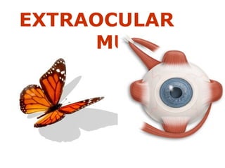
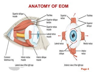
This document provides information about the extraocular muscles:
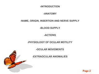
- There are 6 extraocular muscles that control eye movement: the superior, inferior, medial, and lateral rectus muscles, and the superior and inferior oblique muscles.
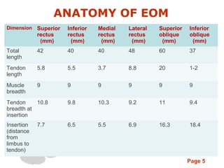
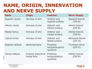
- The muscles originate from the annulus of Zinn and insert into different parts of the eyeball. They are innervated by different cranial nerves.
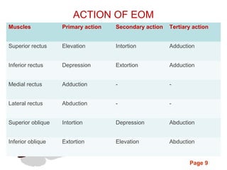
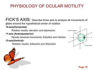
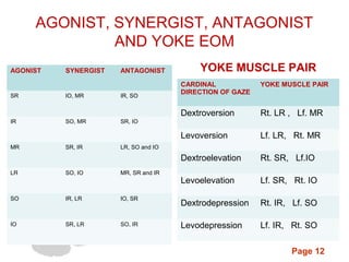
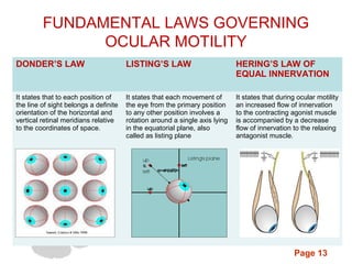
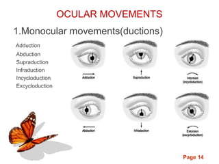
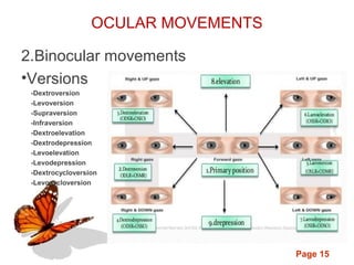
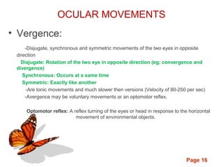
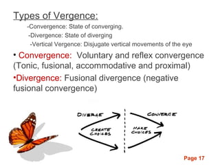
- The muscles work together or antagonistically to enable movements like elevation, depression, adduction, and abduction. Fundamental laws like Listing's law and Hering's law govern their coordinated function.
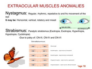
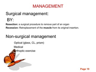
- Common anomalies include nystagmus and strabismus. Surgical procedures like resection and recession can correct muscle