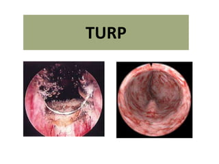
2. TURP
• TURP was the first successful, minimally
invasive surgical procedure of the modern era.
• To this day, it remains the criterion standard
therapy for obstructive prostatic hypertrophy
and is both the surgical treatment of choice
and the standard of care when other methods
fail.
3. Anatomy …
• The prostate is thinnest and most narrow
anteriorly (the 12-o’clock position when viewed
through a cystoscope).
• Care should be taken when operating in this
area to avoid perforating the prostatic capsule,
especially if this portion of the prostate is
resected early in the operation.
• Abundant venous blood vessels are located in
the area just anterior to the prostatic capsule,
which can cause significant bleeding that
cannot be easily controlled if the vessels are
damaged during resection
4.
5.
6. • The proximity of the ureteral orifices to the
cephalad margin of the hypertrophied
prostate varies, particularly in patients with an
enlarged median lobe. This distance should be
frequently assessed throughout surgery.
10. Internal Urethrotomy
• 24 CH instruments normally can be inserted through
the urethra without problems and should be used for
resection of even large glands.
• However, if the anterior urethra is too narrow to
accommodate the instrument, a perineal
urethrostomy can be performed to insert the
instrument. As an alternative, blind urethrotomy over
the narrow segment of the urethra may be performed.
• With larger instruments (28 CH), prophylactic blind
urethrotomy is recommended to prevent ischemic
damage and consecutive urethral stricture.
11. Resectoscope
• Resectoscope is a combination of a cystoscope and
electrosurgical instrument, which enables the
resection of the prostate with an electrical
activated wire loop.
• In low pressure resection, the irrigation fluid is
drained via a suprapubic trocar. Alternatively, a
two-channel resectoscope can be used, one
channel for irrigation and the other channel for the
drainage. Disadvantages are however the larger
diameter of the instrument and less reliable
drainage of the irrigation fluid.
12. Coaxial continuous-flow rectoscopes
• In 1975, Jose Iglesias de la Torre reported a reliable
external spring-loaded continuous-flow rectoscope
that is the most popular resectoscope working
element style used today.
• The Iglesias working element uses the thumb and
the spring to do the actual cutting, while the older
Stern-McCarthy model allows the resection to be
controlled by the thumb and first two fingers using
a rack-and-pinion mechanism, which provides finer
motor control and excellent tactile sensory
feedback.
15. • The main disadvantages are the tendency of the
resected chips to sometimes flow toward the
telescope and interfere with vision; the slightly
reduced wire loop size available because of the
coaxial nature of the instrument; and the lack of
any definitive study that proves they actually
save time, reduce blood loss, or decrease
absorption of irrigation fluid intraoperatively.
• Nevertheless, most urologists find continuous-
flow instrumentation convenient and beneficial.
17. Suprapubic trocars
• Offers several distinct advantages over the more popular
single coaxial continuous-flow instruments.
• First advantage is that chips and irrigation flow away
from the telescope toward the drainage tube in the
bladder, which improves visualization.
• Second is that a larger wire loop can be used with the
same caliber resectoscope sheath.
• A third advantage is that the suprapubic trocar can keep
the bladder fluid pressure at or below only 8 cm water,
which is well below the 10-15 cm water pressure of the
pelvic veins and periprostatic venous system; this keeps
fluid absorption down. When compared directly to a
coaxial continuous-flow system, the suprapubic trocar
technique has been found to allow shorter operating
times with lower intravesical pressures and less fluid
absorption.
18. Role of suprapubic trocar in today’s era..
• Preferable to use a suprapubic trocar for
establishing continuous flow when trying to
resect larger prostates (>80 g).
– Saves time.
– Reduces complication
– Allows larger resectoscopes to be used.
– Additional postop safety drainage.
19. Electrocautery
• In standard TURP monopolar current is used, with
different programs for cutting and coagulation,
comparable to open surgery.
• The coagulation effect is produced by the current
flow from the resection loop through the prostate
tissue to the return electrode. The irrigation solution
used in transurethral resection with monopolar
current must be salt-free to prevent current flow
through the irrigation fluid.
• The salt-free irrigation fluid harbors a risk for a TUR
syndrome, if large amounts of fluid enters the
circulation.
20. • Transurethral resection requires a powerful
diathermy machine which can both cut tissue
and stop bleeding under water. If your budget is
limited, economize on the resectoscope rather
than the diathermy.
• Many of us take diathermy for granted: When it
does not work we ask the nurse to turn up the
current. This is often exactly wrong!!
21. • When an electric current passes between two
contacts on the body there is always a certain
increase in temperature in the tissues through
which the current flows. The stronger the current,
the greater the rise in temperature.
• When a direct current is switched on or off, nerves
are stimulated and the muscles will twitch. If
switching on and off is rapid - ‘tetanic contraction.
• If the frequency of the alternating current is
increased beyond a certain critical level, there is no
time for the cell membrane of nerve or muscle to
become depolarized and nerves and muscles are
no longer stimulated.
Mechanism and physics - electrocautery
22. • And so AC (300 kHz to 5 MHz) is used in
clinical practice today.
• With frequencies as great as this a very large
current can be passed through the patient
without exciting nerves or muscles, and it is
then possible to exploit the heating effect at
the points of contact.
23. • If one contact is made
large, the heat is
dissipated over a wide
area and the rise of
temperature is
insignificant.
• Hence diathermy loops
are kept deliberately thin
so that the heating effect
is maximum.
24. • The effect of heat on tissues is well known to us from
everyday experience in the kitchen: when cooking an
egg, at first the albumen turns white and shrivels as
it coagulates. Then the egg fries, blackens and (in air)
may smoke, crackle and eventually catch fire.
• It is the drying, coagulation and distortion of small
blood vessels and plasma proteins which seals them.
This requires only ‘white coagulation’. Blackening and
smoke are unnecessary and cause needless tissue
necrosis.
25. • If the current is increased to raise the temperature still
further there is an explosive vaporization of
intracellular water in the tissue. In transurethral
resection this additional rise in temperature is
achieved by a spark, the result of ionization of the
water between the electrode and the tissue.
• The electrode does not actually need to touch the
tissue. The sparks explode the cells into steam, but
their energy does not reach the deeper layers, so the
cut is a clean one, and the blood vessels underneath
are not sealed.
The cutting current is a pure sine-wave current
26. • Coagulation is achieved in general with short
bursts of sine waves which give longer sparks,
but with intervals between them to allow the
tissue to cool: the result is sustained heating
which leads to poaching rather than explosion
of the tissue.
27. • By designing the solid-state generator to deliver a
mixture of pure sine-wave ‘cutting’ and interrupted
bursts of sine-wave currents for ‘coagulation’ a current
can be designed to allow a combination of cutting and
coagulation—the ‘blended’ current.
• If the current does not seem to be stopping
the bleeding, do not make the common
mistake of asking for the current to be
increased. The problem may be that it is
sparking and causing explosion (cutting) of
the underlying tissue. Turn it down.
28. Troubleshooting !!
• If the electrosurgery (cautery) unit does not appear
to be functional, inadvertent use of normal saline
(isotonic sodium chloride) irrigation is one of the
first things to check besides the grounding pad,
power switch, and cord connections.
• If normal saline is accidentally used, no cutting or
coagulating will occur. It will appear as though the
electrosurgical unit is not working.
Accordingly, solutions that do not conduct electricity, such as sterile water, glycine, and
sorbitol/mannitol, must be used instead of isotonic sodium chloride solution during TURP.
29. Irrigating solutions
• Sterile water is rarely used because, when
absorbed in large quantities during the
procedure, it causes hyponatremia,
intravascular hemolysis, and hyperkalemia.
• Therefore, nonhemolyzing solutions of
sorbitol/mannitol or glycine are used most
often. These relatively isotonic agents protect
against hemolysis but cannot prevent dilutional
hyponatremia because their intravascular
absorption increases fluid volume without
adding any sodium.
30. • However, as noted by Collins et al, a 5%
glucose solution may be a reasonable and
economical substitute for the much more
expensive glycine irrigation fluid in developing
countries, where it would be less hemolyzing
and safer than sterile water.
31. Glycine
• Currently, glycine is probably the most popular
irrigation media used for TURP surgery, with an
osmolality of approximately 200 mOsm/kg
(compared to 290 mOsm/kg for normal serum).
• Though not truly isotonic, it is close enough to
be essentially nonhemolyzing. The metabolism
of glycine into glycolic acid and ammonia has
been postulated as a contributing factor to TUR
syndrome.
32. Position on table
• The important thing is that the legs are kept in
the correct position with the thighs making an
angle of no more than 45° with the plane of the
table. To have the legs in this almost flat position
puts less strain on the heart.
• The so-called lithotomy position, as used in
operations on the anus, produces an awkward
angulation of the prostate as well as sometimes
causing backache afterwards.
35. RESECTION TECHNIQUES
Although several different techniques of
transurethral resection have been described, their
aim is essentially the same, to remove all the
adenomatous tissue from the inner zone, leaving
the compressed outer zone intact: the so-called
‘surgical capsule’.
36. • The various techniques of transurethral
resection differ only in the order in which the
bulk of tissue is removed.
• The important thing is that you should have a
plan and stick to it, or else you will certainly get
lost. Try each of these methods and choose the
one which suits you best.
37. • In all methods there are three stages
1. Establishing the landmarks.
2. Removing the main bulk of tissue.
3. Tidying up
38.
39. • Make sure that you have seen the sphincter:
bring the resectoscope out beyond it, cut off
the water flow and see it contract like the anus
in its characteristic way.
46. Mauermayer’s technique, 1985
• First, the middle lobe is resected and an
excavation between five and seven o'clock up
to the surgical capsule is formed. After that,
now with good irrigation speed made
possible, the side lobes and ventral parts of
the gland are resected. The apical parts of
gland are resected last.
47. • TURP is divided
into four steps:
1. mid-lobe
resection,
2. paracollicular
transurethral
resection
(TUR),
3. resection of
lateral lobes
and ventral
parts, and
4. apical
resection.
48. Nesbit technique, 1951
• Starts with the ventral parts of the gland
(between 11 and 1 o’clock), followed by both
lateral lobes, the mid-lobe, and finishing with
the apex.
49. • Demonstration of the Nesbit technique. First cut at
the 12-o'clock position, intravesical portion.
50. Extravesical (second) and apical (third) portions. Inserting a
finger in the rectum and tilting the resectoscope help
expose tissue for removal.
51. • The Iglesias resectoscope used for the median lobe resection.
Note resection along the inside margin of the capsule, which
seals the perforating blood vessels and makes the rest of the
resection relatively bloodless.
52. Milner technique
The Milner technique is started with a deep incision directly into the lateral lobe at
the 3 or 9-o'clock position and proceeds until the surgical capsule is reached.
Further resection is then performed from this starting point.
53. Flocks and Culp technique
• Flocks and Culp preferred to start with the
mid-lobe then segmented the lateral lobes at
9 and 3 o’clock.
54. Barne’s method
• Median lobe – lateral lobe – ventral part –
apical lobe.
• Most commonly used.
60. Establishing a rhythm..
• When resecting the bulk of the lateral lobes of
the prostate, once the landmarks have been
established, time is saved by making sure that
every stroke removes the maximum amount of
tissue, i.e. the depth of the chip should be at least
that of the loop and its length as long as that of
the lateral lobe even if this means moving the
sheath outwards, always making sure that you
know the exact situation of the verumontanum.
61. • If the electrode does not spark cleanly it will
not cut, but will coagulate or char the tissue.
This is most likely to occur if you press the
loop into the prostate instead of letting the
sparks do the work.
• A crust of carbonized tissue may cover the
loop. Clean it and start again.
63. If the loop does not cut at all, do not respond by
asking for the current to be increased.
Instead, carry out the following checks:
1. Make sure that the loop is sitting firmly in its holder. A ‘click’
can be felt and heard as the loop fits into the holder.
2. Check that the loop is not broken.
3. Check that the diathermy plate is securely attached to the
thigh.
4. Check that the diathermy lead is attached to the machine.
5. Check that the wire within the diathermy lead has not
worked loose at either end.
6. Check the irrigating fluid: a common mistake is for the
theatre team to hang a bag of saline instead of glycine.
64. If all these items have been
checked and the loop still
does not cut, you must
change the diathermy
machine. You cannot resect
with a loop which merely
chars: it drags in the tissues,
makes it difficult to cut
cleanly, and worse, risks
producing a deep burn in the
underlying tissues which may
damage the sphincter.
65.
66. Median lobe
resection.
Loop without
current is used to
gently lift the
posterior flap of the
median lobe tissue
now lying on the
bladder surface.
Resection can now
be performed
without risk of
bladder injury.
67. A Common site for perforation.
• Perforation at the inferior bladder neck can easily occur if the line
of resection is too straight or is not carved to fit the shape of the
prostate in this area.
68. • Filling the bladder
usually helps
locate the
bleeding vessels
on the interior
bladder neck.
Limited resection
of a covering lip
of tissue is
sometimes
necessary, as
illustrated here.
Identifying the bleeder at the BN
69. Inserting a finger in
the rectum (A) and
applying external
suprapubic
pressure (B) can
assist in locating
and coagulating
bleeding vessels by
altering the
orientation of their
flow and bringing
the bleeding sites
into view.
71. Beware of resecting folds of tissue that may build up in front of the edge of the beak of
the resectoscope because this may cause a perforation. Additional urethral dilation or
use of a smaller resectoscope sheath can help prevent this from occurring.
73. • Resection of the prostate at the bladder
neck.
– With a finger in the rectum for guidance, the
loop without current can be used to lift a flap
of prostatic tissue prior to cutting. This helps
avoid perforation and subtrigonal tunneling.
74. • At the end
• Slow withdrawal of the
resectoscope at the end
of the case sometimes
helps demonstrate a
flap of tissue hanging
down from the roof.
These should be
carefully resected to
avoid a ball-valve effect.
75. Hemostasis
• Most of the light oozing which occurs during a
resection comes from small veins which are cut
as you resect the adenoma. This type of bleeding
is minimized by using a continuous flow Iglesias
irrigating system, but it should be stopped as you
go along in order to keep a clear view. Any
arterial bleeder should be controlled as soon as
you see it by touching it with the loop.
• There should be no charring or burning, only
cessation of bleeding and a little whitening of the
tissue.
77. • Another common source of confusion is the
artery which is shooting out straight at the
telescope. All you can see is a uniform red
haze. The trick is to advance the resectoscope
beyond the bleeder, angulate it to compress
the vessel, and then slowly withdraw the
sheath until the opening of the artery is
betrayed by the emergence of a puff of blood.
78.
79. Prophylactic coagulation
• Sometimes it is obvious from the moment you pass the
cystoscope that the resection is likely to be bloody. One
can save oneself trouble by making a prophylactic
attempt to control the main arteries before one start to
resect.
• Using the roly-ball coagulate the prostate at 2, 5, 7 and
10 o’clock where the main arteries enter the gland. This
simple measure minimizes subsequent bleeding, and
may be repeated later
on during the resection
should bleeding recur.
80. Veins
• Veins are more difficult to detect than arteries.
• You may see no venous bleeding at all during the
resection, but as soon as the handpiece is removed
there is a copious flow of blood.
• Having sealed off all the arteries, the trick in
finding the little veins is to slow down the inflow of
irrigating fluid.
• It is worth taking time to go over the entire inner
surface of the capsule at the end of the operation
to seal them all. Time spent on this manoeuvre is
time well spent.
81. • Even so, there are some patients in whom, despite
prolonged and patient haemostasis, there is still a
copious ooze of venous blood. Here tamponade is
effective.
• It compresses the neck of the bladder where most of the
offending veins are situated.
• It is far easier and faster to sort the bleeding out while
the patient is still on the operating table, anaesthetized,
and the equipment still available, than to bring him back
from recovery and start all over again.
• If the bleeding fails to stop, never hesitate to reinsert
the resectoscope - Briskly bleeding arteries just inside
the bladder neck at roughly the 12 o’clock position can
easily be missed, so look here in particular.
82. Evacuation of the chips
• Whenever one breaks the rhythm of resection
to remove chips, time is wasted so keep the
number of evacuations to the minimum, i.e.
when the chips begin to fall back into the
empty prostatic fossa and get in the way of
the loop.
– Elliks evacuator: It must be used gently: if used
roughly it is possible to rupture the bladder
(particularly in old ladies with thin bladders who
have undergone bladder tumour resection).
– Some surgeons prefer a wide nozzle hand syringe
(called by some a Toomey syringe) to the Ellik.