Diabetic Retinopathy_Dr. Bastola.pptx
•Download as PPTX, PDF•
0 likes•44 views

Diabetic Retinopathy Dr. Pradeep Bastola, MBBS, MD, MPH Professor, Department of Ophthalmology, Chitwan Medical College, Nepal

Recommended
More Related Content
What's hot
What's hot (20)
Similar to Diabetic Retinopathy_Dr. Bastola.pptx
Similar to Diabetic Retinopathy_Dr. Bastola.pptx (20)
Recently uploaded
Recently uploaded (20)
Diabetic Retinopathy_Dr. Bastola.pptx
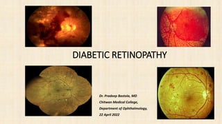
- 1. Dr. Pradeep Bastola, MD Chitwan Medical College, Department of Ophthalmology, 22 April 2022 DIABETIC RETINOPATHY
- 2. Learning Objectives • Recognize the importance of diabetic retinopathy as a public health problem • Discuss diabetic retinopathy as a leading cause of blindness in developed countries • Identify the risk factors for diabetic retinopathy • Describe and distinguish between the stages of diabetic retinopathy • Understand the role of risk factor control, and annual dilated eye exams in the prevention of vision loss
- 3. Diabetes Mellitus Diabetes Mellitus is a group of diseases characterized by high blood glucose levels. Diabetes results from defects in the body's ability to produce and/or use insulin. • Type 1 diabetes is usually diagnosed in children and young adults, and was previously known as juvenile diabetes. In type 1 diabetes, the body does not produce insulin. 5% of people with diabetes have this form of the disease. • In Type 2 diabetes, either the body does not produce enough insulin or the cells ignore the insulin. This is the most common form of diabetes. http:/www.diabetes.org/diabetes-basics/?loc=GlobalNavDB
- 4. Diabetic Retinopathy (DR) Definition • Progressive dysfunction of the retinal blood vessels caused by chronic hyperglycemia. • DR can be a complication of diabetes type 1 or diabetes type 2. • Initially, DR is asymptomatic, if not treated though it can cause low vision and blindness. http://www.mdconsult.com/das/book/pdf/282715756-3/978-0-323-04332-8/4-u1.0-B978-0-323-04332-8..00092-5..DOCPDF.pdf?isbn=978-0-323-04332-8&eid=4-u1.0-B978-0-323-04332-8..00092-5..DOCPDF
- 5. What is THE Retina? • The retina is a multilayered, light sensitive neural tissue lining the inner eye ball. Light is focused onto the retina and then transmitted to the brain through the optic nerve. • The macula is a highly sensitive area in the center of the retina, responsible for central vision. The macula is needed for reading, recognizing faces and executing other activities that require fine, sharp vision.
- 6. RETINA
- 7. Healthy Retina Diabetic Retinopathy
- 8. Diabetic Retinopathy - Epidemiology • The total number of people with diabetes is projected to rise from 285 million in 2010 to 439 million in 2030. • Diabetic retinopathy is responsible for 1.8 million of the 37 million cases of blindness throughout the world . • Diabetic retinopathy (DR) is the leading cause of blindness in people of working age in industrialized countries. http://www.who.int/bulletin/volumes/82/11/en/844.pdf http://www.ncbi.nlm.nih.gov/pubmed/19896746
- 9. 0 2 4 6 8 10 12 14 16 18 20 Causes of global blindness in millions of people (WHO 2002) A. Foster, S.Resnikoff. The impact of vision 2020 on global blindness. Eye 2005; 19:1133-1135
- 10. Diabetic Retinopathy Epidemiology • The best predictor of diabetic retinopathy is the duration of the disease • After 20 years of diabetes, nearly 99% of patients with type 1 diabetes and 60% with type 2 have some degree on diabetic retinopathy • 33% of patients with diabetes have signs of diabetic retinopathy • People with diabetes are 25 times more likely to become blind than the general population. Ophthalmology Myron Yanoff MD and Jay S. Duker Basic and Clinical Science Course, Section 12: Retina and Vitreous AAO http://www.aao.org/eyecare/news/upload/Eye-Health-Fact-Sheet.pdf -
- 11. Prevalence of diabetic retinopathy after 20 years of diagnosis
- 13. Diabetic retinopathy symptoms Diabetic retinopathy is asymptomatic in early stages of the disease As the disease progresses symptoms may include • Blurred vision • Floaters • Fluctuating vision • Distorted vision • Dark areas in the vision • Poor night vision • Impaired color vision • Partial or total loss of vision
- 14. Risk factors • Duration of diabetes • Poor Blood Sugar control • HTN • Hyperlipidemia • Barriers to care http://jama.ama-assn.org/content/304/6/649.short?rss=1
- 15. The Effect of Intensive Diabetes Treatment On the Progression of Diabetic Retinopathy In Insulin-Dependent Diabetes Mellitus The Diabetes Control and Complications Trial The Diabetes Control and Complications Trial Research Group Intensive control reduced the risk of developing retinopathy by 76% and slowed progression of retinopathy by 54%; intensive control also reduced the risk of clinical neuropathy by 60% and albuminuria by 54%. Arch Ophthalmol. 1995; 113:36-51
- 16. Risk factors Diabetic Retinopathy Duration of diabetes is a major risk factor associated with the development of diabetic retinopathy The severity of hyperglycemia is the key alterable risk factor associated with the development of diabetic retinopathy http://one.aao.org/CE/PracticeGuidelines/PPP_Content.aspx?cid=d0c853d3-219f-487b-a524-326ab3cecd9a
- 17. HOW DIABETES CAUSES VISION LOSS Preclinical changes Macular edema Proliferative DR Diabetes Background DR Clinical significant macular edema Vitreous hemorrhage and/or Retinal detachment and/or neovascular glaucoma Preproliferative DR Vision loss
- 18. Pathophysiology Diabetic Retinopathy is a microvasculopathy that causes: • Retinal capillary occlusion • Retinal capillary leakage
- 19. Microvascular Occlusion Microvascular occlusion is caused by: • Thickening of capillary basement membranes • Abnormal proliferation of capillary endothelium • Increased platelet adhesion • Increased blood viscosity • Defective fibrinolysis Retina in systemic disease : a color manual of ophthalmoscopy / Homayoun Tabandeh, Morton F. Goldberg 2009
- 20. Cotton – wool spot Neovascularization Ischemia Neovascular glaucoma Microvascular Occlusion Fibrovascular bands Vitreous hemorrhage Increased VEFG Tractional retinal detachment Retina in systemic disease : a color manual of ophthalmoscopy / Homayoun Tabandeh, Morton F. Goldberg 2009 Infarction
- 21. Pathogenesis of diabetic retinopathy
- 22. Consequences of retinal ischaemia
- 23. Consequences of chronic leakage
- 24. Location of lesions in background diabetic retinopathy
- 25. Signs of background diabetic retinopathy Microaneurysms usually temporal to fovea Intraretinal dot and blot haemorrhages Hard exudates frequently arranged in clumps or rings Retinal oedema seen as thickening on biomicroscopy
- 26. Classification Modified Airlie House classification and Early Treatment and Diabetic Retinopathy Study (ETDRS) Non proliferative DR • Mild non proliferative diabetic retinopathy (NPDR) : Presence of at least one retinal micro aneurysm, but hemorrhages and micro aneurysm less than those in ETDRS standard photograph No. 2A. • Moderate NPDR: Hemorrhages or micro aneurysms or both greater than and equal to those pictured in ETDRS standard photograph No. 2A. Soft exudates, venous beading, intra retinal micro vascular anomaly (IRMA) are definitely present in mild degree. • Severe NPDR (4:2:1 rule): Hemorrhages or micro aneurysms in all four quadrants of the retina Venous beading in at least two quadrants IRMA in at least one quadrant • Very severe NPDR: Any two or more of the findings listed in severe NPDR reflects very severe NPDR
- 27. Proliferative diabetic retinopathy (PDR) • Diabetic retinopathy marked by neo vascularization of the optic disc (NVD) or neo vascularization elsewhere (NVE) in the retina or pre retinal or vitreous hemorrhage by fibrous tissue proliferation is designated as PDR. PDR again was classified as following in the study subjects. • Early PDR: NVD <1/3 or NVE <1/2 dis area • High risk PDR: NVD =>1/3 or NVE =>1/2 disc area or NVD greater than ETDRS standard photograph 10A approximately, with pre retinal hemorrhage or vitreous hemorrhage • Advanced PDR (Advanced diabetic eye disease): Fibrous tissue proliferation in the form of tractional retinal detachment, epiretinal membrane, new vessels in the anterior chamber angle or iris, neo vascular glaucoma (NVG), phthisis bulbi and or absolute blind eye
- 28. Clinically significant macular edema (CSME) • Thickening of retina at or within 500 microns from the centre of the macula or • Hard exudates with thickening of the adjacent retina located at or within 500 microns from the centre of the macula or • A zone of retinal thickening, >1 disc area located at or within 1 disc area from the centre of the macula
- 29. International Council of Ophthalmology (ICO) guidelines and American Academy of Ophthalmology (AAO) guidelines for outline of Management Category of the patients Interventions Severe/Very severe NPDR Early pan retinal photocoagulation (PRP) High risk PDR/Advanced PDR Urgent PRP CSME involving Centre of macula Anti VEGFs CSME not involving centre of the macula Focal/Grid LASER treatment Dense non clearing vitreous hemorrhage Pars plana vitrectomy (PPV) Tractional retinal detachment (TRD) involving or threatening macular involvement PPV Combined tractional and rhegmatogenous retinal detachment (RRD) PPV Significant recurrent vitreous hemorrhage despite maximal PRP PPV
- 30. Focal diabetic maculopathy • Circumscribed retinal thickening • Associated complete or incomplete circinate hard exudates • Focal leakage on FA • Focal photocoagulation • Good prognosis
- 31. Diffuse diabetic maculopathy • Diffuse retinal thickening • Generalized leakage on FA • Guarded prognosis • Grid photocoagulation • Frequent cystoid macular oedema • Variable impairment of visual acuity
- 32. Ischaemic diabetic maculopathy • Macula appears relatively normal • Capillary non-perfusion on FA • Poor visual acuity • Treatment not appropriate
- 33. Clinically significant macular oedema Hard exudates within 500 m of centre of fovea with adjacent oedema which may be outside 500 m limit Retinal oedema one disc area or larger any part of which is within one disc diameter (1500 m) of centre of fovea Retinal oedema within 500 m of centre of fovea
- 34. Microvascular leakage Microvascular leakage is caused by: • Impairment of endothelial tight junctions • Loss of pericytes • Weakening of capillary walls • Elevated levels of vascular endothelial growth factor (VEGF) Retina in systemic disease : a color manual of ophthalmoscopy / Homayoun Tabandeh, Morton F. Goldberg 2009
- 35. Edema Retinal hemorrhage Hard exudates Microvascular Leakage Retina in systemic disease : a color manual of ophthalmoscopy / Homayoun Tabandeh, Morton F. Goldberg 2009.
- 36. RECOMMENDED EYE EXAMINATION SCHEDULE Diabetes Type Recommended Time of First Examination Recommended Follow-up* Type 1 3-5 years after diagnosis Yearly Type 2 At time of diagnosis Yearly Prior to pregnancy (type 1 or type 2) Prior to conception and early in the first trimester No retinopathy to mild moderate NPDR every 3-12 months Severe NPDR or worse every 1-3 months. *Abnormal findings may dictate more frequent follow-up examinations h ttp://one.aao.org/CE/PracticeGuidelines/PPP_Content.aspx?cid=d0c853d3-219f-487b-a524-326ab3cecd9a
- 37. International Clinical Diabetic Retinopathy Disease Severity Scale Proposed Disease Severity Level Findings Observable upon Dilated Ophthalmoscopy No apparent retinopathy No abnormalities Mild nonproliferative diabetic retinopathy Microaneurysms only Moderate nonproliferative diabetic retinopathy More than just microaneurysms but less than severe NPDR Severe nonproliferative diabetic retinopathy Any of the following: More than 20 intraretinal hemorrhages in each of four quadrants Definite venous beading in two or more quadrants Prominent IRMA in one or more quadrants and no signs of proliferative retinopathy. Proliferative diabetic retinopathy One or both of the following: Neovascularization Vitreous/preretinal hemorrhage Proposed international clinical diabetic retinopathy and diabetic macular edema disease severity scales Ophthalmology Volume 110, Number 9, September 2003
- 38. No retinopathy
- 39. MILD NONPROLIFERATIVE DIABETIC RETINOPATHY Characteristics • Microaneurysms only
- 40. MILD NONPROLIFERATIVE DIABETIC RETINOPATHY Microaneurysms
- 41. Moderate Nonproliferative Diabetic Retinopathy (NPDR) Characteristics • More than just microaneurysms but less than severe NPDR but less than severe NPD
- 42. Moderate Nonproliferative Diabetic Retinopathy (NPDR) Hard exudates Flamed shaped hemorrhage Microaneurysm
- 43. Moderate Non-proliferative Diabetic Retinopathy (NPDR) Hard exudates microaneurysm
- 44. Severe Nonproliferative Diabetic Retinopathy (NPDR) Any of the following: • More than 20 Intraretinal hemorrhages in each of four quadrants • Definite venous beading in two or more quadrants • Prominent Intraretinal Microvascular Abnormalities (IRMA) in one or more quadrants • And no signs of proliferative retinopathy
- 45. Severe Nonproliferative Diabetic Retinopathy (NPDR) Venous beading
- 46. Proliferative Diabetic Retinopathy (PDR) Characteristics • Neovascularization • Vitreous/preretinal hemorrhage
- 47. PROLIFERATIVE DIABETIC RETINOPATHY Neovascularization Neovascularization Hard exudate Cotton-wool spot Blot hemorrhage
- 48. High-Risk Proliferative diabetic retinopathy At risk for serious vision loss Any combination of three of the following four findings • Presence of vitreous or preretinal hemorrhage. • Presence of new vessels (neovascularization, NV) • Location of NV on or near the optic disc. • Moderate to severe extent of new vessels. Basic and Clinical Science Course, Section 12: Retina and Vitreous AAO
- 49. Diabetic macular edema • Diabetic macular edema is the leading cause of legal blindness in diabetics. • Diabetic macular edema can be present at any stage of the disease, but is more common in patients with proliferative diabetic retinopathy.
- 50. Meta analysis and review on the effect on bevacizumab id diabetic macular edema Graefes Arch Clin Exp Ophthalmol(2011) 249:15-27
- 51. Why is Diabetic macular edema so important? • The macula is responsible for central vision. • Diabetic macular edema may be asymptomatic at first. As the edema moves in to the fovea (the center of the macula) the patient will notice blurry central vision. • The ability to read and recognize faces will be compromised. Macula Fovea
- 53. Clinically significant macular edema (CSME) • Thickening of the retina at or within 500 µm of the center of the macula. • Hard exudates at or within 500 µm of the center of the macula, if associated with thickening of the adjacent retina. • Area of retinal thickening 1 disc area or larger, within 1 disc diameter of the center of the macula. Early treatment diabetic retinopathy study (ETDRS)
- 54. International Clinical Diabetic Macular Edema Disease Severity Scale Proposed disease severity level Findings observable upon dilated Ophthalmoscopy DME apparently absent DME apparently present DME present No apparent retinal thickening or hard exudates in posterior pole Some apparent retinal thickening or hard exudates in posterior pole Mild DME (some retinal thickening or hard exudates in posterior pole but distant from the center of the macula) Moderate DME (retinal thickening or hard exudates approaching the center of the macula but not involving the center) Severe DME (retinal thickening or hard exudates involving the center of the macula) Proposed International Clinical Diabetic Retinopathy and Diabetic Macular Edema Disease Severity Scales Ophthalmology Volume 110, Number 9, September 2003
- 55. Imaging of macular edema with optical coherence tomography
- 56. PREVENTION http://www.aao.org/newsroom/release/20091030.cfm 90 percent of diabetic eye disease can be prevented simply by proper regular examinations, treatment and by controlling blood sugar.
- 57. Primary prevention Strict glycemic control Blood pressure control Secondary prevention Annual eye exams Tertiary prevention Retinal Laser photocoagulation Vitrectomy
- 58. DIABETIC RETINOPATHY TREATMENT The best measure for prevention of loss of vision from diabetic retinopathy is strict glycemic control
- 59. Laser Photocoagulation Laser Photocoagulation is recommended for eyes with: • Clinical significant macular edema CSME • High risk Proliferative diabetic retinopathy
- 60. DIABETIC RETINOPATHY TREATMENT Once DR threatens vision treatments can include: Laser therapy to seal leaking blood vessels (focal laser) Laser therapy to reduce retinal oxygen demand (scatter laser) Surgical removal of blood from the eye (vitrectomy)
- 61. DIABETIC RETINOPATHY TREATMENT NEWER DEVELOPMENTS The use of anti-vascular endothelial growth factor antibodies has been shown to be useful in the treatment of DR Anti-VEGF antibody treatment appears to be useful for both macular edema and proliferative retinopathy Studies to determine the exact role of anti- VEGF treatment in relation to laser treatment in specific situations are underway. http://drcrnet.jaeb.org
- 62. CONCLUSIONS Diabetic Retinopathy is preventable through strict glycemic control and annual dilated eye exams by an ophthalmologist.
- 63. "Alone we can do so little, together we can do so much.” Helen Keller
- 64. DIABETIC RETINOPATHY 1. Adverse risk factors 2. Pathogenesis 5. Clinically significant macular oedema 6. Preproliferative diabetic retinopathy 3. Background diabetic retinopathy 4. Diabetic maculopathies • Focal • Diffuse • Ischaemic 7. Proliferative diabetic retinopathy
- 65. Adverse Risk Factors 1. Long duration of diabetes • Obesity • Hyperlipidaemia 2. Poor metabolic control 3. Pregnancy 4. Hypertension 5. Renal disease 6. Other • Smoking • Anaemia
- 66. Treatment of clinically significant macular oedema • For microaneurysms in centre of hard exudate rings located 500-3000 m from centre of fovea Focal treatment • Gentle whitening or darkening of microaneurysm (100-200 m, 0.10 sec) • For diffuse retinal thickening located more than 500 m from centre of fovea and 500 m from temporal margin of disc Grid treatment • Gentle burns (100-200 m, 0.10 sec), one burn width apart
- 67. Preproliferative diabetic retinopathy Treatment - not required but watch for proliferative disease • Cotton-wool spots • Venous irregularities • Dark blot haemorrhages • Intraretinal microvascular abnormalities (IRMA) Signs
- 68. Proliferative diabetic retinopathy • Flat or elevated • Severity determined by comparing with area of disc Neovascularization Neovascularization of disc = NVD • Affects 5-10% of diabetics • IDD at increased risk (60% after 30 years) Neovascularization elsewhere = NVE
- 69. Indications for treatment of proliferative diabetic retinopathy NVD > 1/3 disc in area Less extensive NVD + haemorrhage NVE > 1/2 disc in area + haemorrhage
- 70. • Spot size (200-500 m) depends on contact lens magnification • Gentle intensity burn (0.10-0.05 sec) • Follow-up 4 to 8 weeks • Area covered by complete PRP • Initial treatment is 2000-3000 burns Laser panretinal photocoagulation
- 71. Assessment after photocoagulation • Persistent neovascularization • Haemorrhage Poor involution • Re-treatment required • Regression of neovascularization • Residual ‘ghost’ vessels or fibrous tissue Good involution • Disc pallor
- 72. Indications for vitreoretinal surgery Retinal detachment involving macula Severe persistent vitreous haemorrhage Dense, persistent premacular haemorrhage Progressive proliferation despite laser therapy
- 73. • The role of anti vascular endothelial growth factor (ANTI VEGF) has been proved and approved for use in Diabetic Retinopathy with ischaemic retina, proliferative DR, Clinically significant macular edema (CSME) causing significant visual impairment. • Lipid lowering agents • Intravitreal Triamcinolone Injections • Micropulse diode LASER treatment • Frequency doubled nd: YAG LASER treatment
- 74. Poor prognostic factors • Significant macular ischemia • Foveal exudates • Diffuse macular edema • Cystoid macular edema • Severe retinopathy at presentation • Uncontrolled systemic Hypertension (HTN) • Smoking • Renal Disease • Poorly controlled glucose levels in the body
- 75. Advanced Diabetic Eye Disease (DED) Serious vision threatening complications in a DR patients who have had inadequate or unsuccessful previous treatment attempts.
- 76. Signs: • Hemorrhage in the eye, pre-retinal, vitreous, intragel or combined • Tractional Retinal detachment Treatment: Pars plana vitrectomy (PPV) and Vitreo-retinal interventions (70% achieve good vision, 10% worsen and rest are unchanged.
- 77. References • Retina in systemic disease : a color manual of ophthalmoscopy / Homayoun Tabandeh, Morton F. Goldberg 2009 • Goyal S, Laavalley M, Subramanian ML, Meta analysis and review on the effect on bevacizumab in diabetic macular edema, Graefes Arch Clin Exp Ophthalmol(2011) 249:15-27 • C. P. Wilkinson, MD,1 Frederick L. Ferris, III, MD,2 Ronald E. Klein, MD, MPH,3 Paul P. Lee, MD, JD,4 Carl David Agardh, MD,5 Matthew Davis, MD,3 Diana Dills, MD,6 Anselm Kampik, MD,7 R. Pararajasegaram, MD,8 Juan T. Verdaguer, MD,9 representing the Global Diabetic Retinopathy Project Group, Proposed International Clinical Diabetic, Retinopathy and Diabetic Macular Edema Disease Severity Scales Ophthalmology Volume 110, Number 9, September 2003 Proposed international clinical diabetic retinopathy and diabetic macular edema disease severity scales • Preferred Practice Patterns, Diabetic retinopathy, America Academy of Ophthalmology 2008. http://one.aao.org/CE/PracticeGuidelines/PPP_Content.aspx?cid=d0c853d3-219f-487b-a524- 326ab3cecd9a • Brett J. Rosenblatt and William E. Benson Diabetic Retinopathy Yanoff & Duker: Ophthalmology, 3rd ed. http://www.mdconsult.com/das/book/pdf/282715756-3/978-0-323-04332- 8/4-u1.0-B978-0-323- 04332- 8..00092-5..DOCPDF.pdf?isbn=978-0-323-04332-8&eid=4-u1.0-B978-0-323-04332-8..00092- 5..DOCPDF • Resnikoff S, Pascolini D, Etya'ale D, Kocur I, Pararajasegaram R, Pokharel GP, Mariotti SP. Global data on visual impairment in the year 2002. Bull World Health Organ. 2004 Nov;82(11):844-51. Epub 2004 Dec 14. • Basic and Clinical Science Course, Section 12: Retina and Vitreous AAO, 2011-2012. • The Effect of Intensive Diabetes Treatment On the Progression of Diabetic Retinopathy In Insulin-Dependent Diabetes Mellitus, The Diabetes Control and Complications Trial Research Group, Arch Ophthalmol. 1995; 113:36-51 • http://www.ncbi.nlm.nih.gov/pubmed/19896746 • http://www.aao.org/eyecare/news/upload/Eye-Health-Fact-Sheet.pdf • http://www.who.int/bulletin/volumes/82/11/en/844.pdf • http://jama.ama-assn.org/content/304/6/649.short?rss=1 – http://www.aao.org/newsroom/release/20091030.cfm – http://www.diabetes.org/diabetes-basics/?loc=GlobalNavDB – http://www.ophed.com/group/2205
- 78. THANK YOU