
Recommended
More Related Content
What's hot
What's hot (20)
Similar to Structure and Functions of Cell.pdf
Similar to Structure and Functions of Cell.pdf (20)
More from Nithya Murugan
More from Nithya Murugan (20)
Recently uploaded
Recently uploaded (20)
Structure and Functions of Cell.pdf
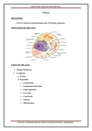
- 1. STRUCTURE AND FUNCTIONS OF CELL M.NITHYA, M.PHARM, DEPARATMENT OF PHARMACOLOGY, JKKMIHSCP. CELL DEFINITION: Cell is a structural and functional unit of all living organisms. STRUCTURE OF THE CELL: PARTS OF THE CELL Plasma Membrane Cytoplasm a. Cytosol b. Organelles Cytoskeleton Endoplasmic Reticulum Golgi Apparatus Lysosome Centrosome Nucleus Mitochondria
- 2. STRUCTURE AND FUNCTIONS OF CELL M.NITHYA, M.PHARM, DEPARATMENT OF PHARMACOLOGY, JKKMIHSCP. PLASMA MEMBRANE Structure of Plasma Membrane The Cell membrane also known as the Plasma membrane. It is a Biological Membrane that separates the interior of all cells from the outside environment. It consists of a lipid bilayer with embedded proteins. The Lipid layer made up of three types of lipid molecules such as Phospholipids, Cholesterols and Glycolipids. The bilayer arrangement occurs because the lipid are amphipathic molecule (Both Polar and Nonpolar parts) Phospholipids – Phosphate (Polar) – Head – Hydrophilic Lipid (Non Polar) – Tail – Hydrophobic Cholesterols – Slightly Amphipathic Glycolipids – Carbohydrate (Polar) – Head Lipid (Non Polar) – Tail Functions of Plasma Membrane: Acts as a barrier separating inside and outside of the cell. Controls the flow of substances into and out of the cell. Helps identify the cell to other cells (e.g., immune cells). Participates in intercellular signalling.
- 3. STRUCTURE AND FUNCTIONS OF CELL M.NITHYA, M.PHARM, DEPARATMENT OF PHARMACOLOGY, JKKMIHSCP. CYTOPLASM Cytoplasm consists of all the cellular contents between the plasma membrane and the nucleus and has two components. a) Cytosol b) Organelles a) Cytosol: (pH - 7) The Cytosol (Intracellular fluid) is the fluid portion of the cytoplasm that surrounds organelles. Cytosol is 75 – 90% of water plus various dissolved and suspended components. Among these are different types of ions, glucose, amino acid, fatty acid, protein, lipid, ATP and waste products. The cytosol is the site of many Chemical reactions for a cell existence. b) Organelles Cytoskeleton Endoplasmic Reticulum Golgi Apparatus Lysosome Centrosome Nucleus Mitochondria CYTOSKELETON: The cytoskeleton is a network of protein filaments. It Consists of three types of filament proteins 1. Microfilament 2. Intermediate filament 3. Microtubules 1. Microfilament Microfilaments are the thinnest elements of the cytoskeleton. Diameter – 6nm They are composed of protein Actin and Myosin. Most Prevalent at the edge of a cell.
- 4. STRUCTURE AND FUNCTIONS OF CELL M.NITHYA, M.PHARM, DEPARATMENT OF PHARMACOLOGY, JKKMIHSCP. Functions of Microfilament: They help generate movement and provide mechanical support. Microfilaments are involved in muscle contraction, cell division and cell locomotion. The Mechanical support that is responsible for the basic strength and shape of cells. 2. Intermediate filaments Several different proteins such as keratin, collagen can compose intermediate filament. Diameter – 10 nm Functions of Intermediate filaments: They help stabilize the position of organelles such as the nucleus. 3. Microtubules Largest cytoskeletal components. Diameter – 25 nm Unbranched hollow tubes composed mainly of the protein tubulin. The assembly of microtubules begins in an organelle called centrosome. Functions of Microtubules Microtubules help determine Cell shape. They also function in the movement of organelles such as secretory vesicles, chromosomes during cell division.
- 5. STRUCTURE AND FUNCTIONS OF CELL M.NITHYA, M.PHARM, DEPARATMENT OF PHARMACOLOGY, JKKMIHSCP. ENDOPLASMIC RETICULUM (ER) Endo – In, Plasmic – Cytoplasm, Reticulum – Network Endoplasmic Reticulum is a network of membrane in the form of flattened tubules. The ER extends from the nuclear envelope throughout cytoplasm. Normally cell contains two types of ER: Rough Endoplasmic Reticulum Smooth Endoplasmic Reticulum Rough Endoplasmic Reticulum Is continuous with the nuclear membrane Usually is folded into a series of flattened sacs. The outer surface of rough ER is containing Ribosomes. Functions of Rough Endoplasmic Reticulum Synthesizes glycoprotein and phospholipid Thus Rough ER produces secretory protein and membrane protein and many organellar proteins. Smooth Endoplasmic Reticulum Extend from the Rough ER to form a network of membrane tubules. It lack of ribosome and does not synthesize proteins. Functions of Smooth Endoplasmic Reticulum Synthesize fatty acids and steroids. Inactivates or Detoxifies drugs. Remove phosphate group from Glucose 6 phosphate. Stores and release calcium ion in muscle cells.
- 6. STRUCTURE AND FUNCTIONS OF CELL M.NITHYA, M.PHARM, DEPARATMENT OF PHARMACOLOGY, JKKMIHSCP. GOLGI APPARATUS Also known as Golgi complex, Golgi body. Present near to the cell nucleus. Shape – Cup like Shape. Made of series of compartment and is a collection of fused, flattened membrane and enclosed disks known as Cisternae. Cisternae broken down into Cis, Medial, Trans. Two main networks: 1. Cis Golgi Network (CGN) 2. Trans Golgi Network (TGN) Functions of Golgi complex The Golgi apparatus is a major collection of protein received from endoplasmic reticulum. Post translational modification of protein. The Golgi bodies act as a post office.
- 7. STRUCTURE AND FUNCTIONS OF CELL M.NITHYA, M.PHARM, DEPARATMENT OF PHARMACOLOGY, JKKMIHSCP. LYSOSOME Lyso – Dissolving and Some – Bodies Also known as suicidal bag. Small spherical, oval bodies surround single layer, pH - 5 It contains a variety of hydrolytic enzymes. Functions of Lysosome Lysosome breakdown bacteria and cell debris engulfed by the cell. The damaged intracellular organelles are also broken down and digested by lysosomes. It contains 60 kinds of digestive and hydrolytic enzymes and lysosomal enzymes best working in acidic pH. CENTROSOME The centrosome located near the nucleus. It consists of two components 1. Pair of Centrioles 2. Pericentriolar materials The two centrioles are cylindrical structure each composed of nine clusters of three microtubules (triplets) arranged in a circular pattern.
- 8. STRUCTURE AND FUNCTIONS OF CELL M.NITHYA, M.PHARM, DEPARATMENT OF PHARMACOLOGY, JKKMIHSCP. Surrounding the centrioles is Pericentriolar material which contains hundreds of ring shaped complexes composed of the protein tubulin. Function of Centrosome These tubulin complexes are the organizing centres for growth of miotic spindle, which play a role in cell division. During the cell division centrosome replicate so that succeeding generation of cells. NUCLEUS It is a largest structure present almost in the centre of cell. Spherical or Oval Shape. It has double membrane envelope separate the nucleus from the cytoplasm. The nuclear envelope also lipid bilayer similar to plasma membrane. Outer layer of nuclear envelope is continuous with rough endoplasmic reticulum. Nucleoli Inside the nucleus are one or more spherical bodies called Nucleoli. That functions is producing Ribosomes. Each nucleolus is simply a cluster of protein, DNA and RNA. It is not enclosed by a membrane. Nucleoli are the sites of synthesis of rRNA and assembly of rRNA and protein into ribosomal subunits. Nucleoli disperse and disappear during cell division and reorganize one new cells are formed. Within the nucleus are most of the cell’s hereditary units called genes. Which control cellular structure and direct cellular activities. Genes are arranged along chromosomes. Human somatic cells have 46 chromosomes, 23 inherited from each parent.
- 9. STRUCTURE AND FUNCTIONS OF CELL M.NITHYA, M.PHARM, DEPARATMENT OF PHARMACOLOGY, JKKMIHSCP. Functions of nucleoli Nuclear pores control the movement of substances between the nucleus and cytoplasm. Nucleoli produce ribosome. Chromosome consists of genes that control cellular structure and direct cellular functions. MITOCHONDRIA The Mitochondria referred the Power houses of the cell. Rod shaped structure. Mitochondria are a double membrane bound organelle. It has the outer membrane and inner membrane. The membranes are made up the phospholipids and proteins. 0.5 to 1 micrometer in diameter. The mitochondria generate energy source in the form of Adenosine Tri Phosphate (ATP). Outer membrane: It is smooth and is composed of equal amount of phospholipids and proteins. It has a large number of special proteins known as porins. The porins are integral membrane proteins and they allow the movement of molecules that are of 5000 daltons or less in weight to pass through it. The outer membrane is freely permeable to nutrient molecules, ions, energy molecules like ATP and ADP molecules. Inner membrane: Inner membrane of mitochondria is more complex in structure. It is folded into a number of fold many times and is known as the Cristae.
- 10. STRUCTURE AND FUNCTIONS OF CELL M.NITHYA, M.PHARM, DEPARATMENT OF PHARMACOLOGY, JKKMIHSCP. This folding help to increase the surface areas of mitochondria inside the organelle. The cristae and the proteins of the inner membrane aids in the production of ATP molecules. Various chemical reactions take place in the inner membrane of the mitochondria. The inner membrane is strictly permeable, it is permeable only to oxygen and ATP. Intermembrane space It is the space between the outer and inner membrane of the mitochondria, it has the same composition as that of the cells cytoplasm. Matrix The fluid filled cavity is called matrix. The matrix of the mitochondria is a complex mixture of proteins and enzymes. The matrix contains ATP molecules, mitochondrial ribosomes, tRNA and mitochondrial DNA. Functions of mitochondria Generate of ATP through the reactions of aerobic cellular respiration. Maintain the concentration of calcium ion within the cell. It can be synthesize proteins, enzymes and hormones like Testosterone and Estrogen etc. The liver cells mitochondria have some enzymes that detoxify the ammonia. It involved in the apoptosis or programmed cell death.