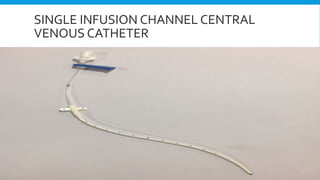
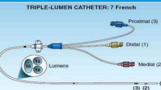
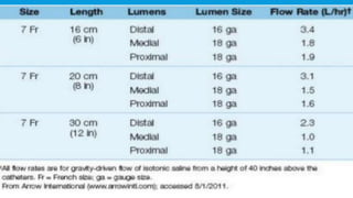
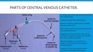
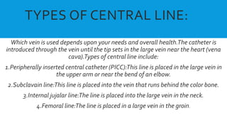
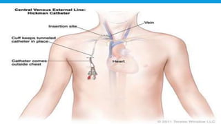
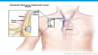
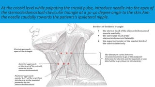
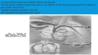
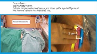
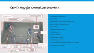
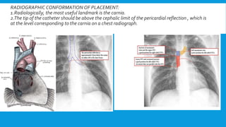
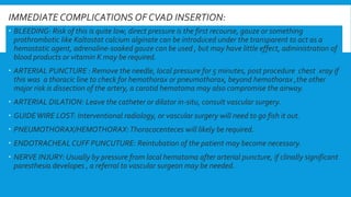
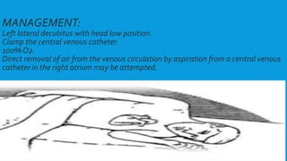
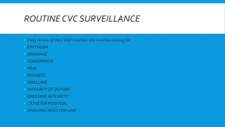
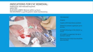
2. DEFINITION:
A central line is a type of a catheter that is placed ina large vein that allows multiple
IV fluids to be given and blood to be drawn.When compared to a typical IV line, a
central line is larger, can stay in place longer,can deliver a greater volume of fluids
and allows blood to be drawn easily.
Central venous catheters are often necessary for reliable vascular access in critically
ill patients
They are typically 15 to 30 cms (6 to 12 inches) in length and have single or multiple (
2 to 4 infusion channels).
6. kjsssdsMultilumen catheters are favoured in the critically ill patients that require a
multitude of parenteral therepies eg fluids,drugs and nutrient mixtures.
Multilumen catheters make it possible to deliver these therapies using a single
venipuncture.
Triple –lumen catheters are favourite for central venous access.
These catheters are variable in sizes of 4 to 9 French.
7. Size 7 French (OD =23mm)triple lumen catheter is a popular choice in adults,which
typically have one 16 gauge channel and two smaller 18 gauge channels.
To prevent mixing of infusate solutions, the three outflow ports are separated.
8.
9.
10. PARTS OF CENTRALVENOUS CATHETER.
10 ml/sec max flow rate through proximal
and medial ports.
5ml/sec through the distal lumen.
Pressure injection information marked
clearly on the hubs.
Soft, contoured tip design minimizes the
risk of vessel trauma.
Staggered exit ports reduce the risk of
mixing incompatible drugs and solutions
that may create precipitate.
Optimal length:15 cm to fit most
needs;cm markings for trimming to
shorter lengths.
Tapered end for easy advancement
,minimizes risk of vessel damage.
11. TYPES OF CENTRAL LINE:
Which vein is used depends upon your needs and overall health.The catheter is
introduced through the vein until the tip sets in the large vein near the heart (vena
cava).Types of central line include:
1.Peripherally inserted central catheter (PICC):This line is placed in the large vein in
the upper arm or near the bend of an elbow.
2.Subclavain line:This line is placed into the vein that runs behind the color bone.
3.Internal jujalar line:The line is placed into the large vein in the neck.
4.Femoral line:The line is placed in a large vein in the groin.
12.
13. A BRIEF DESCRIPTION ABOUT PERIPHERALLY
INSERTED CENTRAL CATHETERS:
Peripherally inserted central catheters (PICCs) are inserted in the basilic or cephalic vein in the arm (just
above the antecubital fossa) and advanced into the superior vena cava.
Useful in patients with concern for the adverse cosquences of central venous cannulation
(e.g.,pneumothorax, arterial puncture, poor patient acceptance).
PICCs are used primarily when traditional central venous access are considered risky (e.g ., severe
thrombocytopenia) or are difficult obtain (eg., morbid obesity).
Major distinction between PICCs and CVCs is their length; i.e., the length of the catheters (50 cm to70 cm)
is atleast double the length of the triple lumen catheters.
Because of this added length there is reduction in flow capacity in PICC.
Flow is particularly sluggish in the double lumen PICCs because of the smaller diameter of the infusion
channels.
The flow limitation of PICCs (especially the double lumen catheters0 makes them ill-suited for aggressive
volume therapy.
14. EXTERNAL CENTRALVENOUS CATHETERSVS
SUBCUTANEOUS CENTRALVENOUS CATHETER
External catheters also calledTunneled central line (known as BROVIAC , HICKMAN) are
surgicially placed in a major vessel of the body,most often in the chest, they are called external
because a portion of the catheter is exposed which allows usage without a needle stick.A dressing
is required to cover the place where the catheter comes through the skin all the time.
Usage of heparin time o time is important to keep it functioning and free from infection.
Subcutaneous ports (mediport, port-a-catheter infera port), these devices are surgically placed
and are totally implanted into subcutaneous tissue, most often the chest, they have an attached
catheter that is inserted into a major vessel.They are used for long term i.e, months or years.They
are single or double lumened.
A special “huber” (90 degree) needle is inserted through the skin and into the post before the part
is used.When it is “accessed” a dressing is required similar to an external catheter when not being
used,it needs no special care unlike external catheters.
15.
16.
17. INDICATIONS:
When peripherally venous access is difficult to obtain (e.g in obese or
intravenous drugs abusers) or difficult to maintain (e.g in agitated patients).
For the delivery of vasoconstricter drugs (e.g dopamine, norepinephrine),
hypertonic solutions (e.g parentral nutrition formulas), or multiple parentral
medication.
For prolonged parentral drug therapy (i.e more than a few days).
For specialized tasks such as hemodialysis,transvenous cardiac pacing, or
hemodynamic monitering ( e.g with pulmonary artery catheters).
IVC filter placement.
Venous stenting.
Catheter guided thrombolysis.
Repeated blood Sampling.
18. CONTRAINDICATIONSTO CENTRALVENOUS
ACCESS:THERE IS NO ABSOLUTE
CONTRAINDICATION.
Obstructed vein ( e.g clot).
Stenosis of the vein.
Raised ICP. (IJ line ).
Severe coagulopathy.
Respiratory failure with high FiO2.
Contaminated site.
Traumatised site (e.g clavicle fracture and subclavian line).
Burned site.
Uncooperative awake ptient.
19. FACTORS INVOLVED IN SITE SELECTION
FOR CENTRAL LINE INSERTION:
Obesity
Bleeding diathesis:
1. Platelets less than 50
2. INR over 1.5
3.APTT over 50
4. Antiplatelets drugs: clopidogrel and ticagrelor especially.
Hypotension.
Previous surgeries at the proposed site of insertion.
Previous central line at the same site.
Infection at the site.
Presence of LBBB.( Relevant to PA catheters only).
Lymphnode dissection in the same area.
Previous DVT in the limb being drained by the central vein you are looking at.
20. CENTRALVENOUS ACCESS SITES:
SITE:SUBCLAVIAN
ADVANTAGES:
1.Suitable for long term use.(~14 days)
2.Lowest rates of infections.
DISADVANTAGES:
1.Increased risk of subclavian stenosis if used for vas caths (which prevents
a fistula from being formed on that arm).
2.Impossible to control bleeding from arterial puncture or severe
coagulopathy (non compressible).
3.Least suitable for the patients for severe lung pathology ( risk of
pneumothorax).
Requires the patient to be supine and head down for insertion.
5.Insertion interferes with CPR.
6.Arm position can “pinch off” .
21. INTERNAL JUGULAR:
ADVANTAGES:
1.Convenient
2.Easy to access during an operation.
3.Leaast acute complication.
4.Best site for aVas cath
5.Fairly unlikely to result in a pneumothorax or hemothorax.
6.NO “pinch off” phenomonan.
DISADVANTAGES:
1.Only short term use is optimal.
2.May be better to save it for when a vas cath , PA catheter or ECMO cannula are required.
3.May get in the way of future tunnelled central line.
4.Require patient to be supine and head down for insertion.
5.May cause increase un ICP if the patient developes an IJ thrombosis.
6.Cannot be an option if the C- supine collar needs to remain in-situ.
7.Not an ideal choice if tracheostomy is planned (will get in the way).
22. FEMORAL SITE:
ADVANTAGES:
1.Zero risk of pneumothorax or hemothorax
2.Easily compressible site.
3.No need for supine or head down position, suitable for patients who are in
respiratory distress.
4.No need for chest x ray conformation.
DISADVANTAGES:
1.Risk of infection is greatest.
2.A poor choice for morbidly obese patients with extensive pannus.
3.Risk of retroperitoneal hematoma.
4.Unsuitable for CVP monitoring.
5.Circulation of drug is comparatively delayed.
6.Impairs patients mobilization.
23. PICC:
ADVANTAGES:
1.Low risk of serious complications.
2.Suitable for prolonged use,(upto 6 weeks in some cases).
3.No need to position the patient supine.
DISADVANTAGES:
1.Higher risk thromboses.
2.More difficult to assure correct tip position.
3.Unsuitable for high volume or highly viscous infusions (too much
resistance to flow).
4.Usually unsuitable for CVP monitoring or central venous blood
sampling (high risk of lumen obstruction and poor wave form fidelity).
24. RIGHT VS LEFT SIDED LINES
The decision to insert a line on left or right depends upon a few considerations:
1.It is easier to insert a right subclavian.
2.It is easier to insert a left PICC.
3.It is better o insert a vas cath into a right IJ.
4.It is better to save right IJ for vas cath if the patient requires ECMO or dialysis at some stage in near
future.
5.The right IJ vein is bigger than the left about 3 to 4 mm difference in their diameter on average.
6.Femoral vessels are bilaterally symmetrical however the right femoral artery is favoured site for arterial
angiography access, and in general he femoral arteries are ideal for this, so if a coronary angiogram ,IABP
or ECMO are planned one may wish to leave the groins alone.
6.In a patient with some sort of severe one sided lung pathology, the IJ or subclavian line should be placed
on the affected site sp that the pneumothorax or the hemothorax of je good lung is not caused.
7.Left sided lines ( IJ and subclavian) tend to have higher chance of damaging the thoracic duct.
25. APPROPRIATE PROCESS OF CONSENT :
An appropriate consent procedure needs to include the following important matters:
1.Description of the procedure.
2.Risks and complications like pain during insertion, pneumothorax, hemothorax, need for chest drain,
cardiac or greater vessel injury, air embolism and death.
3.Minor but common complications like need to reposition the device and arrhythmias.
4.Possible latter complications i.e, the line may get blocked, infection of wound or blood stream, chronic
pain from injured inter coastal nerves.
5.The patient must be legally capable of giving consent.
6.Consent must be informed.
7.Consent must be specific.
8.Consent must be freely given.
9.Consent must cover that what is actually done.
26. THE CORRECT ENVIRONMENT FOR CVC
INSERTION.
You need to have access to:
1.Adequate lightening.
2.Adequate space around the patient.
3.Some means of assuring aseptic technique
(i.e. a trolley and drapes).
Immediate access to cardiac resuscitation
equipment and drugs.
5.Skilled assistants.
6.”Electrical safety support”.
27. PREPARATION AND POSITIONING OFTHE
PATIENT:
INTERNAL JUGULARVEIN:
1.Right side preferred.
2.Tendelenberg position
(10-15 degrees).
3.Head rotated
approximately 15 degree to
the left.
28. At the cricoid level while palpating the cricoid pulse, introduce needle into the apex of
the sternocleidomastoid-clavicular triangle at a 30-40 degree angle to the skin.Aim
the needle caudally towards the patients’s ipsilateral nipple.
29. SUBCLAVIANVEIN:
1.Right side is preferred.
-Supine position, head neutral, arm abducted.
-Trendelenburg position (10-15 degree).
-Shoulders neutral with mid retraction.
30. Junction of the medial and middle third of the clavicle.
The site of the needle insertion lies about 1 cm inferior to the clavicle allowing for the needle to
pass under the clavicle.
Needle should be parallel to the skin.
Aim towards the supraclavicular notch.
32. PICC:
Basilic vein which is approximately midway up the arm (about 12cm medial
to the medial epicondyle of the humerus.
33. Appropriate monitoring for central line insertion:
An ECG monitor.
A pulse oximeter.
A pressure transducer.
34. Sterile tray for central line insertion:
1.Sterile gown and gloves.
2.The CVAD.
3.Antiseptic solution for skin preparation.
4.0.9% sodium chloride solution.
Sterile drapes
6.Sterile procedure trays.
7.Gauze squares.
8.Local anesthetic.
9. Needle and syringes.
10.Suture equipments.
11.Small sterile containers to isolate used sharps.
12.Appropriate dressings.
35. Local and systemic analgesia/anesthesia.
Usually lignocaine without adrenaline is enough-
adrenaline is only useful if we are doing a tunneled line
and want to prevent hemorrhage into the tunnel.
Infiltrate generously but it may distort anatomical land
marks.
Try not to inject any air into the subcutaneous tissues, this
tends to interfere with ultrasound imaging of the vessel.
While performing a subclavian puncture, anesthetize the
clavicular periosteum.
36. SELDINGERTECHNIQUE:
1.Use introducing needle to locate
vein.
2.Wire is threaded through the
needle.
3.Needle is removed.
4.Skin and vessel are dilated.
5.Catheter is placed over the wire.
6.Wire is removed.
7.Catheter is secured in place.
37. Post-Catheter Placement.
Aspirate blood from each port.
Flush with saline or with sterile water.
Secure catheter with sutures.
Cover with sterile dressing (tega-derm).
Obtain chest x ray for IJ and SC lines.
38. THE J-TIPPED SELDINGERGUIDEWIRE: An ideal guide wire should be :
1.Fexible, conforming easily to the vessel.
2.Strong, resistant to lacerations.
3.Kink resistant.
4.Soft tipped ,to prevent damage to vascular structures.
5.Sufficiently stiffed to allow manipulation of its direction.
6.Approximately twice the length of the device it is intended to guide.
39. RADIOGRAPHIC CONFORMATION OF PLACEMENT:
1.Radiologically, the most useful landmark is the carnia.
2.The tip of the catheter should be above the cephalic limit of the pericardial reflection , which is
at the level corresponding to the carnia on a chest radiograph.
40. IMMEDIATE COMPLICATIONS OF CVAD INSERTION:
BLEEDING: Risk of this is quite low, direct pressure is the first recourse, gauze or something
prothrombotic like Kaltostat calcium alginate can be introduced under the transparent to act as a
hemostatic agent, adrenaline-soaked gauze can be used , but may have little effect, administration of
blood products or vitamin K may be required.
ARTERIAL PUNCTURE : Remove the needle, local pressure for 5 minutes, post procedure chest xray if
this was a thoracic line to check for hemothorax or pneumothorax, beyond hemothorax ,the other
major risk is dissection of the artery, a carotid hematoma may also compromise the airway.
ARTERIAL DILATION: Leave the catheter or dilator in-situ, consult vascular surgery.
GUIDEWIRE LOST: Interventional radiology, or vascular surgery will need to go fish it out.
PNEUMOTHORAX/HEMOTHORAX:Thoracocenteces will likely be required.
ENDOTRACHEAL CUFF PUNCUTURE: Reintubation of the patient may become necessary.
NERVE INJURY: Usually by pressure from local hematoma after arterial puncture, if clinally significant
paresthesia developes , a referral to vascular surgeon may be needed.
41. AIR EMBOLISM
DEADLY COMPLICATION ASSOCIATEDWITH CVC’s.
Signs and symptoms are :
Respiratory changes i.e., sudden shortness of breath,
cyanosis.
CVS changes i.e., sudden onset of chest pain , increased
heart rate, decreased BP.
CNS changes i.e., altered neurological signs , dizziness,
confusion, loss of consciousness.
42. MANAGEMENT:
Left lateral decubitus with head low position.
Clamp the central venous catheter.
100% O2.
Direct removal of air from the venous circulation by aspiration from a central venous
catheter in the right atrium may be attempted.
43. TO MINIMIZETHE CHANCE OF AIR ENTERINGTHE SYSTEM INCLUDES:
Ensure the lumen is clamped prior to opening the system.
Position the patient so that the insertion site is at or below the level of heart during
insertion and removal of the catheter.
44. INFECTIONS:
MOST FREQUENT AND SERIOUDS
COMPLICATIONS.
TYPES:
Local infection- cellulites.
Central line associated blood stream
infections (CLABSI).
45. CAUSATIVE ORGANISIMS:
Staph epidermidis 25% to 50%.
Staph aureus 25%.
Candida 5-10%.
RISK FACTORS:
Cutaneous colonization of the insertion site.
Moisture under the dressing
Prolonged catheter time.
Technique of care and placement of the central line.
47. LATE COMPLICATIONS
DEVICE DYSFUNCTION:
Catheter fracture
Catheter tip displacement
Lumen blockage
Fibrin sheath formation
PATIENT-RELATED COMPLICATIONS:
Infection: on average 2.5
infections/1000 catheter days.
Vascular erosion
Vessel stenosis
Thrombosis
Osteomyelitis of clavicle
Nerve injury
Extravasation
48. ROUTINE CVC SURVEILLANCE
Daily review of the CVAD insertion site involves looking for:
ERYTHEMA
DRAINAGE
TENDERNESS
PAIN
REDNESS
SWELLING
INTEGRITY OF DUTURE
DRESSING INTEGRITY
CATHETER POSITION
ONGOING NEED FOR LINE
49. INDICATIONS FOR CVC REMOVAL:
POINTLESS: Not indicated anymore.
INFECTED
BLOCKED LUMEN: Remove within 24 hours.
INCONVINIENT: e.g if the patient needs tracheostomy.
TECHNIQUE:
Asepsis
Supine with head down position
Patient to remain supine for 30 to
60 min.
Airtight dressing on the site for 24
hours.
If CLABSI is suspected, the tip is
sent for culture.
Removal of an intact tip should be
documented.