2ry OAG, OHT, NTG
•Download as PPTX, PDF•
7 likes•289 views

Ocular hypertension Normal Tension Glaucoma 2ry open angle glaucoma

Recommended
More Related Content
What's hot
What's hot (20)
Similar to 2ry OAG, OHT, NTG
Similar to 2ry OAG, OHT, NTG (20)
More from faculty of medicine -benha university
More from faculty of medicine -benha university (19)
Recently uploaded
Recently uploaded (20)
2ry OAG, OHT, NTG
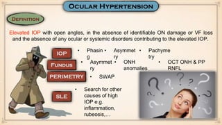
- 1. Elevated IOP with open angles, in the absence of identifiable ON damage or VF loss and the absence of any ocular or systemic disorders contributing to the elevated IOP. • Phasin g • Asymmet ry • Pachyme try • Asymmet ry • ONH anomalies • OCT ONH & PP RNFL • SWAP • Search for other causes of high IOP e.g. inflammation, rubeosis,…
- 2. At 60 months, the cumulative probability of developing POAG was 4.4% in the medication group and 9.5% in the observation group. Thus topical medications were definitely shown to reduce the risk of glaucoma in patients with ocular hypertension; however, most untreated patients did not get worse over a 5-year period.
- 3. • Disturbance of the structural or functional integrity of the optic nerve, in which; IOP constantly equal or < 21 mmHg Glaucomatous ONH damage Characteristic visual field changes Open AC angle Absent signs of 2ry glaucoma or a non glaucomatous cause of optic neuropathy • Phasin g • Pachyme try • Other ONH anomalies mimic glaucoma e.g. neurological, congenital • Other causes of field defects e.g. myopic degeneration
- 4. • More common in old age • More common in Japanese • Siblings, offspring • Abnormal vaso regulation e.g. Raynaud disease, Migraine • Systemic hypotension • Obstructive sleep apnoea • Auto antibodies • More common in females
- 5. • Brimonidine (neuro-protective effect) • Betaxolol (Increase optic nerve blood flow) • Prostaglandin derivatives (great ocular hypotensive effect) • NB; topical beta blockers can have a dramatic effect on blood pressure in some patients & may contribute to nocturnal dips • Systemic: Calcium channel blockers (for vasospasm) • Under trials (Neuro-protective agents); A. Memantine B. Gingko biloba
- 6. 1. Carotid Cavernous Fistula 2. Sturge Weber syndrome 3. Superior vena cava obstruction 1. Pigmentary glaucoma (Pigment) 2. Red cell glaucoma (RBCs) 3. Ghost cell glaucoma (degenerated RBCs) 4. Macrophages & lens protein (phacolytic glaucoma) 5. Pseudo exfoliation glaucoma 1. Neo vascular glaucoma (Fibro vascular tissue) 2. Epithelial ingrowth 3. Irido corneal endothelial syndrome
- 7. • Deposition of amyloid like material on various ocular structure including lens capsule, zonules, TM, iris, … • This substance is derived from abnormal extra cellular matrix metabolism
- 8. • PXF and pigment deposited vertically on the back of the cornea
- 9. • Loss of pupillary ruff with deposition of PXF on pupillary border • Transillumination defects at pupillary border
- 10. • Irregular pigmented band anterior to Schwalbe line
- 11. •The anterior lens capsule typically shows a central disc and a radially indented peripheral layer of PXF material, separated by a clear zone maintained by pupillary abrasion
- 12. • Similar to POAG but failure is common • ALT; More effective than with POAG due to more pigment content in the TM • Same success rates with POAG
- 13. • Pigment shedding is precipitated by rubbing of the posterior pigment layer of the iris against the zonule as a result of excessive posterior bowing of the mid-peripheral portion of the iris.
- 14. • PXF and pigment deposited vertically on the back of the cornea
- 15. • Transillumination defects at (radial, spoke like)
- 17. •Scheie line; pigment granules deposited on anterior lens capsule
- 18. •Acute IOP rise following the release of pigment granules might particularly occur after physical exercise.
- 19. • Similar to POAG but miotics are theoretically of particular benefit as they decrease irido-zonular contact • ALT; More effective than with POAG due to more pigment content in the TM • Laser Peripheral iridotomy, reverse iris concavity & eliminate irido-zonular contact (equilibration of abnormal pressure gradient between AC & PC) • Same success rates with POAG
- 21. + signs of the cause e.g. CRVO Rubeosis irides
- 22. • Angle neo vascularisation
- 23. • Prostaglandin derivatives are used with caution due to their inflammatory promoting effect • Atropine is used topically to resist synechia formation • Topical steroids should be given if significant inflammation is present • Pan retinal photocoagulation (PRP) • Intra vitreal anti VEGF • Cyclodestructive therapy e.g. Cyclo photo coagulation, cyclo diode • SST + MMC • if blind painful eye retro-bulbar alcohol, enucleation
- 25. • Anterior uveitis causes plasmid aqueous. The inflammatory cells clog TM pores increasing resistance to aqueous outflow.
- 26. • KPs
- 27. • AC cells & flare
- 28. • Hyper pigmented TM
- 29. • Prostaglandin derivatives are used with caution due to their inflammatory promoting effect • Atropine is used topically to resist synechia formation • Topical steroids are essential in treatment • SST + MMC • GDD • Cyclodestructive therapy e.g. Cyclo photo coagulation, cyclo diode • if blind painful eye retro-bulbar alcohol, enucleation