SlideShare a Scribd company logo
1 of 105
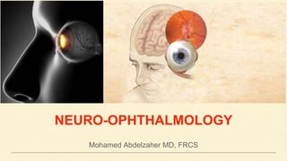
NEURO-OPHTHALMOLOGY
Mohamed Abdelzaher MD, FRCS
Topics
- Pupil
- Ocular motor system
- Visual system
- Visual fields
The involuntary window into our state of emotional thought.
Light Reflex
• Stimulus: Light
• Receptors: Rods & Cones
• Afferent: Optic nerve, chiasm,
anterior 2/3 of optic tract, pre-tectal
nucleus
• Center: Edinger Westphal nucleus
(III)
• Efferent: III nerve
(Parasympathetic root of ciliary
ganglion)
• Response: Miosis
Near Reflex
• Stimulus: changing sight from far to
near object
• Receptors: Rods & Cones
• Afferent: Optic nerve, chiasm, optic
tract, LGB, optic radiation, occipital
cortex, frontal cortex, internal capsule
• Center: Edinger Westphal nucleus (III)
• Efferent: III nerve (Parasympathetic
root of ciliary ganglion + nerve to MR)
• Response: Miosis + Convergence +
Accommodation
How to perform
light reflex:
• Dim room
illumination
• Ask the patient
to look at a far
object
• Use bright light
Afferent Pupillary Defect III nerve palsyEfferent Defect, Parasympathetic
Horner SyndromeEfferent Defect, Sympathetic
Afferent Pupillary Defect
Partial lesion in the afferent pathway;
Optic nerve, chiasm, tract
• Pupil does not respond to light directly
• Pupil respond to light consensually
• Pupils are equal in size (No anisocoria)
• Near reflex is normal
III nerve palsy
Occulomotor palsy; traumatic, (common pupil involvement) stroke (less common pupil involvement)
• Ptosis (sever)
• Outward ocular deviation
• Mydriasis (anisocoria, more in bright light)
Efferent Defect, Parasympathetic
Ipsilateral Pupil initially constricts (Irritation)
Then dilates due to III nerve compression
As the coma deepens,
contralateral pupil dilates
Horner Syndrome
Destruction of sympathetic supply of the iris, e.g. neck surgery, Pancost tumour
• Ptosis (Mild)
• Miosis (anisocoria, more in dim light)
• Anhydrosis (Hemifacial)
Efferent Defect, Sympathetic
• Enophthalmos
• Heterochromia iridis (Congenital Horner)
Ciliospinal Reflex:
dilation of the
ipsilateral pupil in
response to pain
applied to the neck.
Light Near Dissociation (LND)
• Bilateral
• Small irregular pupil
• Pupil does not react to light
• Pupil constrict during near reflex
• Neurosyphilis
• Diabetes
• Encephalitis
Lesion in Intercalated neurons in the Peri
aqueductal area
Fibres subserving light reflex are more ventral
Light Near Dissociation (LND)
• Commonly unilateral
• Large irregular pupil
• Poor response to light
• Slow & tonic response to near reflex
• Late lesions, the pupil becomes miotic (Little old Adie’s)
• Associated absent deep tendon reflexes (Holmes-Adie syndrome)
• Viral
Lesion in the Ciliary Ganglion
Unequal Pupil Size
Check light reflex
CocaineED
Miosis Mydriasis
Physiological
• Light reflex
• Near reflex
• Senile
• Sleep
• 3rd stage of anaesthesia
• Dim Illumination
• Fear, Fight, Fright
• 1st, 2nd & 4th stages of anaesthesia
• Cilio-spinal rflex
Drugs
• Parasympathomimetics: Pilocarpine
• Morphine
• Opium
• Sympathomimetics: Epinephrine
• Parasympatholytics: Atropine,
Cyclopentolate
Neurological
• Argyll Robertson
• Horner
• Hutchinson (Irritative stage)
• Little old Adie’s
• Pontine haemorrhage
• III nerve palsy
• Adie’s tonic pupil
• Hutchinson (Paralytic stage)
Ocular
• Keratitis
• Iritis
• Trauma
• Acute congestive glaucoma
• Trauma
• Nucleus
• Nerve
• Muscle
1. Congenital
2. Trauma
3. Inflammation
4. Vascular
5. Tumor
6. Metabolic
7. Toxic
LMNL
• Diplopia: that increases toward the side of the paralysed muscle
• Ocular deviation
• Abnormal Head Posture
• Contracture
• Contracture
• Underaction
(inhibitional palsy)
False projection (Hess screen)
• Inward
• Abduction
• Increases on looking outwards
• Face turn toward the
paralysed muscle
• Upward
• Down & in
• Increases on looking down & in
• Head tilt to the
opposite shoulder
• Chin depression
• Face turn to the same
side
Parks’ (Bielschowsky) 3 step test
• Right eye hypertropic
• Elevation increases
to the left gaze
• Elevation decreases
in the left head tilt
• Outward
• In all directions except outward
• Treat the cause, if possible e.g. brain tumour, trauma, …
• Temporary measures; (in the 1st 6 months of the insult)
A. Occlusion of the sound eye
B. Relieving prisms
• Surgical treatment;
6th nerve palsy >>> MR recession +/- vertical muscle transposition
4th nerve palsy >>> IO myectomy
3rd nerve palsy >>> LR recession then treat ptosis
Visual
System
1) Optic nerve
2) Optic Chiasm: where nasal fibres
cross to the opposite side
3) Optic Tract: carrying ipsilateral
temporal fibres & contralateral nasal
fibres
4) Lateral Geniculate Body
5) Optic Radiation: The upper
radiation (Parietal lobe) subserve the
lower visual field, while the lower
radiation (Temporal lobe) subserve the
upper visual field
6) Visual Cortex
Visual
System
Optic Nerve Function Tests
1) VA
2) Pupil reactivity
3) Colour vision
4) Visual field
5) VEP
ONH Swelling
Normal Optic Nerve Functions
Crowded, High hypermetropia Tilted
Affected
Optic Nerve
Functions
1) Inflammatory
2) Ischemic
3) Compressive
4) Increased ICT
5) Toxic
6) Vascular; CRVO
Swelling of the optic nerve head secondary to raised intracranial pressure (ICP).
A. Idiopathic Intracranial Hypertension (IIH); the commonest cause
B. Space occupying lesion e.g. tumour
C. Obstruction of the ventricular system
D. Cerebral venous sinus thrombosis
E. Cerebral edema e.g. head trauma
• Raised ICP is transmitted via subarachnoid space acting as a tourniquet around the
optic nerve
• This causes disturbance of the axoplasmic flow along retinal nerve fibres resulting
in stasis swelling of these axons & leakage
1. Headache: more in early morning and may awake the patient from sleep
2. Nausea
3. Vomiting: projectile (sudden & forceful)
4. Deterioration of consciousness
1. Transient visual obscuration (Amaurosis Fugax) : precipitated by bending, cough or
Valsalva manoeuvre
2. Horizontal diplopia: 6th nerve palsy
3. Visual Acuity: Normal in early stages, but shows significant reduction later on
• IIH occurs in obese young adult women.
• Various medications have been implicated especially the Oral Contraceptive Pills
• Limited abduction with inward ocular deviation
• 6th nerve palsy may occur due to stretching of one
or more of the abducens nerve over the petrous
tip.
• It is considered as a false localising sign
• Mild optic disc hyperaemia with
preservation of optic cup
• Indistinct disc margins
• Disappearance of the previously present
spontaneous venous pulsations
• VA is normal or mildly affected
• Severe optic disc hyperaemia with absent
optic cup
• Moderate optic disc elevation
• Indistinct disc margins
• Venous engorgement
• Peripapillary flame shaped haemorrhage &
CWS
• Circumferential retinal folds (Paton lines)
• Enlarged blind spot
• VA is reduced
• Constricted visual field
• Disappearance of haemorrhage & cotton
wool spots
• VA is severely impaired
• Optic disc is pale grey white with few
crossing blood vessels and indistinct
margins
• Papilledema is a bilateral condition
• Except in rare instances as (Foster Kennedy Syndrome)
• Left Olfactory groove tumour causing left optic
atrophy & right papilledema
R
• To measure the Optic Nerve Sheath External Diameter (ONSD) Which is substantially
distended
• To exclude space occupying lesions &/or
enlarged ventricles
• Used also to measure the ONSD
• To measure CSF opening pressure (Normal 10 - 18 cm water) and also to analyse
CSF constituents
• Must not be done except after neuro imaging to avoid herniation of intracranial contents
in case of space occupying lesion
1. Transient ischemic attacks
2. Retinal migraine
3. Hysterical
4. Malingerer
1. Pseudo: Drusen, Hyperopia,Tilted disc
2. True:
A. Ischemic
B. Inflammatory
C. Vascular
• Mainly directed to the cause
1. Weight loss
2. Acetazolamide, Frusemide
3. IV mannitol
4. Lumbar puncture
5. Optic nerve sheath fenestration
(in unresponsive cases)
6. Ventriculo peritoneal shunt
surgery
Occlusion of the blood supply (SHORT
CILIARY VESSELS) of the optic nerve,
either;
• Anterior (AION): 90% of cases, or
• Posterior (PION)
1. AION:
A. Non Arteritic (NAION): DM & HTN are the commonest causes
B. Arteritic (AAION): caused by Giant cell arteritis (GCA)
2. PION:
• Caused by ischemia of the retro laminar portion of the optic nerve head especially post
bloody surgeries. It might be also caused by arteritic or non arteritic causes.
1. NAION: Old age, history of DM, HTN
2. AAION: Old age, Scalp tenderness (with combing hair), headache, jaw claudication
3. PION: History of bloody surgery
1. Sudden loss of vision:
• Painless: NAION
• Painful: AAION
SUPERFICIAL TEMPORAL
ARTERY:
• Tender
• Loss of pulsations
• Thickened
• Nodular
• VA: severely reduced (more in AAION)
• Pupil: RAPD
• Ocular nerve palsies: in AAION
ONH:
• Swollen
• Pale
• Splinter haemorrahge at ONH
margin
• might be associated with
cilioretinal artery occlusion
ONH:
• Swollen (Diffuse or Sectoral)
• Hyperaemic
• Splinter haemorrahge at ONH
margin (Few)
• might be associated with diabetic
or hypertensive retinopathies
• Temporal Artery Biopsy (TAB)
• ESR
• CRP
• HbA1c
• Blood pressure monitoring
• Altitudinal field defect
1. Pseudo: Drusen, Hyperopia,Tilted disc
2. True:
A. Elevated ICP
B. Inflammatory
C. Vascular
1. CRAO
2. CRVO
3. RD
4. Malingerer
• Intravenous methyl prednisolone 1g/day for 3 days followed by oral prednisolone
• Anti platelet therapy e.g. Aspirin to reduce risk of stroke
• Immunosuppressives e.g. Methotrexate
• Aim: to prevent blindness of the fellow eye
• NO DEFINITE treatment
• Anti platelet therapy e.g. Aspirin to reduce risk of stroke
• Strict control of DM & HTN
• Tortuosity & engorgement of
all branches of CRV
• Blot & flame shaped retinal
haemorrhage
• Cotton wool spots
• ONH swelling & hyperaemia
• Macular edema
• Papillitis: Inflammation of the ONH
• Retro Bulbar neuritis: inflammation of the retro laminar part of the optic nerve
A. Demeylinating: most common
B. Infectious: Herpes zoster,
Syphilis
C. Para infectious: following viral
infection or immunisation
D. Non infectious: Sarcoidosis,
Auto immune disease
• Idiopathic demyelinating disease involving the white mater of the central nervous system
Demyelination is a pathological process in
which normally myelinated nerve fibres lose
their insulating myelin layer. The myelin is
phagocytosed by microglia and macrophages,
subsequent to which astrocytes lay down
fibrous tissue in plaques. Demyelinating
disease disrupts nervous conduction within
the white matter tracts of the brain, brainstem
and spinal cord.
1. Spinal cord: weakness, sensory loss, sphincter disturbance
2. Brainstem: Diplopia, Nystagmus, Dysphagia
3. Cerebral: Hemiparesis, Hemianopia
1. Optic neuritis
2. Ocular motor nerve palsies
3. Intermediate uveitis
• Demyelinating Optic neuritis
Young age
Discomfort or pain around
the eye especially on
looking on the field of MR,
SR
VA deteriorates rapidly
within several days to 3
weeks, then begin to
improve
RAPD
Optic disc might appear
normal (Retrobulbar
neuritis), or Swollen
(Papillitis)
Peri ventricular plaques
A. Central scotoma
B. Centro cecal scotoma
C. Nerve fibre bundle defect
D. Altitudinal field defect
• Intravenous methyl prednisolone 1g/day for 3
days followed by oral prednisolone
• Beta interferone
Treatment may speed up recovery by 2–3 weeks and may delay the onset of clinical
MS over the short term. This may be relevant in the patients with poor vision in the
fellow eye or those with occupational requirements, but the limited benefit must be
balanced against the risks of high-dose steroids.
Therapy does not influence the eventual visual outcome and the great majority of
patients do not require treatment.
A. High Alcohol (Ethyl Alcohol), Tobacco consumption
B. Poor diet, deficient in vitamin B complex
particularly, Cyanocobalamin (B12), Thiamine (B2),
Niacin (B3) & Pyridoxine (B6)
C. Strict vegan diet with deficient protein & iron
especially in elderly
• Deficient mitochondrial function especially in the papillo macular bundle (Cyanide
toxicity)
• Bilateral painless central blurring of vision with insidious onset
• Peripheral neurological symptoms e.g. sensory loss, gait disturbance
• History of exposure to the risk factors
• VA: Variable
• Colour vision: central scotoma to red
• ONH: minimal edema, subtle pallor
• Visual field: Bilateral centro cecal scotoma
Bilateral
centro
cecal
scotoma
• Abstention of Tobacco & Alcohol
• IM B12 1gm weekly for several weeks
• Daily multi vitamin preparation plus folate
• In someone who is both folate and
B12 deficient, it is prudent to
correct the B12 deficiency first to
avoid precipitating subacute
combined degeneration of the
cord.
• Quinine is an anti malarial
• Idiosyncrasy to the drug leads to near total blindness, deafness & tinnitus
• Fundus exam shows retinal edema, marked attenuation of retinal vessels & pallor of the
ONH
• Oxidised into formic acid & formaldehyde which are toxic to ganglion cells
• Clinical features: Headache, dizziness, nausea, vomiting, abdominal pain, delirium and
even death with characteristic formaldehyde odour
• Ocular features: ONH edema with marked attenuation of retinal vessels followed by
optic atrophy
• Treatment: Gastric lavage, Sodium bicarb (oral or IV), Ethyl Alcohol, Peritonial dialysis
ONH Swelling
• True vs Pseudo
• Unilateral vs Bilateral
• Young vs Old age
• Death of the retinal ganglion cell axons that comprise the optic nerve with the resulting
picture of a pale optic nerve on funduscopy.
• Optic atrophy is an end stage that arises from myriad causes of optic nerve damage
anywhere along the path from the retina to the lateral geniculate body.
• due to causes outside the eye ball
• due to Optic disc diseases
• due to Chorio retinal diseases
• 2ry to Glaucoma
Optic Nerve Functions
1) VA: diminished
2) Pupil reactivity: RAPD
3) Colour vision: Reuced
4) Visual field: Scotoma
5) VEP: decrease
amplitude,increased latency
• Leber hereditary optic neuropathy
• MS • Neurosyphilis
• Pituitary • ON sheath meningioma
• Colour: Milky White
• Edges: Well defined
• Cup: Shallow
• Lamina: well seen
• Retinal vessels: Normal or slightly attenuated
• Rest of the fundus: Normal
• Optic neuritis
• Papilledema
• Colour: Dirty Greyish White
• Edges: Ill defined
• Cup: Obliterated
• Lamina: Not seen
• Retinal vessels: Attenuated +/- sheathing
• Rest of the fundus: Normal
• Tay Sachs disease
• Colour: Waxy yellow
• Edges: Slightly Ill defined
• Cup: Normal
• Lamina: Not seen
• Retinal vessels: Markedly Attenuated
• Rest of the fundus: Causative lesion
• Colour: Pale
• Edges: Over hanging
• Cup: Deep, Large
• Lamina: Well seen
• Retinal vessels: Bayoneting
• Rest of the fundus: Peri papillary
changes
Neurological Visual Field Defects
• Total Optic nerve destruction
leads to total visual field loss
Neurological Visual Field Defects
• Central Scotoma
DDx
1. Optic neuritis
2. Toxic amblyopia
3. Macular lesion
Neurological Visual Field Defects
• Centrocecal Scotoma
DDx
1. Optic neuritis
2. Toxic amblyopia
Neurological Visual Field Defects
• Altitudinal visual field defect
DDx
1. Optic neuritis
2. AION
3. BRAO
4. BRVO
5. Sectoral RP
Neurological Visual Field Defects
• Bundle visual field defect
(Glaucoma, optic neuritis)
Neurological Visual Field Defects
• Bitemporal hemianopia, due
to affection of the
decussating nasal fibres on
both sides
Neurological Visual Field Defects
• Contralateral Homonymous
Hemianopia, due to affection
of ipsilateral temporal fibres
& contralateral nasal fibres
Neurological Visual Field Defects
• Contralateral Homonymous
Upper Quadrantopia, (Pie in
the sky)
Neurological Visual Field Defects
• Contralateral Homonymous
Lower Quadrantopia, (Pie on
the floor)
Neurological Visual Field Defects
• Contralateral Homonymous
Hemianopia with macular
sparing, WHY?!
(Macula is supplied by the Middle
cerebral artery while the rest of
the visual cortex is supplied by
posterior cerebral artery. Macula
has wide cortical representation
so any incomplete cortical lesion
cannot affect the macula)
Neurological Visual Field Defects
• Contralateral Homonymous
macular defect.
Visualpathways
correlationoflesionsiteandfielddefect
Where is the lesion?
R L
Where is the lesion?
R L
Neuro opthalmology
Neuro opthalmology

More Related Content

What's hot

Introduction to Neuro-ophthalmology
Introduction to Neuro-ophthalmologyIntroduction to Neuro-ophthalmology
Introduction to Neuro-ophthalmologyneurophq8
 
Optic neuritis treatment trial
Optic neuritis treatment trialOptic neuritis treatment trial
Optic neuritis treatment trialVinitkumar MJ
 
Intrastromal Corneal Ring Segment (ICRSs)
Intrastromal Corneal Ring Segment (ICRSs)Intrastromal Corneal Ring Segment (ICRSs)
Intrastromal Corneal Ring Segment (ICRSs)DiyarAlzubaidy
 
Orbital Inflammation
Orbital InflammationOrbital Inflammation
Orbital InflammationDrArpita123
 
Anatomy of optic nerve
Anatomy of optic nerveAnatomy of optic nerve
Anatomy of optic nerveSudheer Kumar
 
Vitreous substitutes
Vitreous substitutesVitreous substitutes
Vitreous substitutesSSSIHMS-PG
 
Aqueous humor anatomy
Aqueous humor anatomyAqueous humor anatomy
Aqueous humor anatomySudheer Kumar
 
Polypoidal choroidal vasculopathy
Polypoidal choroidal vasculopathyPolypoidal choroidal vasculopathy
Polypoidal choroidal vasculopathySujay Chauhan
 
Embryology of angle of anterior chamber
Embryology of angle of anterior chamberEmbryology of angle of anterior chamber
Embryology of angle of anterior chamberMahendar Bathina
 
Anatomy of optic nerve (Optic Nerve Anatomy), Blood Supply & Clinical Signifi...
Anatomy of optic nerve (Optic Nerve Anatomy), Blood Supply & Clinical Signifi...Anatomy of optic nerve (Optic Nerve Anatomy), Blood Supply & Clinical Signifi...
Anatomy of optic nerve (Optic Nerve Anatomy), Blood Supply & Clinical Signifi...Bikash Sapkota
 
PUPIL - ANATOMY, PHYSIOLOGY AND REFLEXES
PUPIL - ANATOMY, PHYSIOLOGY AND REFLEXESPUPIL - ANATOMY, PHYSIOLOGY AND REFLEXES
PUPIL - ANATOMY, PHYSIOLOGY AND REFLEXESDaisy Vishwakarma
 
visual pathways sivateja
visual pathways sivatejavisual pathways sivateja
visual pathways sivatejaSivateja Challa
 
Scleral buckling for rhegmatogenous retinal detachment
Scleral buckling for rhegmatogenous retinal detachmentScleral buckling for rhegmatogenous retinal detachment
Scleral buckling for rhegmatogenous retinal detachmentreboca smith
 
Cscr ( central serous chorioretinopathy )
Cscr ( central serous chorioretinopathy )Cscr ( central serous chorioretinopathy )
Cscr ( central serous chorioretinopathy )Vinitkumar MJ
 
Macular disorders best disease
Macular disorders best diseaseMacular disorders best disease
Macular disorders best diseaseAlexander Al Bayaty
 

What's hot (20)

Introduction to Neuro-ophthalmology
Introduction to Neuro-ophthalmologyIntroduction to Neuro-ophthalmology
Introduction to Neuro-ophthalmology
 
Optic neuritis treatment trial
Optic neuritis treatment trialOptic neuritis treatment trial
Optic neuritis treatment trial
 
Intrastromal Corneal Ring Segment (ICRSs)
Intrastromal Corneal Ring Segment (ICRSs)Intrastromal Corneal Ring Segment (ICRSs)
Intrastromal Corneal Ring Segment (ICRSs)
 
Cyclocryo
CyclocryoCyclocryo
Cyclocryo
 
Orbital Inflammation
Orbital InflammationOrbital Inflammation
Orbital Inflammation
 
Macular hole
Macular holeMacular hole
Macular hole
 
Anatomy of optic nerve
Anatomy of optic nerveAnatomy of optic nerve
Anatomy of optic nerve
 
Exotropia
Exotropia Exotropia
Exotropia
 
Vitreous substitutes
Vitreous substitutesVitreous substitutes
Vitreous substitutes
 
Aqueous humor anatomy
Aqueous humor anatomyAqueous humor anatomy
Aqueous humor anatomy
 
Vitreous substitutes
Vitreous substitutesVitreous substitutes
Vitreous substitutes
 
Retinal artery occlusion
Retinal artery occlusionRetinal artery occlusion
Retinal artery occlusion
 
Polypoidal choroidal vasculopathy
Polypoidal choroidal vasculopathyPolypoidal choroidal vasculopathy
Polypoidal choroidal vasculopathy
 
Embryology of angle of anterior chamber
Embryology of angle of anterior chamberEmbryology of angle of anterior chamber
Embryology of angle of anterior chamber
 
Anatomy of optic nerve (Optic Nerve Anatomy), Blood Supply & Clinical Signifi...
Anatomy of optic nerve (Optic Nerve Anatomy), Blood Supply & Clinical Signifi...Anatomy of optic nerve (Optic Nerve Anatomy), Blood Supply & Clinical Signifi...
Anatomy of optic nerve (Optic Nerve Anatomy), Blood Supply & Clinical Signifi...
 
PUPIL - ANATOMY, PHYSIOLOGY AND REFLEXES
PUPIL - ANATOMY, PHYSIOLOGY AND REFLEXESPUPIL - ANATOMY, PHYSIOLOGY AND REFLEXES
PUPIL - ANATOMY, PHYSIOLOGY AND REFLEXES
 
visual pathways sivateja
visual pathways sivatejavisual pathways sivateja
visual pathways sivateja
 
Scleral buckling for rhegmatogenous retinal detachment
Scleral buckling for rhegmatogenous retinal detachmentScleral buckling for rhegmatogenous retinal detachment
Scleral buckling for rhegmatogenous retinal detachment
 
Cscr ( central serous chorioretinopathy )
Cscr ( central serous chorioretinopathy )Cscr ( central serous chorioretinopathy )
Cscr ( central serous chorioretinopathy )
 
Macular disorders best disease
Macular disorders best diseaseMacular disorders best disease
Macular disorders best disease
 

Similar to Neuro opthalmology

Neurological examination lec 1 vision and ocular system
Neurological examination lec 1 vision and ocular systemNeurological examination lec 1 vision and ocular system
Neurological examination lec 1 vision and ocular systemLobna A.Mohamed
 
Optic Nerve and pupil tutorial
Optic Nerve and pupil tutorialOptic Nerve and pupil tutorial
Optic Nerve and pupil tutorialAhmed Osama Hashem
 
OPTIC NERVE & VISUAL PATHWAY
OPTIC NERVE & VISUAL PATHWAY OPTIC NERVE & VISUAL PATHWAY
OPTIC NERVE & VISUAL PATHWAY MEDICS india
 
Diseases of Autonomic Nervous System I Autonomic Nervous System II Nervous Sy...
Diseases of Autonomic Nervous System I Autonomic Nervous System II Nervous Sy...Diseases of Autonomic Nervous System I Autonomic Nervous System II Nervous Sy...
Diseases of Autonomic Nervous System I Autonomic Nervous System II Nervous Sy...HM Learnings
 
Optic pathway and lesions
Optic pathway and lesionsOptic pathway and lesions
Optic pathway and lesionsUma Chidiebere
 
Some problem solving in neuro imaging updated Oct 2021
Some problem solving in neuro imaging updated Oct 2021 Some problem solving in neuro imaging updated Oct 2021
Some problem solving in neuro imaging updated Oct 2021 VincentBatista2
 
7344134 ophtha-neuro-ophthalmology
7344134 ophtha-neuro-ophthalmology7344134 ophtha-neuro-ophthalmology
7344134 ophtha-neuro-ophthalmologyMaruja Tortolero
 
7344134-ophtha-neuro-ophthalmology-160125202855 (1).pdf
7344134-ophtha-neuro-ophthalmology-160125202855 (1).pdf7344134-ophtha-neuro-ophthalmology-160125202855 (1).pdf
7344134-ophtha-neuro-ophthalmology-160125202855 (1).pdfanju468752
 
Cranial nerve examination(1-6)
Cranial nerve examination(1-6)Cranial nerve examination(1-6)
Cranial nerve examination(1-6)Deepanshu Khanna
 
Some problem solving in neuro imaging - Vincent Batista Lemaire
Some problem solving in neuro imaging  - Vincent Batista LemaireSome problem solving in neuro imaging  - Vincent Batista Lemaire
Some problem solving in neuro imaging - Vincent Batista LemaireNiela Valdez
 
Optic nerve Clinical significance
Optic nerve Clinical significance Optic nerve Clinical significance
Optic nerve Clinical significance KafrELShiekh University
 
Cranial nv applied anatomy
Cranial nv applied anatomyCranial nv applied anatomy
Cranial nv applied anatomyDr. Yogiraj Ray
 
4TH 5TH 6TH NERVE.pptx
4TH 5TH 6TH NERVE.pptx4TH 5TH 6TH NERVE.pptx
4TH 5TH 6TH NERVE.pptxArunCreations
 
Clinical approach to acute vision loss
Clinical approach to acute vision loss  Clinical approach to acute vision loss
Clinical approach to acute vision loss neurophq8
 
Lesions of retina
Lesions of retina Lesions of retina
Lesions of retina Shruti Laddha
 

Similar to Neuro opthalmology (20)

Uvea 1 slideshare
Uvea 1 slideshareUvea 1 slideshare
Uvea 1 slideshare
 
Neurological examination lec 1 vision and ocular system
Neurological examination lec 1 vision and ocular systemNeurological examination lec 1 vision and ocular system
Neurological examination lec 1 vision and ocular system
 
Optic Nerve and pupil tutorial
Optic Nerve and pupil tutorialOptic Nerve and pupil tutorial
Optic Nerve and pupil tutorial
 
Pupil
PupilPupil
Pupil
 
OPTIC NERVE & VISUAL PATHWAY
OPTIC NERVE & VISUAL PATHWAY OPTIC NERVE & VISUAL PATHWAY
OPTIC NERVE & VISUAL PATHWAY
 
Diseases of Autonomic Nervous System I Autonomic Nervous System II Nervous Sy...
Diseases of Autonomic Nervous System I Autonomic Nervous System II Nervous Sy...Diseases of Autonomic Nervous System I Autonomic Nervous System II Nervous Sy...
Diseases of Autonomic Nervous System I Autonomic Nervous System II Nervous Sy...
 
Optic pathway and lesions
Optic pathway and lesionsOptic pathway and lesions
Optic pathway and lesions
 
Some problem solving in neuro imaging updated Oct 2021
Some problem solving in neuro imaging updated Oct 2021 Some problem solving in neuro imaging updated Oct 2021
Some problem solving in neuro imaging updated Oct 2021
 
7344134 ophtha-neuro-ophthalmology
7344134 ophtha-neuro-ophthalmology7344134 ophtha-neuro-ophthalmology
7344134 ophtha-neuro-ophthalmology
 
7344134-ophtha-neuro-ophthalmology-160125202855 (1).pdf
7344134-ophtha-neuro-ophthalmology-160125202855 (1).pdf7344134-ophtha-neuro-ophthalmology-160125202855 (1).pdf
7344134-ophtha-neuro-ophthalmology-160125202855 (1).pdf
 
Cranial nerve examination(1-6)
Cranial nerve examination(1-6)Cranial nerve examination(1-6)
Cranial nerve examination(1-6)
 
Some problem solving in neuro imaging - Vincent Batista Lemaire
Some problem solving in neuro imaging  - Vincent Batista LemaireSome problem solving in neuro imaging  - Vincent Batista Lemaire
Some problem solving in neuro imaging - Vincent Batista Lemaire
 
IIcranial nerve
IIcranial nerveIIcranial nerve
IIcranial nerve
 
Optic nerve Clinical significance
Optic nerve Clinical significance Optic nerve Clinical significance
Optic nerve Clinical significance
 
Cranial nv applied anatomy
Cranial nv applied anatomyCranial nv applied anatomy
Cranial nv applied anatomy
 
The Visual System
The Visual SystemThe Visual System
The Visual System
 
Cranial nerves
Cranial nervesCranial nerves
Cranial nerves
 
4TH 5TH 6TH NERVE.pptx
4TH 5TH 6TH NERVE.pptx4TH 5TH 6TH NERVE.pptx
4TH 5TH 6TH NERVE.pptx
 
Clinical approach to acute vision loss
Clinical approach to acute vision loss  Clinical approach to acute vision loss
Clinical approach to acute vision loss
 
Lesions of retina
Lesions of retina Lesions of retina
Lesions of retina
 

More from faculty of medicine -benha university

More from faculty of medicine -benha university (20)

Rock'n roll with glaucoma
Rock'n roll with glaucomaRock'n roll with glaucoma
Rock'n roll with glaucoma
 
Errors 2
Errors 2Errors 2
Errors 2
 
Errors 1
Errors 1Errors 1
Errors 1
 
Cornea 2
Cornea 2Cornea 2
Cornea 2
 
Cornea 1
Cornea 1Cornea 1
Cornea 1
 
Uveitis
UveitisUveitis
Uveitis
 
Lid 2 slideshare
Lid 2 slideshareLid 2 slideshare
Lid 2 slideshare
 
Lid 1 slideshare
Lid 1 slideshareLid 1 slideshare
Lid 1 slideshare
 
Laser in ophthalmology
Laser in ophthalmologyLaser in ophthalmology
Laser in ophthalmology
 
Eye & systemic diseases 4th year
Eye & systemic diseases 4th yearEye & systemic diseases 4th year
Eye & systemic diseases 4th year
 
Orbit
OrbitOrbit
Orbit
 
Vitreous
VitreousVitreous
Vitreous
 
Conjunctiva
ConjunctivaConjunctiva
Conjunctiva
 
Retina for undergraduate students
Retina for undergraduate studentsRetina for undergraduate students
Retina for undergraduate students
 
Ocula er
Ocula er Ocula er
Ocula er
 
Rvo guidelines
Rvo guidelinesRvo guidelines
Rvo guidelines
 
Psudo & latent squint
Psudo & latent squintPsudo & latent squint
Psudo & latent squint
 
Squint (Anatomy, phsiology)
Squint (Anatomy, phsiology)Squint (Anatomy, phsiology)
Squint (Anatomy, phsiology)
 
Congenital glaucoma
Congenital glaucomaCongenital glaucoma
Congenital glaucoma
 
2ry angle closure glaucoma
2ry angle closure glaucoma2ry angle closure glaucoma
2ry angle closure glaucoma
 

Recently uploaded

mini mental status format.docx
mini    mental       status     format.docxmini    mental       status     format.docx
mini mental status format.docxPoojaSen20
 
ECONOMIC CONTEXT - LONG FORM TV DRAMA - PPT
ECONOMIC CONTEXT - LONG FORM TV DRAMA - PPTECONOMIC CONTEXT - LONG FORM TV DRAMA - PPT
ECONOMIC CONTEXT - LONG FORM TV DRAMA - PPTiammrhaywood
 
“Oh GOSH! Reflecting on Hackteria's Collaborative Practices in a Global Do-It...
“Oh GOSH! Reflecting on Hackteria's Collaborative Practices in a Global Do-It...“Oh GOSH! Reflecting on Hackteria's Collaborative Practices in a Global Do-It...
“Oh GOSH! Reflecting on Hackteria's Collaborative Practices in a Global Do-It...Marc Dusseiller Dusjagr
 
Concept of Vouching. B.Com(Hons) /B.Compdf
Concept of Vouching. B.Com(Hons) /B.CompdfConcept of Vouching. B.Com(Hons) /B.Compdf
Concept of Vouching. B.Com(Hons) /B.CompdfUmakantAnnand
 
A Critique of the Proposed National Education Policy Reform
A Critique of the Proposed National Education Policy ReformA Critique of the Proposed National Education Policy Reform
A Critique of the Proposed National Education Policy ReformChameera Dedduwage
 
Kisan Call Centre - To harness potential of ICT in Agriculture by answer farm...
Kisan Call Centre - To harness potential of ICT in Agriculture by answer farm...Kisan Call Centre - To harness potential of ICT in Agriculture by answer farm...
Kisan Call Centre - To harness potential of ICT in Agriculture by answer farm...Krashi Coaching
 
_Math 4-Q4 Week 5.pptx Steps in Collecting Data
_Math 4-Q4 Week 5.pptx Steps in Collecting Data_Math 4-Q4 Week 5.pptx Steps in Collecting Data
_Math 4-Q4 Week 5.pptx Steps in Collecting DataJhengPantaleon
 
call girls in Kamla Market (DELHI) 🔝 >༒9953330565🔝 genuine Escort Service 🔝✔️✔️
call girls in Kamla Market (DELHI) 🔝 >༒9953330565🔝 genuine Escort Service 🔝✔️✔️call girls in Kamla Market (DELHI) 🔝 >༒9953330565🔝 genuine Escort Service 🔝✔️✔️
call girls in Kamla Market (DELHI) 🔝 >༒9953330565🔝 genuine Escort Service 🔝✔️✔️9953056974 Low Rate Call Girls In Saket, Delhi NCR
 
Enzyme, Pharmaceutical Aids, Miscellaneous Last Part of Chapter no 5th.pdf
Enzyme, Pharmaceutical Aids, Miscellaneous Last Part of Chapter no 5th.pdfEnzyme, Pharmaceutical Aids, Miscellaneous Last Part of Chapter no 5th.pdf
Enzyme, Pharmaceutical Aids, Miscellaneous Last Part of Chapter no 5th.pdfSumit Tiwari
 
Introduction to AI in Higher Education_draft.pptx
Introduction to AI in Higher Education_draft.pptxIntroduction to AI in Higher Education_draft.pptx
Introduction to AI in Higher Education_draft.pptxpboyjonauth
 
microwave assisted reaction. General introduction
microwave assisted reaction. General introductionmicrowave assisted reaction. General introduction
microwave assisted reaction. General introductionMaksud Ahmed
 
BASLIQ CURRENT LOOKBOOK LOOKBOOK(1) (1).pdf
BASLIQ CURRENT LOOKBOOK  LOOKBOOK(1) (1).pdfBASLIQ CURRENT LOOKBOOK  LOOKBOOK(1) (1).pdf
BASLIQ CURRENT LOOKBOOK LOOKBOOK(1) (1).pdfSoniaTolstoy
 
Presiding Officer Training module 2024 lok sabha elections
Presiding Officer Training module 2024 lok sabha electionsPresiding Officer Training module 2024 lok sabha elections
Presiding Officer Training module 2024 lok sabha electionsanshu789521
 
SOCIAL AND HISTORICAL CONTEXT - LFTVD.pptx
SOCIAL AND HISTORICAL CONTEXT - LFTVD.pptxSOCIAL AND HISTORICAL CONTEXT - LFTVD.pptx
SOCIAL AND HISTORICAL CONTEXT - LFTVD.pptxiammrhaywood
 
The basics of sentences session 2pptx copy.pptx
The basics of sentences session 2pptx copy.pptxThe basics of sentences session 2pptx copy.pptx
The basics of sentences session 2pptx copy.pptxheathfieldcps1
 
Alper Gobel In Media Res Media Component
Alper Gobel In Media Res Media ComponentAlper Gobel In Media Res Media Component
Alper Gobel In Media Res Media ComponentInMediaRes1
 
Introduction to ArtificiaI Intelligence in Higher Education
Introduction to ArtificiaI Intelligence in Higher EducationIntroduction to ArtificiaI Intelligence in Higher Education
Introduction to ArtificiaI Intelligence in Higher Educationpboyjonauth
 
Call Girls in Dwarka Mor Delhi Contact Us 9654467111
Call Girls in Dwarka Mor Delhi Contact Us 9654467111Call Girls in Dwarka Mor Delhi Contact Us 9654467111
Call Girls in Dwarka Mor Delhi Contact Us 9654467111Sapana Sha
 

Recently uploaded (20)

Model Call Girl in Tilak Nagar Delhi reach out to us at 🔝9953056974🔝
Model Call Girl in Tilak Nagar Delhi reach out to us at 🔝9953056974🔝Model Call Girl in Tilak Nagar Delhi reach out to us at 🔝9953056974🔝
Model Call Girl in Tilak Nagar Delhi reach out to us at 🔝9953056974🔝
 
mini mental status format.docx
mini    mental       status     format.docxmini    mental       status     format.docx
mini mental status format.docx
 
ECONOMIC CONTEXT - LONG FORM TV DRAMA - PPT
ECONOMIC CONTEXT - LONG FORM TV DRAMA - PPTECONOMIC CONTEXT - LONG FORM TV DRAMA - PPT
ECONOMIC CONTEXT - LONG FORM TV DRAMA - PPT
 
“Oh GOSH! Reflecting on Hackteria's Collaborative Practices in a Global Do-It...
“Oh GOSH! Reflecting on Hackteria's Collaborative Practices in a Global Do-It...“Oh GOSH! Reflecting on Hackteria's Collaborative Practices in a Global Do-It...
“Oh GOSH! Reflecting on Hackteria's Collaborative Practices in a Global Do-It...
 
Concept of Vouching. B.Com(Hons) /B.Compdf
Concept of Vouching. B.Com(Hons) /B.CompdfConcept of Vouching. B.Com(Hons) /B.Compdf
Concept of Vouching. B.Com(Hons) /B.Compdf
 
A Critique of the Proposed National Education Policy Reform
A Critique of the Proposed National Education Policy ReformA Critique of the Proposed National Education Policy Reform
A Critique of the Proposed National Education Policy Reform
 
Kisan Call Centre - To harness potential of ICT in Agriculture by answer farm...
Kisan Call Centre - To harness potential of ICT in Agriculture by answer farm...Kisan Call Centre - To harness potential of ICT in Agriculture by answer farm...
Kisan Call Centre - To harness potential of ICT in Agriculture by answer farm...
 
_Math 4-Q4 Week 5.pptx Steps in Collecting Data
_Math 4-Q4 Week 5.pptx Steps in Collecting Data_Math 4-Q4 Week 5.pptx Steps in Collecting Data
_Math 4-Q4 Week 5.pptx Steps in Collecting Data
 
call girls in Kamla Market (DELHI) 🔝 >༒9953330565🔝 genuine Escort Service 🔝✔️✔️
call girls in Kamla Market (DELHI) 🔝 >༒9953330565🔝 genuine Escort Service 🔝✔️✔️call girls in Kamla Market (DELHI) 🔝 >༒9953330565🔝 genuine Escort Service 🔝✔️✔️
call girls in Kamla Market (DELHI) 🔝 >༒9953330565🔝 genuine Escort Service 🔝✔️✔️
 
Enzyme, Pharmaceutical Aids, Miscellaneous Last Part of Chapter no 5th.pdf
Enzyme, Pharmaceutical Aids, Miscellaneous Last Part of Chapter no 5th.pdfEnzyme, Pharmaceutical Aids, Miscellaneous Last Part of Chapter no 5th.pdf
Enzyme, Pharmaceutical Aids, Miscellaneous Last Part of Chapter no 5th.pdf
 
Introduction to AI in Higher Education_draft.pptx
Introduction to AI in Higher Education_draft.pptxIntroduction to AI in Higher Education_draft.pptx
Introduction to AI in Higher Education_draft.pptx
 
microwave assisted reaction. General introduction
microwave assisted reaction. General introductionmicrowave assisted reaction. General introduction
microwave assisted reaction. General introduction
 
TataKelola dan KamSiber Kecerdasan Buatan v022.pdf
TataKelola dan KamSiber Kecerdasan Buatan v022.pdfTataKelola dan KamSiber Kecerdasan Buatan v022.pdf
TataKelola dan KamSiber Kecerdasan Buatan v022.pdf
 
BASLIQ CURRENT LOOKBOOK LOOKBOOK(1) (1).pdf
BASLIQ CURRENT LOOKBOOK  LOOKBOOK(1) (1).pdfBASLIQ CURRENT LOOKBOOK  LOOKBOOK(1) (1).pdf
BASLIQ CURRENT LOOKBOOK LOOKBOOK(1) (1).pdf
 
Presiding Officer Training module 2024 lok sabha elections
Presiding Officer Training module 2024 lok sabha electionsPresiding Officer Training module 2024 lok sabha elections
Presiding Officer Training module 2024 lok sabha elections
 
SOCIAL AND HISTORICAL CONTEXT - LFTVD.pptx
SOCIAL AND HISTORICAL CONTEXT - LFTVD.pptxSOCIAL AND HISTORICAL CONTEXT - LFTVD.pptx
SOCIAL AND HISTORICAL CONTEXT - LFTVD.pptx
 
The basics of sentences session 2pptx copy.pptx
The basics of sentences session 2pptx copy.pptxThe basics of sentences session 2pptx copy.pptx
The basics of sentences session 2pptx copy.pptx
 
Alper Gobel In Media Res Media Component
Alper Gobel In Media Res Media ComponentAlper Gobel In Media Res Media Component
Alper Gobel In Media Res Media Component
 
Introduction to ArtificiaI Intelligence in Higher Education
Introduction to ArtificiaI Intelligence in Higher EducationIntroduction to ArtificiaI Intelligence in Higher Education
Introduction to ArtificiaI Intelligence in Higher Education
 
Call Girls in Dwarka Mor Delhi Contact Us 9654467111
Call Girls in Dwarka Mor Delhi Contact Us 9654467111Call Girls in Dwarka Mor Delhi Contact Us 9654467111
Call Girls in Dwarka Mor Delhi Contact Us 9654467111
 

Neuro opthalmology

  • 2. Topics - Pupil - Ocular motor system - Visual system - Visual fields
  • 3. The involuntary window into our state of emotional thought.
  • 4. Light Reflex • Stimulus: Light • Receptors: Rods & Cones • Afferent: Optic nerve, chiasm, anterior 2/3 of optic tract, pre-tectal nucleus • Center: Edinger Westphal nucleus (III) • Efferent: III nerve (Parasympathetic root of ciliary ganglion) • Response: Miosis
  • 5. Near Reflex • Stimulus: changing sight from far to near object • Receptors: Rods & Cones • Afferent: Optic nerve, chiasm, optic tract, LGB, optic radiation, occipital cortex, frontal cortex, internal capsule • Center: Edinger Westphal nucleus (III) • Efferent: III nerve (Parasympathetic root of ciliary ganglion + nerve to MR) • Response: Miosis + Convergence + Accommodation
  • 6. How to perform light reflex: • Dim room illumination • Ask the patient to look at a far object • Use bright light
  • 7. Afferent Pupillary Defect III nerve palsyEfferent Defect, Parasympathetic Horner SyndromeEfferent Defect, Sympathetic
  • 8. Afferent Pupillary Defect Partial lesion in the afferent pathway; Optic nerve, chiasm, tract • Pupil does not respond to light directly • Pupil respond to light consensually • Pupils are equal in size (No anisocoria) • Near reflex is normal
  • 9.
  • 10. III nerve palsy Occulomotor palsy; traumatic, (common pupil involvement) stroke (less common pupil involvement) • Ptosis (sever) • Outward ocular deviation • Mydriasis (anisocoria, more in bright light) Efferent Defect, Parasympathetic
  • 11. Ipsilateral Pupil initially constricts (Irritation) Then dilates due to III nerve compression As the coma deepens, contralateral pupil dilates
  • 12. Horner Syndrome Destruction of sympathetic supply of the iris, e.g. neck surgery, Pancost tumour • Ptosis (Mild) • Miosis (anisocoria, more in dim light) • Anhydrosis (Hemifacial) Efferent Defect, Sympathetic • Enophthalmos • Heterochromia iridis (Congenital Horner)
  • 13. Ciliospinal Reflex: dilation of the ipsilateral pupil in response to pain applied to the neck.
  • 14.
  • 15.
  • 16. Light Near Dissociation (LND) • Bilateral • Small irregular pupil • Pupil does not react to light • Pupil constrict during near reflex • Neurosyphilis • Diabetes • Encephalitis Lesion in Intercalated neurons in the Peri aqueductal area Fibres subserving light reflex are more ventral
  • 17.
  • 18. Light Near Dissociation (LND) • Commonly unilateral • Large irregular pupil • Poor response to light • Slow & tonic response to near reflex • Late lesions, the pupil becomes miotic (Little old Adie’s) • Associated absent deep tendon reflexes (Holmes-Adie syndrome) • Viral Lesion in the Ciliary Ganglion
  • 19.
  • 20.
  • 23. Miosis Mydriasis Physiological • Light reflex • Near reflex • Senile • Sleep • 3rd stage of anaesthesia • Dim Illumination • Fear, Fight, Fright • 1st, 2nd & 4th stages of anaesthesia • Cilio-spinal rflex Drugs • Parasympathomimetics: Pilocarpine • Morphine • Opium • Sympathomimetics: Epinephrine • Parasympatholytics: Atropine, Cyclopentolate Neurological • Argyll Robertson • Horner • Hutchinson (Irritative stage) • Little old Adie’s • Pontine haemorrhage • III nerve palsy • Adie’s tonic pupil • Hutchinson (Paralytic stage) Ocular • Keratitis • Iritis • Trauma • Acute congestive glaucoma • Trauma
  • 24. • Nucleus • Nerve • Muscle 1. Congenital 2. Trauma 3. Inflammation 4. Vascular 5. Tumor 6. Metabolic 7. Toxic LMNL
  • 25. • Diplopia: that increases toward the side of the paralysed muscle • Ocular deviation • Abnormal Head Posture
  • 26.
  • 27.
  • 28.
  • 29.
  • 30. • Contracture • Contracture • Underaction (inhibitional palsy)
  • 32. • Inward • Abduction • Increases on looking outwards • Face turn toward the paralysed muscle
  • 33. • Upward • Down & in • Increases on looking down & in • Head tilt to the opposite shoulder • Chin depression • Face turn to the same side
  • 34. Parks’ (Bielschowsky) 3 step test • Right eye hypertropic • Elevation increases to the left gaze • Elevation decreases in the left head tilt
  • 35. • Outward • In all directions except outward
  • 36. • Treat the cause, if possible e.g. brain tumour, trauma, … • Temporary measures; (in the 1st 6 months of the insult) A. Occlusion of the sound eye B. Relieving prisms • Surgical treatment; 6th nerve palsy >>> MR recession +/- vertical muscle transposition 4th nerve palsy >>> IO myectomy 3rd nerve palsy >>> LR recession then treat ptosis
  • 37. Visual System 1) Optic nerve 2) Optic Chiasm: where nasal fibres cross to the opposite side 3) Optic Tract: carrying ipsilateral temporal fibres & contralateral nasal fibres 4) Lateral Geniculate Body 5) Optic Radiation: The upper radiation (Parietal lobe) subserve the lower visual field, while the lower radiation (Temporal lobe) subserve the upper visual field 6) Visual Cortex
  • 38. Visual System Optic Nerve Function Tests 1) VA 2) Pupil reactivity 3) Colour vision 4) Visual field 5) VEP
  • 39. ONH Swelling Normal Optic Nerve Functions Crowded, High hypermetropia Tilted Affected Optic Nerve Functions 1) Inflammatory 2) Ischemic 3) Compressive 4) Increased ICT 5) Toxic 6) Vascular; CRVO
  • 40. Swelling of the optic nerve head secondary to raised intracranial pressure (ICP). A. Idiopathic Intracranial Hypertension (IIH); the commonest cause B. Space occupying lesion e.g. tumour C. Obstruction of the ventricular system D. Cerebral venous sinus thrombosis E. Cerebral edema e.g. head trauma
  • 41. • Raised ICP is transmitted via subarachnoid space acting as a tourniquet around the optic nerve • This causes disturbance of the axoplasmic flow along retinal nerve fibres resulting in stasis swelling of these axons & leakage
  • 42. 1. Headache: more in early morning and may awake the patient from sleep 2. Nausea 3. Vomiting: projectile (sudden & forceful) 4. Deterioration of consciousness 1. Transient visual obscuration (Amaurosis Fugax) : precipitated by bending, cough or Valsalva manoeuvre 2. Horizontal diplopia: 6th nerve palsy 3. Visual Acuity: Normal in early stages, but shows significant reduction later on • IIH occurs in obese young adult women. • Various medications have been implicated especially the Oral Contraceptive Pills
  • 43. • Limited abduction with inward ocular deviation • 6th nerve palsy may occur due to stretching of one or more of the abducens nerve over the petrous tip. • It is considered as a false localising sign
  • 44. • Mild optic disc hyperaemia with preservation of optic cup • Indistinct disc margins • Disappearance of the previously present spontaneous venous pulsations
  • 45. • VA is normal or mildly affected • Severe optic disc hyperaemia with absent optic cup • Moderate optic disc elevation • Indistinct disc margins • Venous engorgement • Peripapillary flame shaped haemorrhage & CWS • Circumferential retinal folds (Paton lines) • Enlarged blind spot
  • 46. • VA is reduced • Constricted visual field • Disappearance of haemorrhage & cotton wool spots
  • 47. • VA is severely impaired • Optic disc is pale grey white with few crossing blood vessels and indistinct margins
  • 48.
  • 49. • Papilledema is a bilateral condition • Except in rare instances as (Foster Kennedy Syndrome) • Left Olfactory groove tumour causing left optic atrophy & right papilledema R
  • 50.
  • 51. • To measure the Optic Nerve Sheath External Diameter (ONSD) Which is substantially distended
  • 52. • To exclude space occupying lesions &/or enlarged ventricles • Used also to measure the ONSD • To measure CSF opening pressure (Normal 10 - 18 cm water) and also to analyse CSF constituents • Must not be done except after neuro imaging to avoid herniation of intracranial contents in case of space occupying lesion
  • 53. 1. Transient ischemic attacks 2. Retinal migraine 3. Hysterical 4. Malingerer 1. Pseudo: Drusen, Hyperopia,Tilted disc 2. True: A. Ischemic B. Inflammatory C. Vascular
  • 54. • Mainly directed to the cause 1. Weight loss 2. Acetazolamide, Frusemide 3. IV mannitol 4. Lumbar puncture 5. Optic nerve sheath fenestration (in unresponsive cases) 6. Ventriculo peritoneal shunt surgery
  • 55. Occlusion of the blood supply (SHORT CILIARY VESSELS) of the optic nerve, either; • Anterior (AION): 90% of cases, or • Posterior (PION)
  • 56. 1. AION: A. Non Arteritic (NAION): DM & HTN are the commonest causes B. Arteritic (AAION): caused by Giant cell arteritis (GCA) 2. PION: • Caused by ischemia of the retro laminar portion of the optic nerve head especially post bloody surgeries. It might be also caused by arteritic or non arteritic causes.
  • 57. 1. NAION: Old age, history of DM, HTN 2. AAION: Old age, Scalp tenderness (with combing hair), headache, jaw claudication 3. PION: History of bloody surgery 1. Sudden loss of vision: • Painless: NAION • Painful: AAION
  • 58. SUPERFICIAL TEMPORAL ARTERY: • Tender • Loss of pulsations • Thickened • Nodular
  • 59. • VA: severely reduced (more in AAION) • Pupil: RAPD • Ocular nerve palsies: in AAION
  • 60. ONH: • Swollen • Pale • Splinter haemorrahge at ONH margin • might be associated with cilioretinal artery occlusion
  • 61. ONH: • Swollen (Diffuse or Sectoral) • Hyperaemic • Splinter haemorrahge at ONH margin (Few) • might be associated with diabetic or hypertensive retinopathies
  • 62. • Temporal Artery Biopsy (TAB) • ESR • CRP • HbA1c • Blood pressure monitoring
  • 64. 1. Pseudo: Drusen, Hyperopia,Tilted disc 2. True: A. Elevated ICP B. Inflammatory C. Vascular 1. CRAO 2. CRVO 3. RD 4. Malingerer
  • 65. • Intravenous methyl prednisolone 1g/day for 3 days followed by oral prednisolone • Anti platelet therapy e.g. Aspirin to reduce risk of stroke • Immunosuppressives e.g. Methotrexate • Aim: to prevent blindness of the fellow eye • NO DEFINITE treatment • Anti platelet therapy e.g. Aspirin to reduce risk of stroke • Strict control of DM & HTN
  • 66. • Tortuosity & engorgement of all branches of CRV • Blot & flame shaped retinal haemorrhage • Cotton wool spots • ONH swelling & hyperaemia • Macular edema
  • 67. • Papillitis: Inflammation of the ONH • Retro Bulbar neuritis: inflammation of the retro laminar part of the optic nerve A. Demeylinating: most common B. Infectious: Herpes zoster, Syphilis C. Para infectious: following viral infection or immunisation D. Non infectious: Sarcoidosis, Auto immune disease
  • 68. • Idiopathic demyelinating disease involving the white mater of the central nervous system Demyelination is a pathological process in which normally myelinated nerve fibres lose their insulating myelin layer. The myelin is phagocytosed by microglia and macrophages, subsequent to which astrocytes lay down fibrous tissue in plaques. Demyelinating disease disrupts nervous conduction within the white matter tracts of the brain, brainstem and spinal cord.
  • 69.
  • 70. 1. Spinal cord: weakness, sensory loss, sphincter disturbance 2. Brainstem: Diplopia, Nystagmus, Dysphagia 3. Cerebral: Hemiparesis, Hemianopia 1. Optic neuritis 2. Ocular motor nerve palsies 3. Intermediate uveitis
  • 71. • Demyelinating Optic neuritis Young age Discomfort or pain around the eye especially on looking on the field of MR, SR VA deteriorates rapidly within several days to 3 weeks, then begin to improve RAPD Optic disc might appear normal (Retrobulbar neuritis), or Swollen (Papillitis)
  • 73. A. Central scotoma B. Centro cecal scotoma C. Nerve fibre bundle defect D. Altitudinal field defect
  • 74. • Intravenous methyl prednisolone 1g/day for 3 days followed by oral prednisolone • Beta interferone Treatment may speed up recovery by 2–3 weeks and may delay the onset of clinical MS over the short term. This may be relevant in the patients with poor vision in the fellow eye or those with occupational requirements, but the limited benefit must be balanced against the risks of high-dose steroids. Therapy does not influence the eventual visual outcome and the great majority of patients do not require treatment.
  • 75. A. High Alcohol (Ethyl Alcohol), Tobacco consumption B. Poor diet, deficient in vitamin B complex particularly, Cyanocobalamin (B12), Thiamine (B2), Niacin (B3) & Pyridoxine (B6) C. Strict vegan diet with deficient protein & iron especially in elderly
  • 76. • Deficient mitochondrial function especially in the papillo macular bundle (Cyanide toxicity) • Bilateral painless central blurring of vision with insidious onset • Peripheral neurological symptoms e.g. sensory loss, gait disturbance • History of exposure to the risk factors • VA: Variable • Colour vision: central scotoma to red • ONH: minimal edema, subtle pallor • Visual field: Bilateral centro cecal scotoma
  • 78. • Abstention of Tobacco & Alcohol • IM B12 1gm weekly for several weeks • Daily multi vitamin preparation plus folate • In someone who is both folate and B12 deficient, it is prudent to correct the B12 deficiency first to avoid precipitating subacute combined degeneration of the cord.
  • 79. • Quinine is an anti malarial • Idiosyncrasy to the drug leads to near total blindness, deafness & tinnitus • Fundus exam shows retinal edema, marked attenuation of retinal vessels & pallor of the ONH • Oxidised into formic acid & formaldehyde which are toxic to ganglion cells • Clinical features: Headache, dizziness, nausea, vomiting, abdominal pain, delirium and even death with characteristic formaldehyde odour • Ocular features: ONH edema with marked attenuation of retinal vessels followed by optic atrophy • Treatment: Gastric lavage, Sodium bicarb (oral or IV), Ethyl Alcohol, Peritonial dialysis
  • 80. ONH Swelling • True vs Pseudo • Unilateral vs Bilateral • Young vs Old age
  • 81. • Death of the retinal ganglion cell axons that comprise the optic nerve with the resulting picture of a pale optic nerve on funduscopy. • Optic atrophy is an end stage that arises from myriad causes of optic nerve damage anywhere along the path from the retina to the lateral geniculate body. • due to causes outside the eye ball • due to Optic disc diseases • due to Chorio retinal diseases • 2ry to Glaucoma
  • 82. Optic Nerve Functions 1) VA: diminished 2) Pupil reactivity: RAPD 3) Colour vision: Reuced 4) Visual field: Scotoma 5) VEP: decrease amplitude,increased latency
  • 83. • Leber hereditary optic neuropathy • MS • Neurosyphilis • Pituitary • ON sheath meningioma • Colour: Milky White • Edges: Well defined • Cup: Shallow • Lamina: well seen • Retinal vessels: Normal or slightly attenuated • Rest of the fundus: Normal
  • 84. • Optic neuritis • Papilledema • Colour: Dirty Greyish White • Edges: Ill defined • Cup: Obliterated • Lamina: Not seen • Retinal vessels: Attenuated +/- sheathing • Rest of the fundus: Normal
  • 85. • Tay Sachs disease • Colour: Waxy yellow • Edges: Slightly Ill defined • Cup: Normal • Lamina: Not seen • Retinal vessels: Markedly Attenuated • Rest of the fundus: Causative lesion
  • 86. • Colour: Pale • Edges: Over hanging • Cup: Deep, Large • Lamina: Well seen • Retinal vessels: Bayoneting • Rest of the fundus: Peri papillary changes
  • 87.
  • 88. Neurological Visual Field Defects • Total Optic nerve destruction leads to total visual field loss
  • 89. Neurological Visual Field Defects • Central Scotoma DDx 1. Optic neuritis 2. Toxic amblyopia 3. Macular lesion
  • 90. Neurological Visual Field Defects • Centrocecal Scotoma DDx 1. Optic neuritis 2. Toxic amblyopia
  • 91. Neurological Visual Field Defects • Altitudinal visual field defect DDx 1. Optic neuritis 2. AION 3. BRAO 4. BRVO 5. Sectoral RP
  • 92. Neurological Visual Field Defects • Bundle visual field defect (Glaucoma, optic neuritis)
  • 93. Neurological Visual Field Defects • Bitemporal hemianopia, due to affection of the decussating nasal fibres on both sides
  • 94. Neurological Visual Field Defects • Contralateral Homonymous Hemianopia, due to affection of ipsilateral temporal fibres & contralateral nasal fibres
  • 95. Neurological Visual Field Defects • Contralateral Homonymous Upper Quadrantopia, (Pie in the sky)
  • 96. Neurological Visual Field Defects • Contralateral Homonymous Lower Quadrantopia, (Pie on the floor)
  • 97. Neurological Visual Field Defects • Contralateral Homonymous Hemianopia with macular sparing, WHY?! (Macula is supplied by the Middle cerebral artery while the rest of the visual cortex is supplied by posterior cerebral artery. Macula has wide cortical representation so any incomplete cortical lesion cannot affect the macula)
  • 98. Neurological Visual Field Defects • Contralateral Homonymous macular defect.
  • 100. Where is the lesion? R L
  • 101. Where is the lesion?
  • 102. Where is the lesion?
  • 103. Where is the lesion? R L