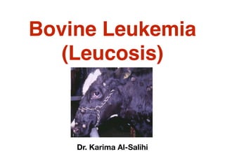
2. (Leucosis)
•It is a malignant state of leucopic disorders. It is
characterized by leucoproliferative changes in bone
marrow along with appearance of abnormal premature
leukocytes in peripheral circulation. It is recorded in dog,
cattle, horse, sheep, pig, and cat.
Causes
•May be:
•(1) Genetic factors.
(2) Carcinogenic factors: virus like particles.
3. There are four forms of bovine
leukocytosis
•
(1) Acute leukemia: predominant blast cells
are in the blood & bone marrow.
(2) Chronic leukemia: Partly mature cells
are in blood & bone marrow.
(3) Leukemic leukemia: Plenty of premature
leucocytes are present in blood & elevation
of totals WBCs more than 15000/C mm
(4) Aleukemic leukemia: Premature cells
are moderate to few in number in the blood.
4. Pathogenesis
(1) The agents activate the cells of the
reticuloendothelial system, causing
subclinical disease which characterized
by lymphomatosis which may last for
months or years or the life.
(2) Course depends on the site, size &
spread of growth of neoplasm
5.
6. Symptoms
(1) Enlargement of superficial lymph nodes.
(2) Enlarged thymus or thoracic lymph nodes.
(3) Exophthalmos: Unilater or bilateral bulging of eye
balls.
(4) Rectal palpation indicates enlargements of inguinal &
iliac lymph nodes & the group of lymph nodes under the
ventral surface of the lumbar pelvic parts of the spine.
(5) Anorexia, weakness, depression, thirsts, polyuria.
(6) Fall of milk yield in cattle.
(7) Jaundice, ascites & cachetic condition.
(8) Dyspnea & coughing.
(9) Abomasal ulcer & hemorrhage.
(10) Posterior paresis.
7.
8.
9. •Diagnosis
Depend on palpation of lymph nodes & blood
changes.
•Treatment
A. Nonspecific treatment
(1) Alkaling agent & folic acid antagonist
(methotrexate).
(2) Repeat blood transfusion.
Control
(1) Prohibition of sale of animal.
(2) Prevention of spread of contagious & contact
with other herds.
10.
11. Leucopenia
•It occurs in many diseases. It
reduces the resistance of the animal
to bacterial infection. The symptoms
depend on the main cause or causes.
Broad spectrum antibiotics are
useful to prevent bacterial invasion.
12.
13. Edema
•It is the excessive accumulation of
body fluid in the tissue space caused by
disturbance in the mechanism of fluid
exchange between capillaries, the
tissue spaces and the lymphatic
vessels. It is accompanied by
endocrine, circulatory, hepatic & renal
changes. It may be local or general.
14.
15. Causes
•
[1] Increased hydrostatic pressure
(1) Hepatic fibrosis as the fibrosis of hepatic cells obstruct the
portal circulation (portal hypertension) which increase the hydrostatic
pressure and help in accumulation of fluid in the tissue space &
peritoneum cavity causing ascites (local edema).
(2) CHF in which venous engorgement & blood stasis occur
resulting in increasing of hydrostatic pressure inside blood capillary
& flow of fluid transudate in the interstitial tissue & body cavities
causing general edema. In traumatic pericarditis edema of the
brisket are more common.
(3) Compression of mammary veins by a large fetus on the
venous & lymphatic drainage results in hypo-proteinemia and a fall
in PCOP. This physiological or mammary edema occurs in udder,
under the belly, vulva & hind legs in late pregnancy or early
parturition. This edema may resolve itself within few days or treated
with diuretic & or protein supplement
16.
17.
18. [2] Decreased plasma osmotic pressure
Hypoproteinemia
(1) Renal diseases causing continuous loss of protein in urine (albumin
urine), occurs in anasarca in the anterior part of the body (head, eye lids,
neck).
(2) Parasites as Fasciola sp & Haemonchus sp in ruminants, Strongylus
in horses, Hook worm in dogs causing loss of osmotic due to protein loss.
(3) Malnutrition due to:
1) Defect in digestion, absorption, metabolism, utilization of protein &
plasma protein.
2) Decrease protein level in diet.
3) Impairment of liver function.
4) Vitamin A deficiency causes edema of legs especially in calves.
5) Copper deficiency in sheep.
(4) Liver damage in heavy parasitic infestation or malnutrition or bacterial,
viral infection or toxicity. They causing failure of protein synthesis.
•
19.
20. [3] Obstruction of blood or lymph or portal circulation due
to tumor, fibrosis, surgical, congenital obstruction in calf,
ulcerative lymphangitis in horse or parasites as filaria.
[4] Allergic condition: In which allerge & histamin like
substance are released causing local liberation of
vasodilators increasing capillary permeability, dilatation,
vascular damage of small vessels and hydrostatic
pressure increasing fluid & protein passage to interstitial
space than that reabsorbed by lymphatic fluid causing
angioedema, urticaria, wheels or purpura hemorrhagica.
[5] Toxines in the course of some infectious diseases as:
Anthrax, Black leg, Malignant edema, Pasteurlosis,
Filariasis, Edematous skin disease as well as equine
infectious anemia, viral arteritis, infectious
rhinopneumonia.
21. Etiological classification of edem
(1) Physiological or mammary edema:
(2) Cardiac edema: It occurs in CHF
(3) Renal edema
(4) Hepatic edema.
(5) Pulmonary edema: due to disturbance of circulatory &
plumonary circulation, nervous system, together with
physiochemical factors regulating fluid exchange in
tissues.
(6) Obstructive edema: due to obstruction of blood or
lymph or portal circulation.
(7) Allergic edema.
(8) Nutritional edema.
22. Pathogenesis
[1] In normal state:
(1) The absorbed water reaches blood to enter
intravascular space (vascular water contains more
protein), interstitial space & intracellular.
(2) There are a constant flow (to & fro) between
vascular & interstitial water which occur between
capillary arterial end (due to higher hydrostatic
pressure & lower osmotic pressure) & capillary
venous end (due to lower hydrostatic pressure &
higher osmotic pressure) & carries nutrient &
metabolites to body tissue.
(3) At capillary venous end the reverse occurs.
23. [2] In diseased condition
When the hydrostatic pressure increased & osmotic
pressure decreased leads to:
(1) An excessive fluid tends to pass into tissue space
at the capillary arterial end as the hydrostatic
pressure of the blood is sufficient to overcome its
osmotic pressure.
(2) An excessive fluid tends to pass into tissue space
(instead of returning to the vascular system) at the
capillary venous end as the position is reversed.
(3) Failure of fluid to return to the capillaries resulting
in accumulation of fluid into tissue space or escape
into serous cavity forming edema.
24. Clinical symptoms (Symptomatical
classification of edema)
(1) Anasarcas: SC formation of transudate in abdominal floor,
sternum, brisket, intermandibular space,pharyngeai and perineal
region. This edema is soft, painless and pit on pressure.
(2) Ascites: Transudate formation in peritoneal cavity causing
enlarged abdomen with pear shape. Percussion of the fluid, thrill
is seen and can be detected on the other side and at top line of
fluid.
(3) Hydropsy: Fluid formation in uterus.
(4) Cerebral edema is manifested by nervous symptoms.
(5) Hydropericardium: fluid in pericardium causing restriction of
cardiac movements and muffled heart sounds occur.
(6) Pulmonary edema: It is accompanied by respiratory
disorders, moist rales and frothy discharge from the nose.
25. Common types of clinical edema
(examples of associated conditions):
(1) Intermandibular edema (parasitic).
(2) Thoraco- abdominal (parasitic/ heart).
(3) Supra-orbital fossa (south AF.H.S./ renal)
(4) Pharyngeal region (pharyngitis/hemorrhagic septicemia).
(5) Perineal area (urticaria).
(6) Buttock region (black leg).
(7) Udder region (physiological/mastitis).
(8) Limbs, whole or with demarcation (heart/renal).
(9) Brisk region (heart).
(10) Dewlap (pericarditis).
(11) Head region (purpura hemorrhagica, swelled head in
rams & blue tongue).
(12) Generalized (systemic).
26. Edema may be:
•
[1] Non inflammatory (cold) edema:
•
(1) May be local or general edema.
(2) Cold and painless swelling contains transudate (serous
fluid).
(3) Causes: Increase in hydrostatic pressure & plasma
colloid osmotic pressure (PCOP).
27. [2] Inflammatory (hot) edema:
•
(1) Local edema.
(2) Hot (local or general fever), red (in unpigmented skin),
firm & painful swelling contains exudate.
(3) Caused by:
1) Increase capillary permeability which aggravated by
blockage lymphatic & capillaries with fibrin clots e.g. boils,
cellulitis, trauma, infection, chemical irritants.
2) Damage of blood vessels due to virus, bacteria,
septicemia, malignant edema...
3) Obstructive by swelling or tumor.
28. •Diagnosis
(1) History of:
1) Diet, its composition, mineral, vitamins and even protein.
2) Onset and duration of edema and number of diseased animal.
3) Renal, hepatic, cardiac, lymphatic, respiratory disorder or
previous diseases.
4) State of appetite.
(2) Physical examination for:
1) Local or general edema.
2) Palpation of external lymph nodes.
3) Palpation of edema: Typical edema is cold, not painful and
pitting (finger impression appears after palpation). Edema may
also be hot and painful.
4) Abnormal cardiac or pulmonary sounds.
5) Jugular, mammary or peripheral veins may be distended,
pulsated and cord like.
29. (3) Laboratory examination include:
•
1) Hepatic and renal function tests.
2) Serum electrolytes.
Differential diagnosis of different
swelling:
(1) Abscess: Pus contents by expiratory puncture.
(2) Hematoma: Blood contents.
(3) Tumor: Biopsy.
(4) Bursitis: Inflammatory swelling of bursa.
(5) Cyst: Soft, painless, doughy swelling, which pit under
finger pressure.
30. Treatment and management of
edema:
•
(1) Treat the real cause.
(2) Cardiac edema: Rest, adequate protein, salt free diet,
diuretics (Potassium citrate and acetate or lasix), digoxin,
restrict water intake or even drainage of the sac in
hydropericardium in equines. Slaughter of cattle in cases
of pericarditis and CHF.
(3) Hepatic coma: Rest, treat the causes (parasite,
infections), dextrose, calcium, liver extract, amino acids,
restrict water intake.
31. NB: Gradually aspirate 2/3 of fluid
in ascites and hydrothorax to
avoid acute dilatation of
splanchenic vessels, which lead
to peripheral circulatory failure.
(4) Renal edema: Rest, sodium
free diet, amino acids,
corticosteroid.
32. (5) Nutritional edema and
hypoproteinemia: whole blood, plasma
expander, amino acids, iron, copper,
cobalt, vitamins B12, A, diet rich in
protein, mineral & vitamins.
(6) Mammary edema: Rest, diuretic,
protein diets.
(7) Allergic edema: Antihistaminic or anti-
inflammatory (corticosteroid)
(8) Obstructive edema: Symptomatic
treatment.