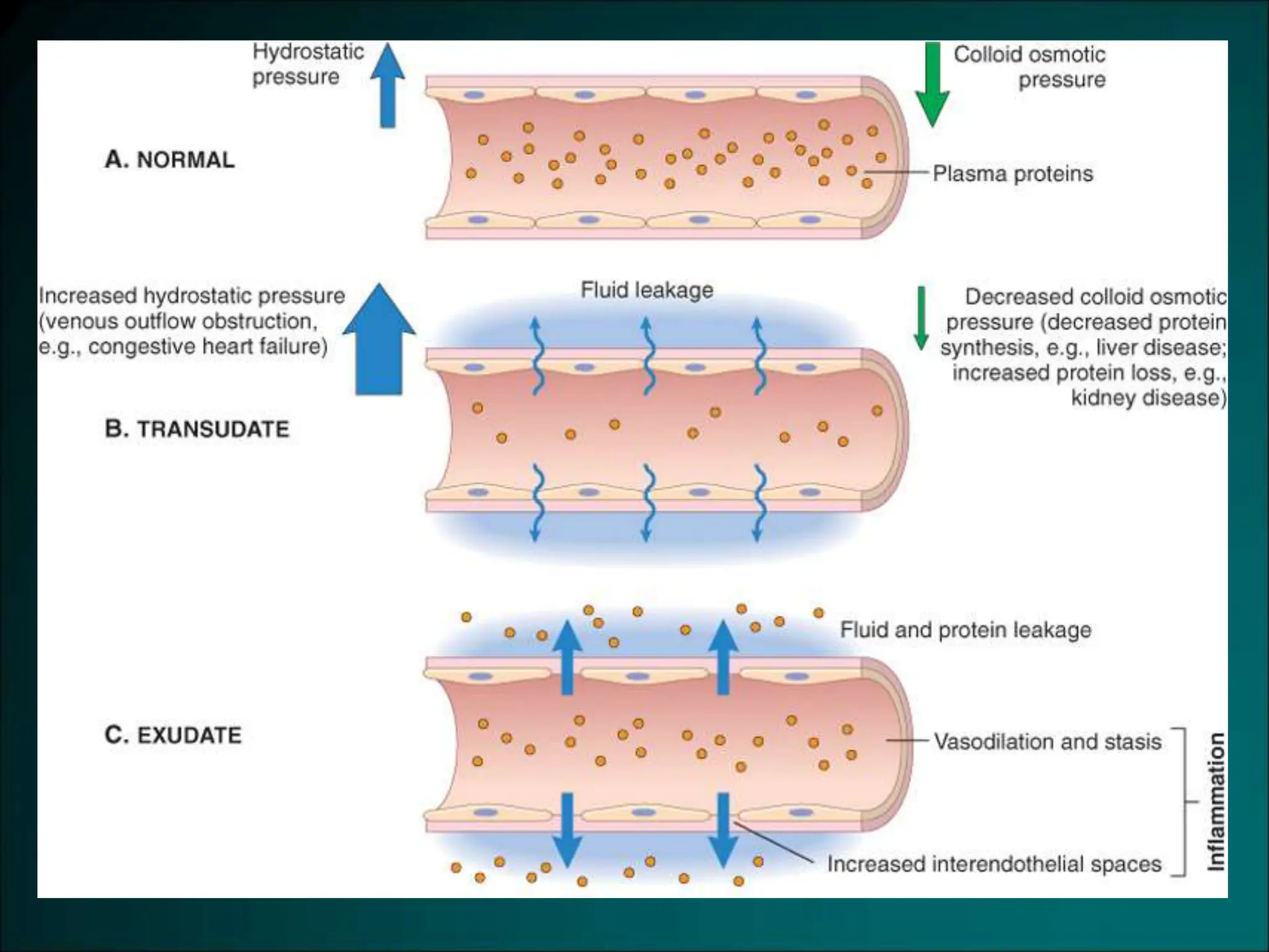
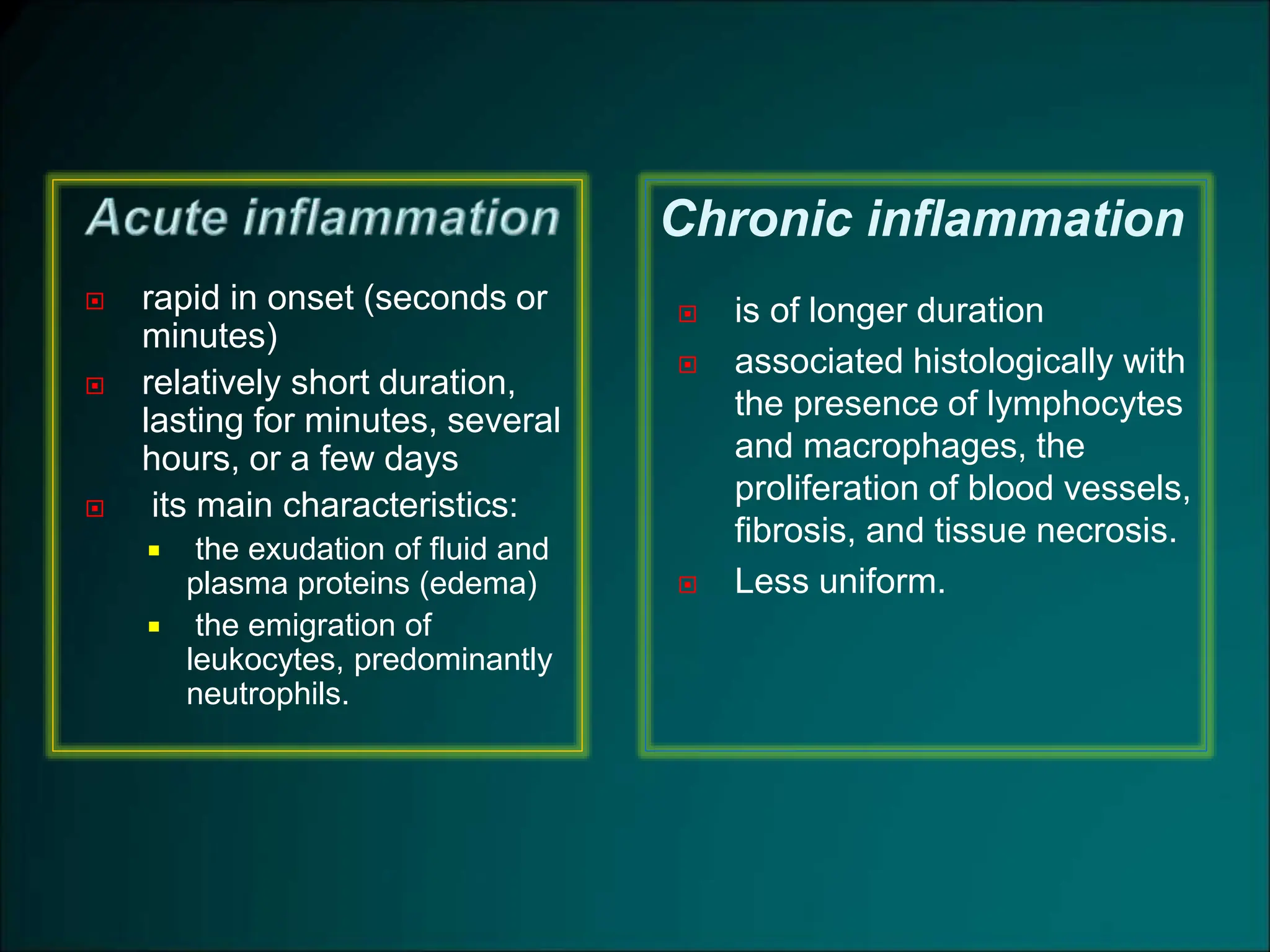
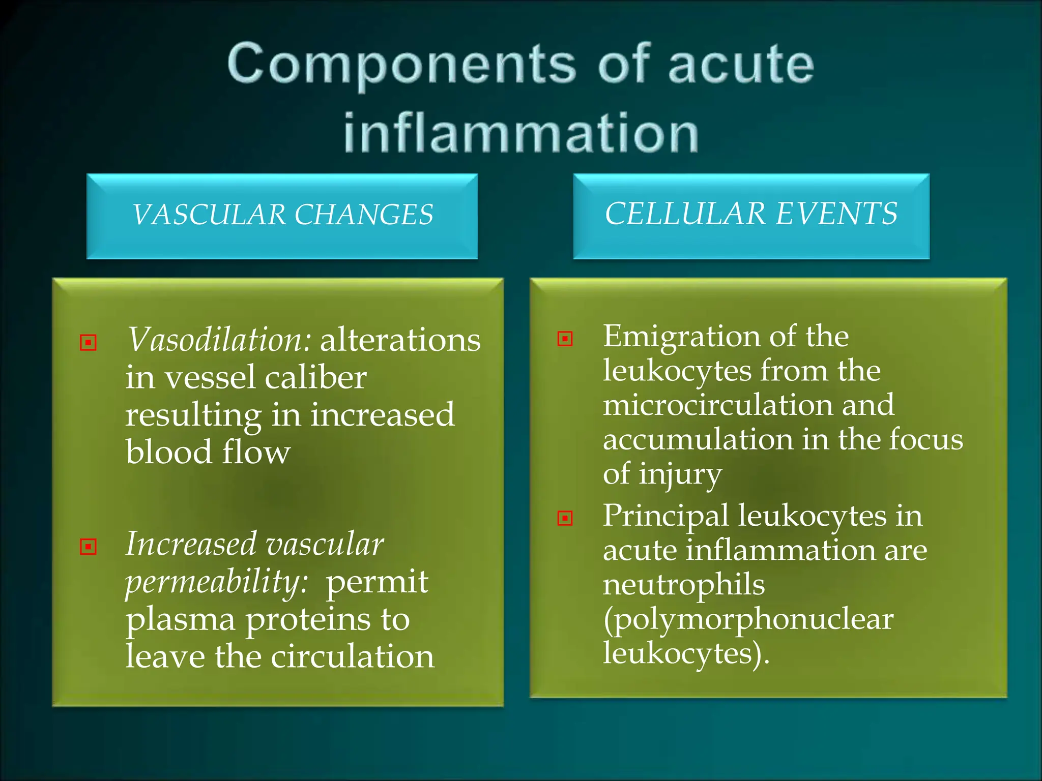
1. Define inflammation, its causes and clinical
appearance.
2. Describe the sequence of vascular changes in acute
inflammation (vasodilation, increased permeability) and
their purpose.
3. Know the mechanisms of increased vascular
permeability.
4. Define the terms edema, transudate, and exudate.
1. Define inflammation, its causes
and clinical appearance.
2. Describe the sequence of vascular changes in acute
inflammation (vasodilation, increased permeability) and
their purpose.
3. Know the mechanisms of increased vascular
permeability.
4. Define the terms edema, transudate, and exudate.
 Inflammation is a protective response intended to
eliminate the initial cause of cell injury as well as
the necrotic cells and tissues resulting from the
original insult
 The reaction of vascularized living tissue to local
injury.
 Inflammation serves to destroy, dilute or isolate
the injurious agent (microbes, toxins) and
eliminate the necrotic cells and tissues.
 Inflammation is part of a broader protective
response (innate immunity )
 It starts a series of events which leads as far as
possible to the healing and reconstitution of
the damaged tissue.
 During repair, the injured tissue is replaced by :
u Regeneration of native parenchyma cells
u Filling of the defect by fibroblastic tissue or both
 Inflammation and repair are protective response
They may induce harm
e.g. anaphylactic reaction
rheumatoid arthritis
atherosclerosis
pericarditis
 The components of the inflammatory reaction that destroy and
eliminate microbes and dead tissues are capable of also
injuring normal tissues.
 This may accompany
entirely normal, beneficial inflammatory reactions, (e.g., when
the infection is severe),
prolonged (e.g., when the eliciting agent resists eradication)
inappropriate (e.g., when it is directed against self-antigens in
autoimmune diseases)
against usually harmless environmental antigens (allergic
disorders)
 Inflammation is terminated when the offending agent is
eliminated and the secreted mediators are broken
down or dissipated.
 There are active anti-inflammatory mechanisms that
serve to control the response and prevent it from
causing excessive damage to the host.
 blood leukocytes
 plasma proteins
 cells of vascular walls
 cells of the surrounding connective tissue
 extracellular matrix (ECM) of the surrounding
connective tissue
The
connective
tissue cells
The circulating cells:
The
extracellular
matrix
Cells and molecules that play important roles in inflammation
Tissues and cells involved in inflammatory response :
The fluid and proteins of plasma, circulating cells, blood vessels and
connective tissue
•The circulating cells: neutrophils, monocytes, eosinophils,
lymphocytes, basophils, and platelets.
• The connective tissue cells are the mast cells, the connective
tissue fibroblasts, resident macrophages and lymphocytes.
•The extracellular matrix, consists of the structural fibrous proteins
(collagen, elastin), adhesive glycoproteins (fibronectin, laminin,
nonfibrillar collagen, tenascin, and others), and proteoglycans
 Inflammationis dividedinto:
u Acute inflammation.
u Chronic inflammation.
 rapid in onset (seconds or
minutes)
 relatively short duration,
lasting for minutes, several
hours, or a few days
 its main characteristics:
 the exudation of fluid and
plasma proteins (edema)
 the emigration of
leukocytes, predominantly
neutrophils.
 is of longer duration
 associated histologically with
the presence of lymphocytes
and macrophages, the
proliferation of blood vessels,
fibrosis, and tissue necrosis.
 Less uniform.
Chronic inflammation
 Phagocytes and other host cells
 Leukocyte
 Endothelium
 Mast cell
 Plasma proteins
 Some of mediators act on small blood vessels
 Promote the efflux of plasma
 Recruitment of circulating leukocytes to the
site where the offending agent is located
 Localclinicalsigns ofacute inflammation:
 Heat
 Redness
 Swelling
 Pain
 Lossoffunction
Redness
Swelling
Swelling
(1) Recognition of the injurious agent
(2) Recruitment of leukocytes
(3) Removal of the agent
(4) Regulation (control) of the response
(5) Resolution
1. Define inflammation, its causes and clinical
appearance.
2. Describe the sequence of vascular
changes in acute inflammation
(vasodilation, increased permeability) and
their purpose.
3. Know the mechanisms of increased vascular
permeability.
4. Define the terms edema, transudate, and exudate.
VASCULAR CHANGES CELLULAR EVENTS
 Vasodilation: alterations
in vessel caliber
resulting in increased
blood flow
 Increased vascular
permeability: permit
plasma proteins to
leave the circulation
 Emigration of the
leukocytes from the
microcirculation and
accumulation in the focus
of injury
 Principal leukocytes in
acute inflammation are
neutrophils
(polymorphonuclear
leukocytes).
 Triggered by a variety of stimuli:
 Infections (bacterial, viral, parasitic) and microbial
toxins
 Trauma (blunt and penetrating)
 Physical and chemical agents (thermal injury,
e.g., burns or frostbite; irradiation; some
environmental chemicals)
 Tissue necrosis (from any cause)
 Foreign bodies (splinters, dirt, sutures)
 Immune reactions (also called hypersensitivity
reactions)
 Acute inflammation is a rapid response to an injurious
agent that serves to deliver mediators of host defense-
leukocytes and plasma proteins-to the site of injury.
 Acute inflammation has three main events:
(1) Hemodynamic changes
(alterations in vascular caliber that lead to an increase in blood flow)
(2) Increased vascular permeability
(structural changes in the microvasculature that permit plasma proteins and
leukocytes to leave the circulation)
(3) Emigration of the leukocytes from the microcirculation
(their accumulation in the focus of injury, and their activation to eliminate the
offending agent)
(1) Hemodynamic changes
(alterations in vascular caliber that lead
to an increase in blood flow)
(2) Increased vascular permeability
u 1. Transient vasoconstriction of arterioles
- It disappears within 3-5 seconds in mild injuries
- It may last several minutes in more severe injury
(burn)
u 2.Vasodilatation:
It involves the arterioles and then results in opening of
new microvasculature beds in the area thus leading to
increasing blood flow (early hemodynamic change and
the cause of inflammation heat and redness).
u Increased blood volume lead to increased local
hydrostatic pressure leading to transudation of
protein -poor fluid into the extravascular space.
u 3.Slowing of the circulation due to increased
permeability of the microvasculature, this leads to
outpouring of protein-rich fluid in the extravascular
tissues.
This results in concentration of the red cells in small
vessels and increased viscosity of the blood leadind to
stasis
(stasis: slow circulation due to dilated small vessels
packed with red cells).
u 4.persistence of stasis
leads to peripheral orientation of leukocytes (mainly
neutrophils) along the vascular endothelium [leukocytic
margination], then they migrate through the vascular
wall into the interstitial tissue [emigration].
A 72-year-old man with severe emphysema has had
worsening right ventricular failure for the past 5
years. He has had fever and increasing dyspnea for
the past 4 days. A chest radiograph shows an
accumulation of fluid in the pleural spaces. Fluid
obtained by thoracentesis has a specific gravity of
1.030 and contains degenerating neutrophils.
The most likely cause of this fluid accumulation is an
increase in which of the following mechanisms?
(A) Colloid osmotic pressure
(B) Lymphatic pressure
(C) Vascular permeability
(D) Renal retention of sodium and water
(E) Leukocytic diapedesis
u A hallmark of acute inflammation
(escape of a protein-rich fluid).
u It affects small & medium size venules,
through gaps between endothelial cells
Slide 3.3
Marked outflow of fluid and its
accumulation in the interstitial
tissue:
• The loss of protein from the
plasma reduces the intravascular
osmotic pressure and increases the
osmotic pressure of the interstitial
fluid
• the increased hydrostatic
pressure
The net increase of extravascular
fluid results in edema
1. Define inflammation, its causes and clinical
appearance.
2. Describe the sequence of vascular changes in acute
inflammation (vasodilation, increased permeability) and
their purpose.
3. Know the mechanisms of increased vascular
permeability.
4. Define the terms edema, transudate, and exudate.
Edema
• denotes an excess of fluid in the
interstitial or serous cavities
• it can be either an exudate or a
transudate.
Exudate
 An inflammatory extravascular fluid that has a high protein
concentration, cellular debris, and a specific gravity above 1.020.
 It implies significant alteration in the normal permeability of small
blood vessels in the area of injury.
Transudate
 A fluid with low protein content (most of which is albumin) and a
specific gravity of less than 1.012.
 It is essentially an ultrafiltrate of blood plasma that results from
osmotic or hydrostatic imbalance
 No an increase in vascular permeability.
Pus (a purulent exudate)
 is an inflammatory exudate rich in leukocytes (mostly neutrophils),
the debris of dead cells and microbes.
1. Define inflammation, its causes and clinical
appearance.
2. Describe the sequence of vascular changes in acute
inflammation (vasodilation, increased permeability) and
their purpose.
3. Know the mechanisms of increased
vascular permeability.
4. Define the terms edema, transudate, and exudate.
(1) an immediate transient response
lasting for 30 minutes or less, mediated mainly by
the actions of histamine and leukotrienes on
endothelium
(2) a delayed response
starting at about 2 hours and lasting for about 8
hours, mediated by kinins, complement products,
and other factors
(3) a prolonged response
that is most noticeable after direct endothelial
injury, for example, after burns.
Phases of Increased Vascular Permeability
A 72-year-old man with severe emphysema has
had worsening right ventricular failure for the
past 5 years. He has had fever and increasing
dyspnea for the past 4 days. A chest
radiograph shows an accumulation of fluid in
the pleural spaces. Fluid obtained by
thoracentesis has a specific gravity of 1.030 and
contains degenerating neutrophils. The most
likely cause of this fluid accumulation is an
increase in which of the following
mechanisms?
(A) Colloid osmotic pressure
(B) Lymphatic pressure
(C) Vascular permeability
(D) Renal retention of sodium and water
(E) Leukocytic diapedesis
Normal fluid exchange and microvascular permeability are critically dependent
on an intact endothelium. How then does the endothelium become leaky in
inflammation?
mechanisms proposed are:
1. Formation of endothelial gaps in venules
- This is the most common mechanism of vascular leakage
- is elicited by histamine, bradykinin, leukotrienes, the neuropeptide
substance P, and many other classes of chemical mediators.
- It occurs rapidly after exposure to the mediator and is usually reversible
and short-lived (15 to 30 minutes)
- it is known as immediate transient response.
- affects venules 20 to 60 μm in diameter, leaving capillaries and
arterioles unaffected
Phases of Increased Vascular Permeability
2. Immediate sustained reactions
 Direct endothelial injury (necrosis and detachment)
 direct damage to the endothelium by the injurious
stimulus, as, severe burns or lytic bacterial
infections.
 Neutrophils may also injure the endothelial cells.
 In most instances, leakage starts immediately after
injury and is sustained at a high level for several
hours until the damaged vessels are thrombosed or
repaired.
 All levels of the microcirculation are affected:
venules, capillaries, and arterioles.
3. Delayed prolonged leakage.
u relatively common type of increased permeability that begins
after a delay of 2 to 12 hours, lasts for several hours or even
days
u involves venules as well as capillaries.
u Such leakage is caused by mild to moderate thermal injury, x-
radiation or ultraviolet radiation, sunburn and certain bacterial
toxins.
u The mechanism of such leakage is unclear. It may result from
the direct effect of the injurious agent, leading to delayed
endothelial cell damage (perhaps by apoptosis), or the effect of
cytokines causing endothelial retraction.
Leukocyte-mediated endothelial injury.
u Leukocytes adhere to endothelium relatively early in
inflammation.
u such leukocytes may be activated in the process, releasing
toxic oxygen species and proteolytic enzymes, which then
cause endothelial injury or detachment, resulting in
increased permeability.
u In acute inflammation, this form of injury is largely
restricted to vascular sites, such as venules and pulmonary
and glomerular capillaries.
Increased transcytosis across the endothelial cytoplasm
 Transcytosis occurs across channels consisting
of clusters of interconnected
 This mechanism of increased permeability is
induced by histamine and most chemical
mediators
Other mechanisms:
 Leakage from new blood vessels.
 Endothelial cell contraction 15-30 min
 Endothelial injury
 immediate sustained response 6-24 hours
 delayed prolonged leakage 12 hours- days
 Leukocyte-mediated endothelial injury
 Transcytosis (occurs via channels
formed by fusion of intracellular
vesicles)
 Leakage from new blood vessels
Mechanisms lead to increased vascular permeability
A 53-year-old woman has had a high fever and
cough productive of yellowish sputum for the past
2 days. Her vital signs include temperature of
37.8°C, pulse 83/mm, respirations 17/mm, and
blood pressure 100/60 mm Hg. A chest radiograph
shows bilateral patchy pulmonary infiltrates. Which
of the following inflammatory cell types is most
likely to be seen in greatly increased numbers in a
sputum specimen?
(A) Macrophages
(B) Neutrophils
(C) Mast cells
(D) Small lymphocytes
(E) Langhans giant cells
 Vascular Reactions in Acute Inflammation
 Vasodilation is induced by chemical mediators such as
histamine , and is the cause of erythema and stasis of
blood flow.
 Increased vascular permeability is induced by histamine,
kinins and other mediators that produce:
 gaps between endothelial cells,
 by direct or leukocyte-induced endothelial injury
 by increased passage of fluids through the endothelium
 increased vascular permeability allows plasma proteins and
leukocytes to enter sites of infection or tissue damage;
 fluid leak through blood vessels results in edema

07-inflammation,wound-repair, regeneration.ppt

  • 2.
    1. Define inflammation,its causes and clinical appearance. 2. Describe the sequence of vascular changes in acute inflammation (vasodilation, increased permeability) and their purpose. 3. Know the mechanisms of increased vascular permeability. 4. Define the terms edema, transudate, and exudate.
  • 3.
    1. Define inflammation,its causes and clinical appearance. 2. Describe the sequence of vascular changes in acute inflammation (vasodilation, increased permeability) and their purpose. 3. Know the mechanisms of increased vascular permeability. 4. Define the terms edema, transudate, and exudate.
  • 5.
     Inflammation isa protective response intended to eliminate the initial cause of cell injury as well as the necrotic cells and tissues resulting from the original insult  The reaction of vascularized living tissue to local injury.
  • 7.
     Inflammation servesto destroy, dilute or isolate the injurious agent (microbes, toxins) and eliminate the necrotic cells and tissues.  Inflammation is part of a broader protective response (innate immunity )  It starts a series of events which leads as far as possible to the healing and reconstitution of the damaged tissue.
  • 8.
     During repair,the injured tissue is replaced by : u Regeneration of native parenchyma cells u Filling of the defect by fibroblastic tissue or both  Inflammation and repair are protective response
  • 9.
    They may induceharm e.g. anaphylactic reaction rheumatoid arthritis atherosclerosis pericarditis
  • 10.
     The componentsof the inflammatory reaction that destroy and eliminate microbes and dead tissues are capable of also injuring normal tissues.  This may accompany entirely normal, beneficial inflammatory reactions, (e.g., when the infection is severe), prolonged (e.g., when the eliciting agent resists eradication) inappropriate (e.g., when it is directed against self-antigens in autoimmune diseases) against usually harmless environmental antigens (allergic disorders)
  • 11.
     Inflammation isterminated when the offending agent is eliminated and the secreted mediators are broken down or dissipated.  There are active anti-inflammatory mechanisms that serve to control the response and prevent it from causing excessive damage to the host.
  • 12.
     blood leukocytes plasma proteins  cells of vascular walls  cells of the surrounding connective tissue  extracellular matrix (ECM) of the surrounding connective tissue
  • 13.
    The connective tissue cells The circulatingcells: The extracellular matrix
  • 14.
    Cells and moleculesthat play important roles in inflammation
  • 15.
    Tissues and cellsinvolved in inflammatory response : The fluid and proteins of plasma, circulating cells, blood vessels and connective tissue •The circulating cells: neutrophils, monocytes, eosinophils, lymphocytes, basophils, and platelets. • The connective tissue cells are the mast cells, the connective tissue fibroblasts, resident macrophages and lymphocytes. •The extracellular matrix, consists of the structural fibrous proteins (collagen, elastin), adhesive glycoproteins (fibronectin, laminin, nonfibrillar collagen, tenascin, and others), and proteoglycans
  • 17.
     Inflammationis dividedinto: uAcute inflammation. u Chronic inflammation.
  • 19.
     rapid inonset (seconds or minutes)  relatively short duration, lasting for minutes, several hours, or a few days  its main characteristics:  the exudation of fluid and plasma proteins (edema)  the emigration of leukocytes, predominantly neutrophils.  is of longer duration  associated histologically with the presence of lymphocytes and macrophages, the proliferation of blood vessels, fibrosis, and tissue necrosis.  Less uniform. Chronic inflammation
  • 21.
     Phagocytes andother host cells  Leukocyte  Endothelium  Mast cell  Plasma proteins
  • 22.
     Some ofmediators act on small blood vessels  Promote the efflux of plasma  Recruitment of circulating leukocytes to the site where the offending agent is located
  • 24.
     Localclinicalsigns ofacuteinflammation:  Heat  Redness  Swelling  Pain  Lossoffunction
  • 25.
  • 26.
  • 27.
  • 29.
    (1) Recognition ofthe injurious agent (2) Recruitment of leukocytes (3) Removal of the agent (4) Regulation (control) of the response (5) Resolution
  • 30.
    1. Define inflammation,its causes and clinical appearance. 2. Describe the sequence of vascular changes in acute inflammation (vasodilation, increased permeability) and their purpose. 3. Know the mechanisms of increased vascular permeability. 4. Define the terms edema, transudate, and exudate.
  • 32.
    VASCULAR CHANGES CELLULAREVENTS  Vasodilation: alterations in vessel caliber resulting in increased blood flow  Increased vascular permeability: permit plasma proteins to leave the circulation  Emigration of the leukocytes from the microcirculation and accumulation in the focus of injury  Principal leukocytes in acute inflammation are neutrophils (polymorphonuclear leukocytes).
  • 34.
     Triggered bya variety of stimuli:  Infections (bacterial, viral, parasitic) and microbial toxins  Trauma (blunt and penetrating)  Physical and chemical agents (thermal injury, e.g., burns or frostbite; irradiation; some environmental chemicals)  Tissue necrosis (from any cause)  Foreign bodies (splinters, dirt, sutures)  Immune reactions (also called hypersensitivity reactions)
  • 35.
     Acute inflammationis a rapid response to an injurious agent that serves to deliver mediators of host defense- leukocytes and plasma proteins-to the site of injury.  Acute inflammation has three main events: (1) Hemodynamic changes (alterations in vascular caliber that lead to an increase in blood flow) (2) Increased vascular permeability (structural changes in the microvasculature that permit plasma proteins and leukocytes to leave the circulation) (3) Emigration of the leukocytes from the microcirculation (their accumulation in the focus of injury, and their activation to eliminate the offending agent)
  • 37.
    (1) Hemodynamic changes (alterationsin vascular caliber that lead to an increase in blood flow) (2) Increased vascular permeability
  • 39.
    u 1. Transientvasoconstriction of arterioles - It disappears within 3-5 seconds in mild injuries - It may last several minutes in more severe injury (burn)
  • 40.
    u 2.Vasodilatation: It involvesthe arterioles and then results in opening of new microvasculature beds in the area thus leading to increasing blood flow (early hemodynamic change and the cause of inflammation heat and redness). u Increased blood volume lead to increased local hydrostatic pressure leading to transudation of protein -poor fluid into the extravascular space.
  • 41.
    u 3.Slowing ofthe circulation due to increased permeability of the microvasculature, this leads to outpouring of protein-rich fluid in the extravascular tissues. This results in concentration of the red cells in small vessels and increased viscosity of the blood leadind to stasis (stasis: slow circulation due to dilated small vessels packed with red cells).
  • 42.
    u 4.persistence ofstasis leads to peripheral orientation of leukocytes (mainly neutrophils) along the vascular endothelium [leukocytic margination], then they migrate through the vascular wall into the interstitial tissue [emigration].
  • 43.
    A 72-year-old manwith severe emphysema has had worsening right ventricular failure for the past 5 years. He has had fever and increasing dyspnea for the past 4 days. A chest radiograph shows an accumulation of fluid in the pleural spaces. Fluid obtained by thoracentesis has a specific gravity of 1.030 and contains degenerating neutrophils. The most likely cause of this fluid accumulation is an increase in which of the following mechanisms? (A) Colloid osmotic pressure (B) Lymphatic pressure (C) Vascular permeability (D) Renal retention of sodium and water (E) Leukocytic diapedesis
  • 44.
    u A hallmarkof acute inflammation (escape of a protein-rich fluid). u It affects small & medium size venules, through gaps between endothelial cells
  • 45.
    Slide 3.3 Marked outflowof fluid and its accumulation in the interstitial tissue: • The loss of protein from the plasma reduces the intravascular osmotic pressure and increases the osmotic pressure of the interstitial fluid • the increased hydrostatic pressure The net increase of extravascular fluid results in edema
  • 46.
    1. Define inflammation,its causes and clinical appearance. 2. Describe the sequence of vascular changes in acute inflammation (vasodilation, increased permeability) and their purpose. 3. Know the mechanisms of increased vascular permeability. 4. Define the terms edema, transudate, and exudate.
  • 47.
    Edema • denotes anexcess of fluid in the interstitial or serous cavities • it can be either an exudate or a transudate.
  • 49.
    Exudate  An inflammatoryextravascular fluid that has a high protein concentration, cellular debris, and a specific gravity above 1.020.  It implies significant alteration in the normal permeability of small blood vessels in the area of injury. Transudate  A fluid with low protein content (most of which is albumin) and a specific gravity of less than 1.012.  It is essentially an ultrafiltrate of blood plasma that results from osmotic or hydrostatic imbalance  No an increase in vascular permeability. Pus (a purulent exudate)  is an inflammatory exudate rich in leukocytes (mostly neutrophils), the debris of dead cells and microbes.
  • 52.
    1. Define inflammation,its causes and clinical appearance. 2. Describe the sequence of vascular changes in acute inflammation (vasodilation, increased permeability) and their purpose. 3. Know the mechanisms of increased vascular permeability. 4. Define the terms edema, transudate, and exudate.
  • 53.
    (1) an immediatetransient response lasting for 30 minutes or less, mediated mainly by the actions of histamine and leukotrienes on endothelium (2) a delayed response starting at about 2 hours and lasting for about 8 hours, mediated by kinins, complement products, and other factors (3) a prolonged response that is most noticeable after direct endothelial injury, for example, after burns. Phases of Increased Vascular Permeability
  • 54.
    A 72-year-old manwith severe emphysema has had worsening right ventricular failure for the past 5 years. He has had fever and increasing dyspnea for the past 4 days. A chest radiograph shows an accumulation of fluid in the pleural spaces. Fluid obtained by thoracentesis has a specific gravity of 1.030 and contains degenerating neutrophils. The most likely cause of this fluid accumulation is an increase in which of the following mechanisms? (A) Colloid osmotic pressure (B) Lymphatic pressure (C) Vascular permeability (D) Renal retention of sodium and water (E) Leukocytic diapedesis
  • 56.
    Normal fluid exchangeand microvascular permeability are critically dependent on an intact endothelium. How then does the endothelium become leaky in inflammation? mechanisms proposed are: 1. Formation of endothelial gaps in venules - This is the most common mechanism of vascular leakage - is elicited by histamine, bradykinin, leukotrienes, the neuropeptide substance P, and many other classes of chemical mediators. - It occurs rapidly after exposure to the mediator and is usually reversible and short-lived (15 to 30 minutes) - it is known as immediate transient response. - affects venules 20 to 60 μm in diameter, leaving capillaries and arterioles unaffected Phases of Increased Vascular Permeability
  • 58.
    2. Immediate sustainedreactions  Direct endothelial injury (necrosis and detachment)  direct damage to the endothelium by the injurious stimulus, as, severe burns or lytic bacterial infections.  Neutrophils may also injure the endothelial cells.  In most instances, leakage starts immediately after injury and is sustained at a high level for several hours until the damaged vessels are thrombosed or repaired.  All levels of the microcirculation are affected: venules, capillaries, and arterioles.
  • 60.
    3. Delayed prolongedleakage. u relatively common type of increased permeability that begins after a delay of 2 to 12 hours, lasts for several hours or even days u involves venules as well as capillaries. u Such leakage is caused by mild to moderate thermal injury, x- radiation or ultraviolet radiation, sunburn and certain bacterial toxins. u The mechanism of such leakage is unclear. It may result from the direct effect of the injurious agent, leading to delayed endothelial cell damage (perhaps by apoptosis), or the effect of cytokines causing endothelial retraction.
  • 61.
    Leukocyte-mediated endothelial injury. uLeukocytes adhere to endothelium relatively early in inflammation. u such leukocytes may be activated in the process, releasing toxic oxygen species and proteolytic enzymes, which then cause endothelial injury or detachment, resulting in increased permeability. u In acute inflammation, this form of injury is largely restricted to vascular sites, such as venules and pulmonary and glomerular capillaries.
  • 63.
    Increased transcytosis acrossthe endothelial cytoplasm  Transcytosis occurs across channels consisting of clusters of interconnected  This mechanism of increased permeability is induced by histamine and most chemical mediators
  • 65.
    Other mechanisms:  Leakagefrom new blood vessels.
  • 66.
     Endothelial cellcontraction 15-30 min  Endothelial injury  immediate sustained response 6-24 hours  delayed prolonged leakage 12 hours- days  Leukocyte-mediated endothelial injury  Transcytosis (occurs via channels formed by fusion of intracellular vesicles)  Leakage from new blood vessels Mechanisms lead to increased vascular permeability
  • 68.
    A 53-year-old womanhas had a high fever and cough productive of yellowish sputum for the past 2 days. Her vital signs include temperature of 37.8°C, pulse 83/mm, respirations 17/mm, and blood pressure 100/60 mm Hg. A chest radiograph shows bilateral patchy pulmonary infiltrates. Which of the following inflammatory cell types is most likely to be seen in greatly increased numbers in a sputum specimen? (A) Macrophages (B) Neutrophils (C) Mast cells (D) Small lymphocytes (E) Langhans giant cells
  • 69.
     Vascular Reactionsin Acute Inflammation  Vasodilation is induced by chemical mediators such as histamine , and is the cause of erythema and stasis of blood flow.  Increased vascular permeability is induced by histamine, kinins and other mediators that produce:  gaps between endothelial cells,  by direct or leukocyte-induced endothelial injury  by increased passage of fluids through the endothelium  increased vascular permeability allows plasma proteins and leukocytes to enter sites of infection or tissue damage;  fluid leak through blood vessels results in edema