
Recommended
Recommended
More Related Content
What's hot
What's hot (20)
Similar to Plant phenotyping systems
Similar to Plant phenotyping systems (20)
More from Michal Slota
More from Michal Slota (19)
Recently uploaded
Recently uploaded (20)
Plant phenotyping systems
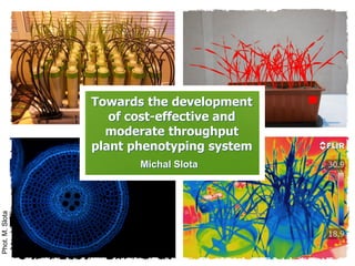
- 1. Towards the development of cost-effective and moderate throughput plant phenotyping system Michal Slota Phot.M.Slota
- 2. Presentation outline Slota M., 2014 Introduction Plant cultivation Image acquisition Data analysis Concept of plant phenotyping Definitions and terminology Historical background Applications Plant cultivation systems Soil-grown cultures Petri plates cultures Hydroponics & aeroponics Image acquisition techniques Digital photography, optical scanners IR, NIR, fluorescence imaging Hyperspectral, 3D imaging Data analysis process Noise reduction and filtering Image segmentation Measurements and data analysis Phenotyping system Design of the system System maintenance Preliminary results System costs and throughput Proposed phenotyping system
- 3. Slota M., 2014 BIOPHYSICSA/ BIOMECHANICSANATOMY/ HISTOLOGY PHENOTPYE PROTEOME/ METABOLOME SIGNALLING GENOTYPE [PuigandPeñarrubia2009;Tardieu2012;WikimediaCommons] Introduction
- 4. Introduction Slota M., 2014 GENOTYPING PHENOTYPING [Pennisi2009;WikimediaCommons]
- 5. Slota M., 2014 PHENOTYPIC VS. GENOTYPIC DATA ACQUISITION 27 000 genes Phenome = gene x environment 34 000 proteins Possible interactions = ? [Integr8 - A.thaliana Genome Statistics] (Furbank and Tester, 2011) Introduction
- 6. Slota M., 2014 PHENOTYPIC VS. GENOTYPIC DATA ACQUISITION High throughput techniques: ✓ next generation sequencing, ✓ microarrays, ✓ bioinformatics. ✓ automated image acqusition ✓ robotics/ automated sensoring ✓ bioinformatics/ image analysis Introduction PHENOTYPING = HIGH THROUGHPUT PLANT PHYSIOLOGY (?)
- 7. Introduction Slota M., 2014 Phenome = Gene x Environment or the expression of the genome as traits in a given environment. Plant phenomics Plant phenomics is the study of plant growth, performance and composition. (Furbank and Tester, 2011)
- 8. Slota M., 2014Introduction Plant phenotyping - the quantitative or qualitative investigation of traits at any organizational level, in a given genomic expression state and a given environment Organizational levels in plant phenotyping (Dhondt et al. 2013)
- 9. Slota M., 2014Concept of plant phenotyping a) Mendel’s garden (Augustinian Abbey, Brno), b) Mendel’s phenotyping instrument: microscope Gregor Mendel (1822-1884) Pisum sativum [www.zlgc.seu.edu.cn;www.mendel-museum.com] Time period: 1856- 1863’ (7 years) Plant material: 29 000 pea plants (Pisum sativum) Plot: 2 ha monastery garden Traits tested: 7 traits (7 different loci, each possesing 2 alleles) (color and seed smoothness, color of the cotyledons, color of the flowers, shape of the pods, color of the unripe pods, position of flowers and pods, height of the plants).(Butler, 2009)
- 10. Slota M., 2014Phenotyping facilitities HIGH THROUGHPUT HIGH RESOLUTION University of Nottingham; MicroCT (High Resolution X-ray micro-Computed Tomograph). » « National Institute for Agricultural Research (INRA) in Montpellier; High Throughput Plant Phenotyping Platform (PPHD).
- 11. Slota M., 2014Concept of plant phenotyping Breeding Agricultural production Biodiversity assessment Functional genomics Horticultural production Climate change IDENTIFICATION OF HERITABLE TRAITS FIELD ENVIRONMENT PLANT GROWTH STATUS (Walter et al., 2009)
- 12. Slota M., 2014Concept of plant phenotyping (Reynolds et al., 2020)
- 13. Slota M., 2014Concept of plant phenotyping GROWTH SYSTEMS (organic/soil, hydroponic, aeroponics etc.) DATA COLLECTING (digital camera images, high resolution scanning, microscope imaging) IMAGE ANALYSIS (noise reduction, segmentation, filtering counting/measuring, statistical analysis) (Furbank and Tester, 2011)
- 14. Slota M., 2014Growth systems IN-VITRO CULTURES SCREENING GROWTH CHAMBER/ GREENHOUSE ASSAYS FIELD EXPERIMENTS ✓ highly controlled conditions, ✓ high throuput, ✓ high capacity, ✓ easy imaging, - adapted only for small plants. ✓ controlled conditions, ✓ high throuput, ✓ adapted for medium and large plants, - high costs, - amount of data. ✓ high capacity, ✓ natural conditions, - high costs, - complex imaging, - extremely high amount of data. COMPARISON OF GROWTH SYSTEMS
- 15. Slota M., 2014Growth systems IN-VITRO CULTURES SCREENING GROWTH CHAMBER/ GREENHOUSE ASSAYS FIELD EXPERIMENTS SCREENING EFFECTIVENESS NATURAL GROWTH PARAMETERS COMPARISON OF GROWTH SYSTEMS
- 16. Slota M., 2014Conclusion EXPERIMENT REPRODUCIBILITY REQUIRES: ✓ data objectively described in a mathematical, easily digitized and searchable format (using ontologies), ✓ information on how the experiment was carried out (plant material, growth conditions), ✓ standardization of the employed phenotyping techniques. (Furbank and Tester 2011) (Poorter et al., 2012) Meta analysis of leaf area of Arabidopsis thaliana Col. plants grown with the same protocol, the same seed stock and the same soil in growth rooms of nine different laboratories. » Experiments reproducibility
- 17. Slota M., 2014Conclusion Experiments reproducibility EXPERIMENT REPRODUCIBILITY REQUIRES: ✓ data objectively described in a mathematical, easily digitized and searchable format (using ontologies), ✓ information on how the experiment was carried out (plant material, growth conditions), ✓ standardization of the employed phenotyping techniques. (Furbank and Tester 2011) (Poorter et al., 2012) Venn diagram of the genes that were differently affected by transfer from Arabidopsis thaliana plants from 20-22°C to chilling temperatures (4-8◦C). Data are from two independent experiments from Vogel et al. (2005) and Usadel et al. (2008). »
- 18. Slota M., 2014Growth systems Aeroponics: nutrient solution is sprayed on roots Petri plates in vitro techniques In vitro techniques In vitro techniques of plant cultivation are widely used for plant phenotyping as a quick and cost-effective screening methods for a large number of plant genotypes. Petri plates screening of a shoot and root phenotype is most applicable for small plants (eg. Arabidopsis) for short term experiments. On the other hand, petri plates assays lack to imitate complex natural conditions and frequently cause stress on its own. Rice seedlings, 6th day after plates transfer grown on the surface (top) within agarose gel (bottom) [Phot. M. Slota]. » A. thaliana root phenotyping on vertical agar plates. http://www.ipk-gatersleben.de/
- 19. Slota M., 2014Growth systems Dynamics of plant traits (Wattetal.2020)
- 20. Slota M., 2014Data collecting COLOUR IMAGES (5) Plant area, volume, mass, structure, phenology Senescence, relative chlorophyll content, pathogenic lesions Seed yield, agronomic traits NEAR IR IMAGING (1-4) Tissue water content Soil water content FAR IR IMAGING Canopy, leaf temperature, water use, salt tolerance HYPERSPECTRAL IMAGING (6) Carbohydrates, pigments and protein CARBON ISOTOPE RATIO Transpiration efficiency, photosynthetic pathway (TDL/MS) FTIR IMAGING SPECTROSCOPY Cellular localisation of metabolites (sugars, protein, aromatics) FLUORESCENCE IMAGING (7) Physiological state of photosynthetic machinery (Fioranietal.2012) SENSORS - Overview
- 21. Fluorescence Infrared imaging Near Infrared imaging Hyperspectral sensoring Visible light 3D reconstruction Slota M., 2014Data collecting Imaging device Sensors Visualized object [http://www.lemnatec.de/products/imaging-modules]
- 22. Slota M., 2014Data collecting (Clarketal.2020)
- 23. Slota M., 2014Phenotyping facilitities RGB imaging consists of the construction of growth profiles of plant shoot by acquiring time series using cameras sensitive to the visible light range (400-700 nm). The RGB imaging characteristics: ▪ projected shoot area is extracted following image preprocessing and segmentation either in the RGB space or in the HSV (hue, saturation, value) space, ▪ imaging setups vary widely depending on the cultivation format, ▪ for cereals, multiple view angles created by rotating the plants are generally used to reduce image occlusions. The calibration of projected shoot area (the projected area of a 3D object onto a plane) based on total leaf area and fresh and dry shoot mass measured destructively throughout the growth trajectory was first performed in Arabidopsis, barley, and tobacco leading to highly significant linear or polynomial correlations. (Fiorani and Schurr, 2013) SENSORS - Visible light RGB imaging Time-lapse images for different cereals. http://www.lemnatec.de/
- 24. Slota M., 2014Data collecting Chlorophyll fluorescence is commonly used from lab to field scales. It offers a rapid way to probe photosystem II status in vivo . Active fluorescence protocols exploiting pulse amplitude modulation of commercial instruments can measure the potential and effective quantum efficiency of photosystem II, the electron transport rate, and the extent of nonphotochemical quenching. Several possible uses of chlorophyll fluorescence have been recently proposed for diagnosing early stress responses to abiotic and biotic factors before a decline in growth can be measure. (Fiorani and Schurr, 2013) SENSORS - Chlorophyl fluorescence Segmentation of various stages of the symptom development of 2 weeks- old bean plants inoculated with mock. A: visible image obtained by scanning. B: Fv/Fm image obtained by chlorophyll fluorescence imaging. (Rousseau et al. 2013)
- 25. Slota M., 2014Data collecting Measurements of leaf and canopy temperature by thermal imaging (3–14 μm spectral range) have been introduced in the lab and in the field to evaluate leaf water status. Canopy temperature depression (the temperature difference between the canopy and the surrounding air) is currently used in cereal breeding programs as a selection trait for drought resistance in dry environments. Direct selection for canopy temperature depression has contributed to yield gains. (Fiorani and Schurr, 2013) SENSORS - Infrared imaging (IR) Thermal images of control and drought-stressed barley plants [A] and a wheat field [B]. (FurbankandTester,2011) (SlotaM.2014)
- 26. Slota M., 2014Data collecting Near-infrared spectroscopy (NIR) is a spectroscopic method that uses the near-infrared region of the electromagnetic spectrum (800-2500 nm). Specific bands of the NIR to mid-infrared region due to the changes in reflectance or transmittance to a range of water content can be applied to estimate tissue water status noninvasively and design screening protocols for plant differential responses to drought. (Fiorani and Schurr, 2013) SENSORS - Near Infrared imaging (NIR) (CIMMYT,2013) NIR images of maize field [A] and wheat drought-stressed plant at elevated temperature [B]. http://www.lemnatec.de/
- 27. Slota M., 2014Data collecting Hyperspectral imaging is focused on a smaller range of electromagnetic spectrum (e.g. 400–1000 nm), but takes images at a spectral resolution between 1 and 10 nm. Multispectral and hyperspectral cameras capable of scanning wavebands of interest at high resolutions, in particular around the peak of green reflectance at 550 nm and the water absorption bands in the near-infrared (NIR). Several indices have been introduced in both field research and breeding programs for large-scale phenotyping and dynamic estimation of biomass, greenness, nitrogen content, pigment composition, photosynthetic status, and water content. (Fiorani and Schurr, 2013) SENSORS – Spectral imaging (ChapmanS.,CSIRO) Plant height data collected by camera on the phenocopter [A] and image acquisition principle [B]. http://www.lemnatec.de/
- 28. Slota M., 2014Data collecting It is anticipated that plant phenotyping research will eventually address the need for 3D reconstructions at different scales, from individual leaves to entire shoots and canopies. It is difficult to precisely estimate the potential impact of high- precision 3D reconstructions of shoot phenotyping for screening purposes, but these approaches will certainly be invaluable for modeling purpose. However, recent work has demonstrated the first applications of stereo camera systems and the simultaneous use of multiple sensors to enable 3D canopy reconstruction. (Fiorani and Schurr, 2013) SENSORS – 3D reconstruction (IPKGatersleben) Software for automatic 3-D model generation by analyzing the structure of object rotation pictures [A] and a 3-D visualisation of maize using X-ray computed tomography [B]. (CPIBNottingham)
- 29. Slota M., 2014Image analysis Object parameters quantification ▪ Object measurements ▪ Object tracking Raw image acquisition Data analysisImage segmentation ▪ Format conversion ▪ Noise reduction ▪ Brightness, color tresholding IMAGE ANALYSIS PROCESS (Phot. M. Slota)
- 30. Slota M., 2014Image analysis Image segmentation process http://prian.lab.imtlucca.it/ Top: time-lapse images of Arabidopsis seedlings; bottom: corresponding segmentation masks obtained manually. Segmentation is the process dividing an image into regions with similar properties. Image segmentation can be carried by using HSV-Segmentation. Images were converted from RGB- to HSV color space and segmented by using a minimum and maximum threshold value for each single channel. Based on the segmented images, the following parameters can be measured: ▪ Projected Leaf Area: The number of pixels A belonging to the plant in the image, given by segmentation. ▪ Plant Height: Height of the plant without pot in pixel or cm in images from side camera. ▪ Area Coverage: Relation of the plant area to the area of their convex hull. Software: FIJI (http://fiji.sc/Fiji)
- 31. Slota M., 2014Image analysis Analysis software Phenotyping methods require new software solutions for data extraction and treatment. These solutions are instrumental in supporting various research pipelines, ranging from the localisation of cellular compounds to the quantification of tree canopies. Number of available systems varies very much with plant organs. In particular, a large proportion of the tools are dedicated to individual leaves, then to the analysis of roots (either root systems or single roots) and cells. Distribution of the tools presented in the plant- image-analysis.org website. A. Number of software by plant organ type. B. Proportion of operating systems by organ type. C. Proportion of license type by organ type. D. Proportion of automation levels by organ type. (Lobet et al. 2013)
- 32. Slota M., 2014Image analysis Analysis software Main search page of the www.plant-image-analysis.org website. Users can browse through the software solutions (A), make a free search (B), or use pre-defined search criteria (C). Here, the list of software was restricted by the application of a filter on the organ type root-system(D). (Lobetetal.2013)
- 33. Slota M., 2014Phenotyping facilitities (Clarketal.2020)
- 34. Slota M., 2014Phenotyping facilitities [http://www.lemnatec.com/] Worldwide distribution of large-scale phenotyping facilities
- 35. Slota M., 2014Phenotyping facilitities Phenoarch, Lemnatec Installation for: medium/large plants Environmental monitoring: temperature, humidity, light, soil water content Parameters: growth, transpiration, growth rate, leaf area transpiration, biomass, 3D architecture Capacity: 1680 plants Experiment duration: 90 days (Neumann et al., 2012)
- 36. Slota M., 2014Phenotyping facilitities Scanalyzer Field Installation for: multitude of crops, small trees Environmental monitoring: CO2, humidity, wind, light, temperature Parameters: plant heigh, coverage, leaf area index, N content, transpiration (FIR) Capacity: 10 x 40 m field Experiment duration: up to 6 months Field growth monitoring instalation in Jülich. (Sirault et al., 2009)
- 37. Slota M., 2014 AIM OF STUDIES: Screening for osmotic component of salinity tolerance in cereals using infrared thermography. PLANT MATERIAL: durum wheat commercial varieties PLATFORM: field trial (5m x 2m plots per genotype) Imaging: ThermaCAM SC660 IR camera (Sirault et al., 2009) Scanalyzer Field Phenotyping facilitities
- 38. Slota M., 2014Phenotyping facilitities - Roots Shoot growth dynamics imaging Root architecture analysis with a use of semihydoponics system University of Nottingham; MicroCT image. UC Louvain aeroponics. NIR root water content imaging LemnaTec scanalyzer. Gel-based growth platform, GIT Atlanta.
- 39. Slota M., 2014References ▪ Butler, J.M. 2009. Fundamentals of Forensic DNA Typing, Elsevier Academic Press, Burlington Clark, N. M., Van den Broeck, L., Guichard, M., Stager, A., Tanner, H. G., Blilou, I., & Sozzani, R. (2020). Novel Imaging Modalities Shedding Light on Plant Biology: Start Small and Grow Big. Annual Review of Plant Biology, 71. ▪ Fiorani, F. , U. Rascher and S. Jahnke. 2012. Imaging plants dynamics in heterogenic environments. Current opinion in biotechnology 23: 227-235 ▪ Fullerton-Smith, J. 2007. The Truth About Food. Bloomsbury Publishing, London. ▪ Furbank, R.T. and M. Tester. 2011. Phenomics--technologies to relieve the phenotyping bottleneck. Trends Plant Sci. 16(12): 635-44 ▪ Karsch-Mizrachi, I., Y. Nakamura, G. Cochrane. 2012. The International Nucleotide Sequence Database Collaboration. Nucleic Acids Res. 40: D33–D37 ▪ Nagel, K.A., B. Kastenholz, S. Jahnke, D. van Dusschoten and T. Aach. 2009. Temperature responses of roots: impact on growth, root system architecture and implications for phenotyping. Functional Plant Biology 36: 947-959 ▪ Neumann, K., N. Stein, A. Graner, C. Klukas, A. Entzian and B. Kilian. 2012. Non-destructive phenotyping using the high-throughput LemnaTec-Scanalyzer 3D platform to investigate drought tolerance in barley, European Cereals Genetics Co-operative Newsletter 158-160. ▪ Poorter, H., F. Fiorani, M. Stitt, U. Schurr, A. Finck, Y. Gibon, B. Usadel, R. Munns, O. Atkin, F. Tardieu and T.L. Pons . 2012. The art of growing plants for experimental purposes: a practical guide for the plant biologist. Funct. Plant Biol. 39(11) 821-838 ▪ Reynolds, M., Chapman, S., Crespo-Herrera, L., Molero, G., Mondal, S., Pequeno, D. N., ... & Saint Pierre, C. (2020). Breeder friendly phenotyping. Plant Science, 110396. ▪ Sirault, X.R.R., R.A. James and R.T. Furbank. 2009. A new screening method for osmotic component of salinity tolerance in cereals using infrared thermography. Functional Plant Biology 36: 970-977 ▪ Watt, M., Fiorani, F., Usadel, B., Rascher, U., Muller, O., & Schurr, U. (2020). Phenotyping: New Windows into the Plant for Breeders. Annual review of plant biology, 71.
- 40. Slota M., 2014 Michal Slota University of Silesia, Faculty of Biology and Environmental Protection, 28 Jagiellońska Street, 40-032 Katowice, Poland