Anatomy of articular cartilage & Osteoarthritis
•Download as PPTX, PDF•
0 likes•284 views

Let's learn about the anatomy of articular cartilage and its clinical importance in osteoarthritis. Happy Learning!

Recommended
More Related Content
What's hot
What's hot (20)
Similar to Anatomy of articular cartilage & Osteoarthritis
Similar to Anatomy of articular cartilage & Osteoarthritis (20)
More from Manoj Khadka
More from Manoj Khadka (20)
Recently uploaded
Recently uploaded (20)
Anatomy of articular cartilage & Osteoarthritis
- 1. Anatomy of ARTICULAR CARTILAGE & Osteoarthritis
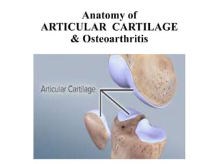
- 2. Articular cartilage • Highly specialized connective tissue covering bone epiphyses in synovial joints • Found at articulating ends of bones within joints of body • Hyaline cartilage and is 2 to 4 mm thick • Function Load-bearing material of the joints Provide excellent lubrication & great wear characteristic • Limited capacity for intrinsic healing and repair Limited potential of chondrocytes for replication Devoid of blood vessels, lymphatics, nerve (Nutrition ?)
- 3. Structure of articular cartilage 4 zones of articular cartilage Superficial zone Middle zone Deep zone Calcified zone Within each zone, 3 regions can be identified Pericellular region Territorial region Interterritorial region
- 9. Take home questions • How is microanatomy of articular cartilage damaged in OA ? • Why more in older age group ? • Is Osteoarthritis only a disease of articular cartilage ? • Why gender differences in OA ? • Immobilization Vs OA
- 10. Why OA ? • Most common chronic musculoskeletal disorder • Most prevalent cartilage degenerative disease • Substantial economic burden • Leading cause of activity limitation & absenteeism among working-age adults • Significant decline in function among older individuals.
- 11. Age related changes • Dissipation of chondrocytes in the superficial region, followed by an increase in number of chondrocytes in the deep layers. • Decrease in the proteoglycan aggregate numbers within the ECM. May be a result of Proteolytic damage to link protein & glycosaminoglycan chains Increase in partially degraded hyaluronan without newly synthesized molecules • Increased mechanical forces exerted on the tissue further lead to subchondral tissue calcification
- 12. Pathogenesis of OA • Trauma causes a microfracture or inflammation • Slight increase in enzymatic activity which allow formation of “ wear” particles, engulfed by resident macrophages. • Production of “wear” particles overwhelms ability of system to eliminate & they become mediators of inflammation, stimulating chondrocyte to release degradative enzymes • Molecules from breakdown of collagen & proteoglycan, also taken up by synovial macrophages, cause release of proinflammatory cytokines, like TNFα, IL-1 and IL-6. • These cytokines bind to chondrocyte receptors leading to further release of metalloproteinases & inhibition of type II collagen production, thus increasing cartilage degradation • Increased water content & decreased proteoglycan content of ECM, weakening of the collagen network due to decreased synthesis of type II collagen & increased breakdown of pre- existing collagen
- 13. Females Vs OA • Anatomical factor Decrease articular cartilage volume than male • Previous history of trauma Women more cartilage wear, increase ACL injury • Hormonal factor Estrogen beneficial effect in articular cartilage Increase proteoglycan in cartilage • Social factors Present for t/t in more advanced stages of OA, so more pain & disability HCW more likely to recommend total joint arthroplasty for male patient
- 14. Is Osteoarthritis only a disease of articular cartilage ? • Initially, osteoarthritis considered disease of articular cartilage • Recent research indicate involvement of entire joint • subchondral bone • synovium • menisci • ligaments, periarticular muscles and nerves
- 15. Subchondral bone • Concomitant increase in levels of cartilage oligomeric matrix protein (COMP) & bone sialoprotein (BSP) in people with early osteoarthritis • progressive increase in subchondral bone plate thickness • formation of new bone at the joint margins – osteophytes • In osteoarthritic bone tissue, ratio of α1 & α2 chains of type I collagen varied between 4:1 & 17:1, and this appears to be responsible for abnormal mineralization pattern In normal bone, type I collagen consist of heterotrimer of α1 & α2 chains at an average ratio of 2.4:1.
- 16. • Synovial membrane of osteoarthritic joints commonly exhibits hyperplasia of lining cell layer occasionally accompanied by focal infiltration of lymphocytes & monocytes in sublining layers • Meniscal degeneration : menisci appear torn, fissured, fragmented, macerated or completely destroyed
- 17. • The loss of articular cartilage thought to be primary change, but a combination of cellular changes & biomechanical stresses causes several secondary changes, including subchondral bone remodeling, formation of osteophytes development of bone marrow lesions change in the synovium, joint capsule, ligaments & periarticular muscles meniscal tears and extrusion
- 18. Immobility Vs OA • Mechanical load necessary for cartilage homeostasis. • Induces fluid movement between cartilage & synovial fluid, that helps in diffusion of molecules across cartilage thus facilitating its nutrition • Decrease protease (MMP-3, ADAMTS-5) expression in human chondrocytes • Increases proteoglycan (aggrecan) • Inhibits IL-1 & TNF-α induced inflammatory & catabolic responses • immobilization leads to joint damage
- 20. Normoxia Vs hyoxia • Since cartilage is an avascular tissue, chondrocytes live in a hypoxic environment • Hypoxia displays a protective effect on cartilage • Lower basal synthesis & release of MMP-1, MMP-13 • Lower generation of type II collagen cleavage fragments • HIF-1α promote chondrocyte function and survival • Gene encoding HIF-1α, Epas1 expressed in cartilage • Deletion of Epas1 gene in cartilaginous growth plate associated with chondrocyte apoptosis • Inhibitor of HIF-1,intraarticular injection promote cartilage degradation & osteophyte formation
- 21. References • Sophia Fox AJ, Bedi A, Rodeo SA. The basic science of articular cartilage: structure, composition, and function. Sports Health. 2009;1(6):461-468. • Houard X, Goldring MB, Berenbaum F. Homeostatic mechanisms in articular cartilage and role of inflammation in osteoarthritis. Curr Rheumatol Rep. 2013;15(11):375.
- 22. THANK YOU