Meniscus: Structure, Role & Injury.
•Download as PPTX, PDF•
19 likes•10,422 views

The presentation investigates the following characteristics of the meniscus; Role of the Meniscus Material Properties Structural Limitations / Failure Limits Mechanism & Treatment of Injuries

Recommended
More Related Content
What's hot
What's hot (20)
Similar to Meniscus: Structure, Role & Injury.
Similar to Meniscus: Structure, Role & Injury. (20)
More from Chris Hattersley
More from Chris Hattersley (16)
Recently uploaded
Recently uploaded (20)
Meniscus: Structure, Role & Injury.
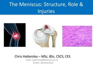
- 1. The Meniscus: Structure, Role & Injuries Chris Hattersley – MSc, BSc, ASCC, CSCS, CES
- 2. Overview Investigate the following characteristics of the meniscus; • Role of the Meniscus • Material Properties • Structural Limitations / Failure Limits • Mechanism & Treatment of Injuries
- 3. Gross Anatomy • C shaped ends attached to tibia, which deepen it’s articular surface and distribute impact forces evenly. • Also provides stability, lubrication and proprioception through its insertional attachments. • Attached by anterior and posterior horns as well as MCL, MFL’s, anterior inter-meniscal ligament & popliteus tendon. • During flexion the menisci translate outwards and posteriorly. The lateral meniscus is more mobile and it’s anterior horn can displace up to 9.5mm posteriorly. • Medial meniscus is more frequently injured than the lateral meniscus with a prevalence of 2:1 (Masouros et al, 2008)
- 4. Fibre Orientation • The menisci are wedge shaped on cross section and have different layers of fibres which run in different angles. • The superficial fibre layer are randomly orientated mimicking hyaline cartilage, this lowers friction between menisci, tibia & femur. • Underneath this, one third of the fibres are distributed in a radial pattern and two thirds are in a circumferential pattern. • The fibre orientation dictates how the meniscus distributes forces and therefore how the fibres can be compromised. (Pereira et al, 2013)
- 5. Blood Supply • Red – red zone is vascularized by the perimeniscal capillary plexus, contains fibroblast like cells and can heal. • Red-white zone is at the border of the vascular area, contains chondrocyte like cells and has a limited capacity to heal. • White zone is avascular, receives nourishment from synovial fluid and can’t heal. • Not as adaptable as other tissues in the body but, when healthy, still has the capacity to remodel in response to loading ‘Mechanotransduction’. (Fox et al, 2014)
- 6. Cellular Structure • The menisci is a stratified fibrocartilage tissue with regional variation in its cellular structure, this dictates it’s material properties. • Chondrocytes construct the extracellular matrix consisting of type l, ll & lll collagen and glycosaminoglycans (GAG). • Collagen provides structure & elasticity, GAG provides structure but also attracts water. • The inner two thirds are more deformable than the outer third. • Chondroitin & glucosamine sulphate are involved in the formation of GAG, so people with cartilage damage sometimes supplement with that. (Pereira et al, 2013)
- 7. Material Properties • Elastic properties of materials are measured by the Young’s modulus. • Young’s modulus = stress x strain • The compressive elastic modulus of the meniscus is very low meaning it is the most likely structure in the knee to deform under axial load. • It’s high water content coupled with its collagen & GAG structure means the meniscus compresses under load, but has the elasticity to return to it’s originally shape. • Under load it is squeezed out of the joint creating a hoop, shear & radial tensile stress. Highly deformable under compression = Effective Shock Absorber (Masouros et al, 2008)
- 8. Shock Absorption • The main role of the meniscus is to evenly distribute impact forces through the knee & to maintain joint stability. • As the femoral condyles roll over the menisci it compresses and increases it’s circumference. • Absorbs 70-99% of the joints load. • If the meniscus is damaged it increases the load per unit area and accelerates the degeneration of cartilage and subchondral bone usually leading to OA. (McDermott et al, 2008)
- 9. Mechanism of Injury • As the knee is placed between 2 large levers it experiences very high forces which can cause injury to the meniscus through repetitive chronic loading or an acute traumatic event. • A meniscus tear is usually caused by twisting or turning quickly. These tears can occur when you lift something heavy or play sports. • Can occur alongside a ligament injury, ACL, MCL etc. • As you get older the meniscus gets worn and it’s water content is reduced, this is natural with aging but can make it tear more easily. (Fox et al, 2014)
- 10. Meniscus Tears
- 11. Meniscus Sources of Stress Tension Fluid Pressure Compression Fluid Flow Hydrostatic Pressure Strain / shear (McNulty & Guilak, 2015) Chronic Loading: Time / Age Acute Loading: Traumatic Injury
- 12. Force Distribution - It is apparent that meniscus tears happen in consistent patterns and this is associated with the inherent way this structure dissipates force. Tissue Loading > Tissue Tolerance = Tissue Failure Hoop Stress Radial Tensile Stress Shear Stress https://www.abdn.ac.uk/ims/research/musculoskeletal/meniscus-1206.php
- 13. How can the tissue fail? Menisci sitting on the tibia, showing how loading forces them outwards to produce a hoop stress around the curve. - The way the meniscus dissipates force corresponds strikingly with the most common tears highlighting weaknesses in its structure. Radial tensile stress from the centre outwards can cause a radial tear. Sheer stress between the inner and outer layer can also cause a tear. https://www.abdn.ac.uk/ims/research/musculoskeletal/meniscus-1206.php
- 14. • History of traumatic injury to the knee • Knee pain on weight bearing activities, walking, sit to stand etc. • Delayed swelling of the knee • Popping , clicking or locking of the knee joint • Stiffness of the knee • Loss of full range of motion • Tenderness on the knee joint line • Pain with squatting (Brukner, 2012) Signs & Symptoms
- 15. Meniscus Injury Assessment - Old McMurray’s Test Apley's Test
- 16. Meniscus Injury Assessment - Current Thessaly's Test • McMurrays & Apley’s tests can cause unnecessary pain and risk further injury to the meniscus. • It is therefore preferred to make a diagnosis using a patients subjective history & identifying any of the previously mentioned signs & symptoms. • If necessary Thessaly’s test can be used which is less invasive than McMurray’s / Apley’s. (Alexanders et al, 2016)
- 17. Imaging • MRI is the modality of choice when a meniscal tear is suspected. • Grade 0 – Normal intact meniscus • Grade I – Intra-substance globular appearing signal, not extending to the articular surface • Grade II – Linear increased signal patterns, not extending to the articular surface • Grade III – Abnormal signal intersects the superior and/or inferior articular surface (Fox et al, 2014)
- 18. Treatment Options Non surgical Meniscal repair Meniscectomy Graft / Implant
- 19. Rehabilitation - Reduce pain and inflammation. - Improve joint range of motion. - Strengthen leg muscles,. - Improve alignment , biomechanics and proprioception. - Rehabilitation normally around 6-8 weeks. Non-Surgical Treatment: - Preferred option - Aims to heal or reduce symptoms Surgical Treatment: - May be required for large tears or if a fragment breaks away - Short term relief but long term problems i.e. OA. (Brukner, 2012)
- 20. References • Aspden., R. (1985). ‘A model for the function and failure of the meniscus’. Proc.Instn.Mech.Engrs.[H], J.Eng.Med. 14:119-122. • Brindle,. et al. (2001). ‘The Meniscus: Review of Basic Principles With Application to Surgery and Rehabilitation’. Journal of Athletic Training, v.36(2), pp.160-169 • Brukner, P.,( 2012). Brukner & Khan's clinical sports medicine. North Ryde: McGraw-Hill. • Fox, A., Wanivenhaus, F., Burge, A., & Rodeo, S. (2014), ‘The human meniscus: A review of anatomy, function, injury, and advances in treatment: The Meniscus: Anatomy, Function, Injury and Treatment’ ,Clinical Anatomy, 28, pp 1-19. • Masouros, S., Mcdermott, I., Amis, A., & Bull, A (2008), ‘Biomechanics of the meniscus-meniscal ligament construct of the knee’ , Knee Surgery, Sports Traumatology, Arthroscopy, 12, 1121-1132. • McDermott, I.D., Masouros, S.D. and Amis, A.A., (2008). ‘Biomechanics of the menisci of the knee’. Current Orthopaedics, 22(3), pp.193-201. • Mcnulty, A., & Guilak, F. (2015), ‘Mechanobiology of the meniscus’, Journal of Biomechanics, 48, pp 1469-1478. • Periera, H., Silva-Correia., J., Oliveira, J., & Reis., L. (2013), ‘ The Meniscus: Basic Science’, Meniscal Transplantation. Springer, Berlin, Heidelberg. • Sanchez-Adams, J., & Athanasiou, K. (2012) ‘Biomechanics of meniscus cells: regional variation and comparison to articular chondrocytes and ligament cells’ Biomechanics and Modelling in Mechanobiology, 11, pp 1047–1056.
Editor's Notes
- Can break away and block the joint
- Hoop stress = it is forced out but resisted creating the hoop stress. Most efficient way to distribute force, long term leads to degeneration through normal aging. Radial = a force from the centre outwards. Shear = caused between the inner and outer layer Radial / Sheer = more likely to cause a tear
- Ligament injury will swell quickly 0-2 hours, meniscus is much slower than this
- Autologous chondrocyte Implantation