
Recommended
More Related Content
What's hot
What's hot (20)
Similar to meristmatic tissue.pptx
Similar to meristmatic tissue.pptx (20)
More from manoj Joshi
More from manoj Joshi (20)
Recently uploaded
Recently uploaded (20)
meristmatic tissue.pptx
- 1. Meristems or Meristimatic tissue Dr Manoj Joshi
- 2. • Meristematic tissue is group of immature cells that has capacity of division and re-division. • The term meristem was coined by Nageli (1858). Meristemsin plants are found in apex of stem, root, leaf primordia, vascular cambium, cork cambium, etc.
- 3. Characteristics of Meristematic Tissue: • 1. They are composed of immature cells. • 2. Absence of intercellular spaces. • 3. Cells are oval, rounded or polygonal in shape. • 4. Cells are always living and thin walled. • 5. Cells are rich in cytoplasm with minute vacuoles. • 6. Cell is diploid and shows mitotic cell division. • 7. Cell is devoid of reserve food materials, ER and plastids.
- 6. Functions of Meristematic Tissue • 1. Meristems are actively dividing tissues of the plant. • 2. They are responsible for primary (elongation) and secondary (thickness) growth of the plant. • 3. All new organs and their growth occur by the division of meristematic tissue. • 4. Secondary tissues such as, wood, cork are also formed due to activity of meristematic tissue.
- 8. Apical Cell Theory: • Nageli in 1944 advocated this theory. According to this theory the apical meristem consists of a single apical cell (also called apical initial) and this cell is interpreted as the ‘structural and functional unit of apical meristem’. The cell is very large and is shaped like an inverted pyramid. • The apical cell is tetrahedral in shape and has three or four cutting faces among which single face is directed upward and the rest points downward. The side of apical cell that is directed upward is triangular or square in shape and forms a part of the outer surface of the shoot apex
- 9. • The cutting faces of apical cell divide in an orderly fashion that is in helical succession. The cell divides by an asymmetric division; as a result a narrow and flat cell is formed. The next division of the apical cell is also asymmetric. This type of asymmetric division is repeated in the downwardly pointed faces of the apical cell. • As a result all cutting faces have their daughter cells. The daughter cells also divide and form large packet of cells. The packet of cells differentiates and forms different segments of shoot. So the apical cell is regarded as ‘a reserve of one genetically sound cell’. • A single apical cell composing an apical meristem is present in vascular cryptogams. After the discovery of solitary apical cell in vascular cryptogams, it was supposed that such apical meristem might exist in higher plants as well. • Later extensive investigations refuted the universal occurrence of solitary apical cell in a meristem. In higher plants the apical cell theory was replaced by the concept that the different parts of a plant body have independent origin. So the apical cell theory was later superseded by histogen theory.
- 10. Histogen Theory: • Hanstein in 1868 put forward histogen theory (histogen means tissue builder). • According to this theory the tissues of a plant body originate from a mass of meristem where the following three (histogens) can be distinguished • (a) Dermatogen: • (In Greek meaning skin). It is the outermost layer of the meristem. It gives rise to epidermises of root and stem. • (b) Periblem: • (In Greek meaning clothing). This region occurs internal to dermatogen but peripheral to plerome. This histogen is destined to form cortex of root and shoot and inner tissues of leaves. It surrounds plerome. • (c) Plerome: • (In Greek meaning that this fills). This region gives rise to vascular cylinder of stem and root including pith. It is the central core of stem and root and the cells composing this zone are very irregular. This region is enveloped by a variable number of mantle-like layers which are represented by dermatogen and periblem.
- 12. • According to Hanstein dermatogen, periblem and plerome arise from independent initials of the apical meristem. • Later investigations reveal that the sub-divisions — dermatogen, periblem and plerome have no universal application due to the following two reasons: (i) In gymnosperm and angiosperm there exists no clear distinction between periblem and plerome. (ii) The respective roles of the three histogens cannot be demonstrated. • The main weakness of Hanstein’s concept was to assign specific destinies of histogens. The histogens — dermatogen, periblem and plerome are committal and respectfully give rise to epidermis, cortex and stele. Later this theory was superseded by tunica- corpus theory because the zones are noncommittal.
- 13. • Though histogen theory is rejected it is still regarded as ‘classic’ due to the fact that Hanstein regarded shoot and root apex as a composite unit consisting of different groups of histogens and the histogens differed fundamentally from each other producing different tissues. It is to note that Hanstein visualized this long before when the concept of gene and DNA was established. • The present-day concept is that the different zones in the shoot apex are fundamentally same and capable of producing all tissues. It is the position in the meristem that determines the destiny of derivatives. This is due to the fact that in zones some genes are activated and others are repressed resulting in the production of different tissues.
- 14. Tunica-Corpus Theory: • Schmidt in 1924 postulated tunica- corpus theory on the basis of studies of shoot apices of angiosperm. This theory is concerned with planes of cell division in the apex. In contrast to apical cell theory and histogen theory tunica-corpus theory is applicable only to shoot apex and not to root. Schmidt distinguishes two tissue zones in the shoot apex and termed them as tunica and corpus. • Majority of angiosperm shoot apex exhibits tunica consisting of two layers of cells and corpus . Researchers designate the layers as L1, L2 and L3 to denote respectively outer layer of tunica, inner layer of tunica and corpus. • Plasmodesmata exist between the cells of tunica and corpus. It is thought that plasmodesma controls the gene expression that leads to the formation of protoderm, ground meristem and provascular tissue.
- 16. i. Tunica: Tunica is the peripheral tissue zone of shoot apex. It consists of one or more peripheral layers of cells. Dicotyledons exhibit one to five layers of cells in tunica; two layers of cells are represented by largest number of species. Monocotyledons have one to four layers of cells in tunica; one and two layered tunica predominates in it. One single layered tunica is termed as monostratose. Many layered tunica is termed as multistratose. Xanthorrhoea media shows eighteen layered tunica. Tunica is characterized in having anticlinal plane of cell division, i.e., the division wall is laid down perpendicular to the surface. This division reflects to the surface growth in the apex. As a result tunica grows as a sheet but not in thickness. To increase in thickness a tunica cell will have to undergo cell division with periclinal wall, i.e. the cell wall will have to be laid down parallel to surface (Fig. 7.9B). Normally, periclinal division does not occur in tunica where anticlinal division predominates except at the point of origin of leaf primordium and axillary bud.
- 17. In multistratose tunica each layer arises from separate initials present in the shoot apex. The independence regarding origin among different layers of tunica and between tunica and corpus is strongly supported with the results of investigation by the use of periclinal chimeras. Such chimera in a plant body exhibits layers with different chromosome numbers. Such differences can be traced from promeristem through continuous cell lineages to similar differences in the derivatives of promeristem. Promeristem may be induced to develop cytochimeras by treating the shoot apex with colchicine. Colchicine induces polyploidy in the cell(s) or layer(s) of promeristem and it is possible to relate the polyploid cell(s)/Iayer(s) to their progeny in the mature plant body. Using this technique it was determined that outermost layer of tunica gives rise to epidermis whereas the inner layers donate cells to form other tissues. Cytochimera: Chimaera, also spelt as chimera, refers a plant or plant organ having tissues of varied genetic origin lying adjacent to one another. In some plant the meristematic region consists of at least two populations of cells that differ in their chromosome numbers. The chimera of this plant is recognizable only at the cytological level and termed as cytochimera.
- 18. • Usually two types of chimera are recognized: • (1) Sectoral or mericlinal chimera — where the two populations of cells are arranged in sectors on the circumference around the axis of the meristem. • (2) Periclinal chimera — where the two populations of cells are arranged in different layers parallel to the surface of apical meristem. L1, that is the outer layer of tunica, is commonly destined to form the epidermis of all the leaves and stem formed by the shoot and it is revealed on analyzing the periclinal chimera. • Shoot apices are induced to form cytochimeras to study the function of L1, L2 and L3 layers. Treating the shoot apex with colchicine does this. The alkaloid colchicine, a chemical mutagen, is obtained from the plant Colchicum autumnale. Colchicine has the property to disrupt the spindle microtubules formed during mitosis. The drug colchicine is applied to the shoot tips by slow trickling or as paint. • The treatment temporarily blocks mitosis. The cells that are in metaphase stage cannot complete chromosome movement due to the failure of spindle mechanism but the other events of mitosis do continue. As a result nuclei are formed with tetraploid chromosome numbers. Then colchicine was washed out and the meristems were allowed to continue cell division. • Staining properties of nucleus can recognize the descendents of tetraploid cell. Using this technique it becomes possible to find that the LI layer, i.e. the outermost layer gives rise to the epidermis of a leaf and the inner layers donate cells to form mesophyll and vascular tissues.
- 19. • In stems LI gives rise to epidermis and the rest forms the cortex and vascular tissue. In Datura the shoot apices were experimentally induced to develop cytochimera. The apices exhibited that the cells of LI, L2 and L3 layers had nuclei with chromosome number respectfully 8n, 2n and 2n; 4n, 2n and 4n, and 2n, 8n and 4n]. • It is the anticlinal plane of cell division that characterizes tunica. In maize, certain grasses like Agropyron repens and Chlorogalum pomeridianum (Liliaceae) etc. periclinal divisions are observed in tunica. This led anatomists to modify the ‘strictness of definition of the tunica’. One view regards that the tunica should include only those layer(s) that exhibit- anticlinal plane of division. If any inner layer(s) of tunica show periclinal division they are to be assigned to corpus. • Other workers treat tunica in a very loose sense and regard that the inner layer(s) may divide periclinally. So to avoid discrepancies the term mantle was proposed in a loose sense instead of tunica. Corpus was replaced by the term core. Mantle overarches core. Popham and Chan (1950) advocated the mantle and core concept in the shoot apex and they did not take into account to the planes of cell division. • Cytologically two zones are recognized in the tunica though all cells exhibit the same anticlinal cell division. The first zone is central apical zone and the second occurs between the central apical zone and leaf primordium. • The central apical zone consists of one or few initial cells that are larger and contain large nuclei and vacuoles than the cells of the second zone. The cells of the second zone are small and more darkly staining than the cells of the first zone. In contrast to first zone periclinal division may occur in the second zone close to the leaf primordium in addition to anticlinal division. • The main function of tunica is to give rise epidermis. Sometimes the inner layers of tunica form cortex and vascular tissue.
- 20. • ii. Corpus: • Corpus is the inner tissue zone of shoot apex. It consists of cells that are several cell layers deep. Tunica overarches corpus. Meristematic tissues composing this zone are larger than tunica. The initial cells of corpus occur below the tunica. They are orderly arranged in contrast to haphazardly arranged cells in the mass of corpus. So the initials of corpus are difficult to differentiate from the initials of tunica. • The initials arise independently and not related to tunica. The initial cells divide periclinally and the derivatives divide to form the core of the shoot apex. In the division of derivative cells there is no definite orientation of cell wall formation, i.e. cells divide in all planes. As a result the shoot apex increases in volume. Generally corpus is destined to give rise cortex and vascular tissue. • Usually corpus is not homogeneous. It consists of several zones. The zones are illustrated by Popham (1952) in the usual angiosperm type. In this type the meristem is composed of tunica and corpus. The tunica consists of one or more layers of cells that overlie the corpus. • The corpus is composed of three zones: • (a) Central mother cells —the uppermost zone of corpus. • (b) Pith-rib meristem that occurs below the central mother cell zone. • (c) Flank meristem (also called peripheral meristem) that surrounds both central mother cell zone and pith-rib meristem. The peripheral zone is shaped like a truncated hollow cone. • The component cells of tunica and corpus differ in size, shape, plane of cell division and topography. Ultrastructurally each zone is composed of cells that have characteristic architectures as is revealed by quantitative techniques.
- 21. Merits of tunica-corpus theory: i. It deals with one thing, i.e. planes of cell division. As a result the description of meristem becomes precise. ii. It has topographical value in the studies of development of different tissue system in plants. iii. The destiny of derivatives of corpus is not predetermined. iv. The derivatives of the zones are not rigid like histogen theory. v. It explains clearly the growth pattern in the shoot apex of angiosperm. vi. It enables to understand the development of leaves as they arise close to apex. vii. The specific variation in the number of tunica layers may be of taxonomic significance, e.g. grasses. Tunica-corpus organization of shoot apical meristem is observed in angiosperm only. Cryptogams and majority of gymnosperm do not exhibit tunica- corpus organization. They do not have any stable surface layer that divides only by anticlinal division. Araucaria and Ephedra have a single stable surface layer that divides only by anticlinal division. Thus they exhibit tunica-corpus pattern. Tunica-corpus theory is noncommittal regarding the ultimate destiny of cell-lineages. In present-day concept all cells in the shoot apex with tunica-corpus organization are fundamentally same and have equal potentiality. In corpus the cells of peripheral zone and pith-rib meristem are also same as they owe their origin from central mother cell zone.
- 22. But during differentiation some genes are repressed and others are activated resulting in the production of different tissues. It is thought that if some how or means the cells of peripheral meristem zone of corpus is transferred to the location of pith-rib meristem, the cells will produce pith cells. In conclusion it is to mention that tunica-corpus theory provides a flexible description of cell arrangement in the shoot apex and this is not rigid like histogen theory. It has served well in understanding the structure of shoot apical meristem and the origin and development of leaf. The shoot apices of angiosperm exhibit cytohistological zonation in addition to tunica corpus zonation (Fig. 7.10). Four zones are recognized namely: (1) Tunica initials-that consist of an apical group of cells, (2) Corpus initials-that occur below apical initials and are similar to central mother cells, (3) A peripheral zone and (4) A rib meristem. Tunica initials contribute cells to central mother cell zone and to peripheral meristem. The central mother cell zone donates cells to the rib meristem and pith. The peripheral meristem is highly meristematic. Leaf primordia originate from this layer.
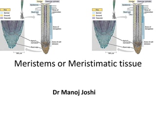
- 24. Quiescent Centre or Quiescent Zone: • Clowes (1959) studied the root tips of Zea mays and observed an inactive centre in between root cap and meristematic region. He called it as Quiescent Centre. 1. It is a biconvex structure and made up of thousands of inactive cells. 2. In this region the rate of cell division is very slow as compared to surrounding cells. 3. This region carries less amount of DNA, RNA, ER, mitochondria and ribosomes. 4. This zone is the site of auxin synthesis. • Functions: • 1. It acts as reservoir of cells. • 2. It is more resistant to injury and irradiation. • 3. If root growth is stopped or root tip is damaged then this zone restores the growth.