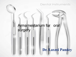
Hello friends this is ur Dr.Lx brought you with best of oral surgery instruments at one place. Dear readers I would like to inform that the techniques for use of forceps and elevators were mentioned in my previously uploaded ppt. EXODONTIA so watch it to gain maximum knowledge .Hope you learn and enjoy this ppt.
2. content
Introduction
Classification of instruments.
Instrument for Transferring Sterile Instrument.
Instrument for Incising Tissue.
Instrument for Elevating Mucoperiosteum.
Instrument for Retracting Soft Tissue.
Instrument for Controlling Hemorrhage.
Instrument for Grasping Tissue.
Instrument for Removing Bone.
Instrument for Removing Pathology Tissue.
3. Cont..
Instruments for Suturing Mucosa.
Instrument for Opening Mouth.
Instrument for Suctioning.
Instrument for Irrigation.
Instruments for Holding Towels And Drapes In Position
Instrument for Extraction Of Teeth
-Local Anesthesia
-Dental Elevators
-Extraction Forceps
Instrument Trays
Conclusion
References
6. CHEATLE FORCEPS
Long handles
Long angulated beaks
serrated
Beaks dipped in antiseptic
solution lift up sterile
instruments from
autoclave/drum
TRANSFER FORCEP:
HEAVY , RIGHT angled-heavy
jaws
7. SWAB HOLDING FORCEP:
Long handles ,straight beaks
fenestrated ends
Rings end of handles
Working end :inner aspect serrated
USES:
Pick up sterile gauze transfer to tray.
Hold gauze dipped in antiseptic
solution-scrub the surgical field
9. SCALPEL:
Handle No.3,No.7
Differently shaped
Disposable ,sterile sharp blade.
No.15 most commonly used
For Mucoperiosteum
No.10 similar to No.15
For large incisions
No.11 sharp pointed, small stab
incision for abcess
No.12 Hooked, Mucogingival
surgery. posterior aspect of
teeth, tuberosity
10. Pen grasp(allows maximum control)
Hold mobile tissue firmly
Press down firmly
Single pt. use
Always use no blade because it gets dulled
easily and thus gives dull incision
Blade loaded
Pen grasp
Blade removed
12. Mucosa and periosteum reflected in single
layer:
Periosteal elevator
NO.9 MOLT PERIOSTEAL ELEVATOR:-
Sharp pointed end reflects papillae from
between teeth, loosen soft tissues via
gingival sulcus.
Broader end elevating the tissues from
bone
Thin, sharp cutting edge clean separation
of periosteum from bone.
ROUNDED ENDED MOLT PERIOSTEAL
ELEVATOR:-
Single/double ended
REFLECTION OF SOFT TISSUE-3 METHODS
Prying motion
Push motion
Pull/scrape motion
PRYING
13. WOODSON PERIOSTEAL ELEVATOR:-
Small and delicate
Loosen soft tissues via gingival sulcus
HOWARTH’S PERIOSTEAL ELEVATOR:-
Double ended
One ended flat, broad ,spatulate -sharp edge
Other end rugine end: flat and rectangular
small tip-sharp perpendicular
Reflection and retraction mucoperiosteal flaps
Reflection periosteum
MOON’S PROBE:-
Right angled narrow working edge
Flat handle and blade blade perpendicular to
handle.
Narrow working edge blunt and rounded tip
Mucoperiosteal elevation prior to extraction
15. It is done for good vision and access
Cheeks, tongue and mucoperiosteal
flaps .
MOUTH MIRROR:-
Commonly used
WEIDER RETRACTOR:-
Broad ,heart shaped
Serrated on one side firmly engage
tongue , retract it medially and
inferiorly.
Don’t position posteriorly causes
gagging.
AUSTIN RETRACTOR:-
It is right an right angle L-shaped
no handle
Retraction of small intraoral flaps ,
removal of impacted teeth.
16. MINNESOTA RETRACTOR:-
Both austin and minnesota retract
cheek and mucoperiosteal flap
simultaneously.
Before flap retractor held loosely in
the cheek.
After flap reflection retractor
placed on the bone and used to
retract the flap.
SELDIN RETRACTOR:-
Similar to a periosteal elevator.
Leading edge dull, shouldn’t
reflect periosteum.
PERIOSTEAL ELEVATOR:-
Primary instrument for retraction.
Positioned on the bone and held to
reflect tissue
17. LANGENBACK’S RETRACTOR:-
‘L’ Shaped retractor long handle
Retraction of flap edges improved
visualization of deeper layers and
structures.
Different sizes handle length and
blade width
TONGUE DEPRESSOR:-
‘L’ shaped no handle
Broad flat ,flat ,rounded blade.
Retraction and depression of tongue
Improve visibility posterior
pharyngeal wall and tonsillar region ,
lingual side of mandible.
Removal of throat pack.
As cheek retractor
18. TOWEL CLIP:-
Hold the tongue.
BIOPSY performed on
the posterior aspect; by
holding the anterior
tongue.
Profound L/A
20. Arteries and veins bleeding pressure
not enough.
HAEMOSTAT
Variety of shapes
Small or delicate/large.
straight/curved
Curved hemostat :-
Long ,delicate beak to grasp tissue and looking
handle.
Locking handle clamps onto a vessel; then
let go and remains clamped onto tissue.
Removes granulation tissue.
Picks up root tips, pieces of calculus,
fragments of amalgam restorations, any other
small particles dropped into the mouth.
Small hemostat MOSQUITO forceps.
Eg. Crile, Spencer Wells , Halstead mosquito
artery forceps.
22. Soft tissue stabilization pass suture
needle.
ADSON’S TISSUE FORCEP/PICKUPS:-
Delicate forceps.
Small teeth.
Gently hold tissue and stabilize.
Don’t grasp too tightly crushing
Non toothed.
TISSUE HOLDING FORCEPS:-
Toothed periosteum, muscle,
aponeurosis.
Non toothed fascia, mucosa, pathological
tissues.
STILLIES FORCEPS:-
Longer but similar Adson’s
7”-9” long
Easy grasp of tissue in the posterior part,
with enough part protruding beyond the lips
23. COTTON FORCEPS:-
Angled
Small fragments of
tooth/amalgam/foreign material.
Placing/removing gauze packs
ALLIS TISSUE FORCEPS:-
Locking handles
- Proper placement
-Held by asst. necessary tension
Teeth which will firmly grip the tissue
Removal of large amounts of fibrous
tissue epulis fissuratum
Never let tissue to be left in mouth
crushing injury
24. RUSSIAN TISSUE FORCEPS:-
Large rounded end
Teeth elevated from sockets
Round end positive grip, avoids
spillage; unlike hemostat
Placement of gauze isolation
BABCOCK’S TISSUE HOLDING FORCEP:-
Non toothed blades
Long beaks-broad working edge
Smooth, non serrated edges
Rings-locking mechanism
Hold delicate tissues lymph nodes,
mucosa
Holding cyst lining during enucleation
26. RONGEUR FORCEPS:-
Most commonly used
Sharp blades squeezed together,
cutting/pinching through bone.
Leaf spring between the handle
instrument opens when hand pressure is
released.
Repeated cuts without manually reopening
2 major designs:
Side-cutting
Side-cutting and end cutting/
BLUMENTHAL RONGEURS
-most dentoalveolar surgical procedures
- inserted into sockets: interradicular bone
-sharp edges of bone
Large amounts of bone, quickly and
efficiently
Do not remove large amount of bone in
single bites
27. CHISEL:-
Monobevel chiesel bone is
removed
Bibevel chisel teeth
Success sharpness before
sterilization
Carbide tips use more than once,
before sharpening.
Cylindrical handle serrated with
flat end struck with mallet
Flat and rectangular cutting edge
in different sizes
Single bevel cutting edge
Transalveolar extraction/removal of
impacted tooth
Bevel faces bone to be cut
Cutting edge perpendicular to
bone.
28. OSTEOTOME:-
Splitting bone
Cylindrical handle serrated for good
grip
Flat end tapped with mallet
Flat and rectangular blade
Bibevel cutting edge converge to a
sharp edge
BONE FILE :-
Final smoothing of bone before
suturing of mucoperiosteal flap
Double-ended small and large
Removes bone pull stroke
Avoids push motion burnishing and
crushing the bone
29. SURGICAL MALLET:-
Cutting bone with osteotome/ chisel
Stainless steal strong cylindrical handle
Tapped ‘pull-back’ action force from
wrist
Tapped with controlled force ; made to spring
back from chisel/osteotome
For jaw surgery inadvertent force.
BUR AND HANDPIECE:-
Surgical removal of teeth.
High speed + sharp carbide burs = cortical
bone removal
No.557 ,703 fissure burs ;No.8 round bur
Large bone bur : acrylic bur – large bone
removal (torus)
Relatively high speed and torque :rapid bone
removal and efficient sectioning
Avoid high speed turbine drills used inn
restorative dentistry causes tissue
emphysema
31. PERIAPICAL CURETTE:-
Angled, double ended
Removal of granulation /small cysts
from periapical lesions
Small amounts of granulation tissue
debris from tooth sockets.
HILTONS SINUS FORCEPS:-
Handles with rings at the end .
No lock/ratchet
Narrow , long , slender beaks
Inner surface traverse striations
:close to the tip
Draining pus from an abcess
Inserted by blunt dissection and
opened up.
No lock blind insertion and close
injure structures
33. Flap returned to its original position and
held by suture
NEEDLE HOLDER:-
Instrument with a locking handle, short,
stout, beak
I/O use: 6” or 15 cm recommended
Beak shorter and stronger than hemostat
Face of the beak cross hatched, positive
grasp ,unlike hemostat.
Thumb and ring finger through the rings
Index finger along the length of the holder
Second finger aids in controlling the
locking mechanism
COMPARISON:
HAEMOSTAT :Beaks smaller than sinus
forceps, longer than needle holder; traverse
striation; ratchet
NEEDLE HOLDER: Criss - cross striations
;ratchet
SINUS FORCEP: Striations only near the tip;
no ratchet
34. SUTURE NEEDLE:-
Mucosal closure :1/2 circle or 3/8 circle.
Curved pass through limited space
;twisted wrist
Large variety of shapes
Very small to very large
Cutting needle pass through
mucoperiosteum more easily than a tapered
needle
1/3 cutting remaining round
Tapered vascular, ocular
Should always be cut through tissues lateral
to the track
Suture material :usually swaged on
Held 2/3rd -> between the tip and the base.
-enough exposed to pass through the tissue
- grasp in the strong portion to prevent
bending
35. SCISSORS:-
Short cutting edges
Long handles
Thumb and ring fingers
Held same as needle holder
DEAN SCISSORS:-
Slightly curved handles
Serrated blades
IRIS SCISSORS and METZENBAUM
SCISSORS:-
Straight or curved blades
IRIS SCISSORS : very fine skin sutures, small,
sharp pointed delicate
METZENBAUM SCISSORS :undermining soft
tissue and cutting ;sharp or rounded tips.
Don’t cut sutures causes dulling of the
edges –less effective and more traumatic
37. Soft, rubber like block where pt.
rests his teeth
Pt. opens to comfortably wide
position –block inserted: holds in the
position
Protects pt, TMJ ,while mandibular
teeth
Various sizes – for various pt. and
varying degrees of opening
Fergusson ackland
mouth gag
45. Bone removal with steady stream of
irrigation sterile saline or water
Cools the bur
Prevents bone damaging heat build
up
Increase efficiency of bur
-Washes away bone chips
-Lubrication
Completion of procedure : before
suturing
LARGE PLASTIC SYRINGE + BLUNT 18-
GAUGE NEEDLE
49. LOCAL ANESTHETIC
INSTRUMENTS:-
LENGTH:32mm and 20mm needles
27/30 gauge : commonly used
25 gauge: preferred for high risk of
positive aspiration
30 gauge: not specific; local
infiltration
PARTS OF NEEDLE
54. DENTAL ELEVATORS
GRIPS:-
Palm grip:-heavy forces; handle rests
against heel of palm
Finger grip:-delicate applications
TYPES:-
1. BASED ON THEIR SHAPE AND SIZE:-
Straight
Triangle/pennant-shape
pick
2. BASED ON THEIR FORM
Straight
Angular
Crossbar
55. 3. BASED ON THEIR USE:-
Remove roots broken at the
gingival line
Remove entire tooth
Remove mucoperiosteum
Remove roots broken half
way to the apex
Remove the apical third of the root