

Recommended
More Related Content
What's hot
What's hot (20)
Similar to Cracked tooth syndrom
Similar to Cracked tooth syndrom (20)
More from NUHA ELKADIKI
Recently uploaded
Recently uploaded (20)
Cracked tooth syndrom
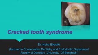
- 1. Cracked tooth syndrome Dr. Nuha Elkadiki (lecturer in Conservative Dentistry and Endodontic Department ,Faculty of Dentistry ,University Of Benghazi )
- 2. Cracked tooth syndrome (CTS) is where a tooth has incompletely cracked but no part of the tooth has yet broken off. Sometimes it is described as a greenstick fracture. Tooth crack in the upper first molar tooth in a patient who suffers from bruxism.
- 3. Classification and definition Cracked tooth syndrome could be considered a type of dental trauma and also one of the possible causes of dental pain. Cracked tooth syndrome is "a fracture plane of unknown depth and direction passing through tooth structure that, if not already involving, may progress to communicate with the pulp and/or periodontal ligament."
- 4. Signs and symptoms Sharp pain when biting on a certain tooth, which may get worse if the applied biting force is increased. Sometimes the pain on biting occurs when the food being chewed is soft with harder elements, e.g. seeded bread. "Rebound pain" i.e. sharp, fleeting pain occurring when the biting force is released from the tooth, Sharp pain when drinking cold beverages or eating cold foods, lack of pain with heat stimuli.
- 5. Signs and symptoms Pain when eating or drinking sugary substances. Sometimes the pain is well localized, and the individual is able to determine the exact tooth from which the symptoms are originating, but not always. If the crack propagates into the pulp, irreversible pulpitis, pulpal necrosis and periapical periodontitis may develop, with the respective associated symptoms.
- 6. Pathophysiology CTS is typically characterized by pain when releasing biting pressure on an object. This is because when biting down the segments are usually moving apart and thereby reducing the pressure in the nerves in the dentin of the tooth. When the bite is released the "segments" snap back together sharply increasing the pressure in the intradentin nerves causing pain. If untreated, CTS can lead to severe pain, possible pulpal death, abscess, and even the loss of the tooth.
- 7. Pathophysiology If the fracture propagates into the pulp, this is termed a complete fracture, and pulpitis and pulp death may occur. If the crack propagates further into the root, a periodontal defect may develop, or even a vertical root fracture.
- 8. Pathophysiology According to one theory, the pain on biting is caused by the 2 fractured sections of the tooth moving independently of each other, triggering sudden movement of fluid within the dentinal tubules. This activates A-type nociceptors in the dentin-pulp complex, reported by the pulp-dentin complex as pain. Another theory is that the pain upon cold stimuli results from leak of noxious substances via the crack, irritating the pulp. Crack (vertical fracture) of tooth and root # (green arrows) splitting it in two even pieces which has caused a lateral periodontal abscess (blue arrows).
- 9. Diagnosis The diagnosis of cracked tooth syndrome is notoriously difficult even for experienced clinicians. The features are highly variable and may mimic sinusitis, temporomandibular disorders, headaches, ear pain, or atypical facial pain/atypical odontalgia (persistent idiopathic facial pain). A detailed history may reveal pain on release of pressure when eating or sharp pain when consuming cold food and drink. There are a variety of habits which predispose patients to CTS including chewing ice, pens and hard sweets etc. Recurrent occlusal adjustment of restorations due to discomfort may also be indicative of CTS, alongside a history of extensive dental treatment.
- 10. Techniques used for diagnosing CTS Clinical examination: Cracks are difficult to see during a clinical exam which may limit diagnosis. However other , clinical signs which may lead to the diagnosis of CTS includes: 1. Wear faceting indicating excessive forces perhaps from clenching or grinding . 2. Presence of an isolated deep periodontal pocket which may symbolize a split tooth. 3. Removing restorations may help to visualize fracture lines but should only be carried out after gaining informed consent from the patient. 4. Tactile examination with a sharp probe may also aid diagnosis.
- 11. Gentian Violet or Methylene Blue Stains Dyes may be used to aid visualization of fractures. The technique requires 2–5 days to be effective and a temporary restoration may be required. The structural integrity can be weakened by this method, leading to crack propagation. Techniques used for diagnosing CTS
- 12. Transillumination Transillumination of tooth , showing vertical fracture (blue arrows) and inflammation in marginal gingiva at fracture site (green arrow) Transillumination is best performed by placing a fibre optic light source directly onto the tooth and optimal results can be achieved with the aid of magnification. Cracks involving dentine interrupt the light transmission. Techniques used for diagnosing CTS
- 13. Radiographs Radiographs offer little benefit in visualizing cracks( This is due to the fact that cracks propagate in a direction which is parallel to the plane of the film) (Mesio-distal) Techniques used for diagnosing CTS
- 14. Bite Test o Patients bite down followed by sudden release of pressure. o CTS diagnosis is confirmed by pain on release of pressure. o The involved cusp can be determined by biting on individual cusps separately. Tooth Slooth II Techniques used for diagnosing CTS
- 15. Microscopic Detection Experienced clinicians using a clinical microscope have reached a general consensus that ×16 provides an ideal magnification level for the evaluation of enamel cracks, with a range from . Techniques used for diagnosing CTS
- 16. Epidemiology CTS is multifactorial, the causative factors include: previous restorative procedures. occlusal factors; patients who suffer from bruxism, or clenching are prone to have cracked teeth. developmental conditions/anatomical considerations. 1.trauma 2.others, e.g., aging dentition or presence of lingual tongue studs.
- 17. Treatments There is no universally accepted treatment strategy, but, generally, treatments aim to prevent movement of the segments of the involved tooth so they do not move or flex independently during biting and grinding and so the crack is not propagated. Stabilization (core buildup) (a composite bonded restoration placed in the tooth or a band is placed around the tooth to minimize flexing) Fractured tooth (blue arrows) viewed in the mouth (left) and after extraction (right).
- 18. Treatments Crown restoration (to do the same as above but more permanently and predictably) Root Canal therapy (if pain persists after above) Extraction