Muscles of mastication
•Download as PPTX, PDF•
100 likes•49,580 views

BRIEF DISCUSSION ABOUT MUSCLES OF MASTICATION

Recommended
More Related Content
What's hot
What's hot (20)
Similar to Muscles of mastication
Similar to Muscles of mastication (20)
More from Dr Khushboo Sinhmar
Recently uploaded
Recently uploaded (20)
Muscles of mastication
- 1. MUSCLES OF MASTICATION KHUSHBOO MDS IST YEAR Department of Paedodontics and Preventive dentistry
- 2. CONTENTS • Introduction • Development of muscles • Classification • Properties • Muscles of mastication • Primary muscle of mastication • Palpation • Accessory muscles of mastication • Clinical considerations • Conclusion • References
- 3. DEFINITIONS • Muscle:- an organ that by contraction produces movements of an animal; a tissue composed of contractile cells or fibres that effect movement of an organ or part of the body. • Mastication :- is the mechanical process which breaks up larger food particles into smaller pieces which • -make it easier for the food to be swallowed • -mixes the food with the secretions of salivary glands to soften it. • -increases the surface area of food particles thus helping in subsequent digestion of food.
- 4. • Mastication is a sensory motor activity aimed at the preparation of food for swallowing. It is complex process involving activities of facial elevator and suprahyoid muscles and tongue. • Rhythmic mandible movements , food manipulation and crushing of food between the Teeth. Saliva facilitates mastication, moistens the food particles, makes a bolus and assists swallowing. A. Van der Bilt et al/physiology and behavior 89(2006) 22-27
- 5. INTRODUCTION • Muscle refers to a group of muscle fibres bound together by connective tissue. • Muscle generates force and movements used in the regulation of the internal environment. • By controlling the activity of these muscles the human mind ultimately expresses itself.
- 6. DEVELOPMENT OF MUSCLES • The muscular system develops from mesoderm of first brachial arch • But the posterior belly of digastric muscle develops from 2nd brachial arch and supplied by facial nerve
- 8. CLASSIFICATION OF MUSCLES • Depending upon striations 1) Striated muscles • 2) Non- Striated muscles • Depending upon control 1) Voluntary muscles • 2) Involuntary muscles • Depending upon functions 1) Skeletal muscles 2) Cardiac muscles 3) Smooth muscles
- 9. • Skeletal – most are attached to the skeleton and form ‘somatic musculature’ • Shows well developed cross striations thus called striated muscles. • Generally under voluntary control • Help to maintain body posture and position. • Cardiac-present in heart • Well developed cross striations • Involuntary control • Maintain arterial B.P. • Smooth-mostly in hollow viscera • Lack cross striations also called plain muscle. • Involuntary • responsible for maintaining vegetative function of day to day living i.e. its action favors digestion, respiration, excretion and reproduction.
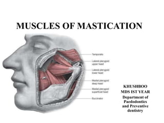
- 10. MUSCLES OF MASTICATION Muscles of mastication- originate skull and insert mandible, only mandible moves during mastication and other activities. Four muscles are the primary participants in mastication, other accessory muscles. Each of these primary muscles of mastication is paired.
- 11. Primary muscles of mastication • Temporalis • Masseter • Lateral pterygoid • Medial pterygoid
- 12. ACCESSORY MUSCLES OF MASTICATION • SUPRAHYOID INFRAHYOID • Digastric Sternohyoid • Stylohyoid Thyrohyoid • Mylohyoid Omohyoid • Geniohyoid
- 14. Functionally the muscle of mastication are classified as • Jaw elevators- • Masseter • Temporalis • Medial Pterygoid • Jaw depressors- • Lateral Pterygoid • Anterior digastric • Geniohyoid • Mylohyoid
- 15. Temporalis Muscle It is a powerful fan shaped chewing muscle which feels temporal fossa. Origin Temporal fossa from infratemporal lines excluding -Zygomatic bone -Temporal fascia Insertion -Margins and deep surface of coronoid process of mandible -Anterior border of ramus of mandible Blood supply- Superficial temporal artery branch of Maxillary artery -Superficial temporal vein and middle temporal vein Nerve supply- Deep Temporal branches from anterior division of mandibular nerve
- 18. FUNCTIONS:- • Elevation of mandible • Retraction of mandible • Crushing of food between the molars • Posterior fibres draw the mandible backwards after it has been protruded • It is also a contribute side to side grinding movement. PALPATION:- - To locate the muscle, have the patient clench. - Apply two pounds of pressure. - The anterior region is palpated above the zygomatic arch and anterior to the TMJ. - The middle region palpated directly above the TMJ and superior to the zygomatic arch. - The posterior region is palpated above and behind the ear. APPLIED ANATOMY: • Sudden reflex contraction of temporalis in times of trauma can cause the fracture of coronoid process
- 20. Masseter Muscle • The second most efficient masticatory muscle. ARRANGEMENT OF FIBRES: Superficial: Origin: thick aponeurosis. From zygomatic process of maxilla and anterior 2/3 of lower border of zygomatic arch, pass downward and backwards at an angle of 45 degree. Insertion: into lower part of lateral surface of ramus of mandible. Middle Layer: origin: anterior 2/3 of the deep surface and poterior 1/3 lower border of the zygomatic arch. Insertion: Middle part of ramus. Deep Layer: origin: deep surface of zygomatic arch. Insertion: upper part of the ramus and into the coronoid process.
- 22. FUNCTIONS • The main function of masseter muscle is • Elevation of mandible • Lateral movements of the mandible for efficient chewing and grinding of the food. • Unilateral chewing • Retraction of mandible BLOOD SUPPLY:- supplied by massetric artery branch of maxillary artery - Venous drainage through masseteric vein NERVE SUPPLY :- Massetric nerve branch of anterior division of mandibular nerve
- 23. Palpation • The patient is asked to clench their teeth and using both hands, the practitioner palpates the masseter muscles on both sides extraorally, making sure that the patient continues to clench during the procedure. • Palpate the origin of the masseter bilaterally along the zygomatic arch and continue to palpate down the body of the mandible where the masseter is attached. • Palpate multiple areas of masseter muscle.
- 25. RELATIONS • SUPERFICIAL- • Skin • Platysma • Risorius • Zygomaticus major parotid gland and duct • Temporal branch of the facial nerve • DEEP- • Temporalis • Ramus of mandible
- 26. LATERAL PTERYGOID MUSCLE • It is short thick conical muscle with definite upper and lower heads. • ORIGIN: • Upper head- infratemporal surface and crest of greater wing of sphenoid bone • Lower head- lateral pterygoid plate • INSERTION: • Fibres run backwards and laterally and converge into the Pterygoid fovea on the anterior surface of neck of mandible. • Anterior margin of articular disc and capsule of TMJ • NERVE SUPPLY: Pterygoid branch of Trigeminal nerve. • BLOOD SUPPLY:- Perygoid branch of maxillary artery - Ascending palatine artery
- 28. FUNCTIONS • Depress the mandible to open mouth • Lateral and medial pterygoid of both sides contract to protrude the mandible. • When contract alternatively producing chewing movements PALPATION Placing the forefinger, or the little finger, over the buccal area of the maxillary third molar region and exerting pressure in a posterior, superior, and medial direction behind the maxillary tuberosity
- 29. RELATIONS • SUPERFICIAL • Masseter • Ramus of mandible • Tendon of temporalis • Maxillary artery • DEEP • Mandibular nerve • Deep head of medial pterygoid • Middle meningeal artery • Sphenomandibular ligament
- 30. MEDIAL PTERYGOID MUSCLE • This is quardrilateral muscle having a small superficial head and a large deep head which forms the major part of muscle • ORIGIN and INSERTION: • superficial head from the maxillary tuberosity., deep head from medial surface of lateral pterygoid plate and part of palatine bone • Insertion is seen on the medial angle of the mandible • BLOOD SUPPLY: By facial artery • by lingual vein • NERVE SUPPLY:branch of main trunk of mandibular nerve
- 32. FUNCTIONS • Elevates the mandible • Closes the jaw • Helps in side to side movement PALPATION -Gently palpate them on the medial aspect of the jaw. -simultaneously from both inside and outside the mouth. APPLIED ANATOMY - During improper inferior alveolar nerve block, the needle may prick and irritate the muscle causing spasm and consequently Trismus
- 34. RELATIONS • SUPERFICIAL- • Upper part of muscle is separated from the lateral pterygoid muscles by; • LATERAL PTERYGOID PLATE • LINGUAL NERVE • INFERIOR ALVEOLAR NERVE • Lower part of muscle is separated from the • RAMUS OF MANDIBLE • MAXILLARY ARTERY • SPHENOMANDIBULAR LIGAMENT
- 35. • DEEP • Superior constrictor of pharynx • Styloglossus • Stylopharyngeous
- 36. ACCESSORY MUSCLES OF MASTICATION • SUPRAHYOID INFRAHYOID • Digastric Sternohyoid • Stylohyoid Thyrohyoid • Mylohyoid Omohyoid • Geniohyoid
- 37. DIGASTRIC • Two bellies united by Tendon • Origin anterior belly- digastric fossa present lateral to the symphysis menti on lower border of mandible. • Posterior belly- mastoid notch on temporal bone • INSERTION: Intermediate tendon ( hyoid bone) • ARTERY: Anterior belly- Sub mental branch of facial artery • Posterior belly- occipital artery • NERVE: • anterior belly- mandible division of the trigeminal nerve via the mylohyoid nerve; • Posterior belly - by facial nerve
- 39. STYLOHYOID • Thin muscle sheet • It accompanies the posterior belly of digastric • ORIGIN: middle of the posterior surface of styloid process. • INSERTION: junction of the body and greater cornu of hyoid bone anteriorly. • ACTION: draws the hyoid bone upwards and backwards. • NERVE SUPPLY: Facial nerve
- 40. GENIOHYOID MUSCLE Short and narrow muscle lies above mylohyoid ORIGIN: Inferior mental spine INSERTION: Anterior surface of body of hyoid bone ARTERY: Facial Artery NERVE: C1 via hypoglossal nerve • ACTION: - Geniohyoid elevates the hyoid bone and draws it forward, thus acting as a partial antagonist to stylohyoid. • -when the hyoid bone is fixed it depresses the mandible
- 41. MYLOHYOID MUSCLE Flat triangular muscle ORIGIN: Mylohyoid line of mandible INSERTION: Middle and anterior fibres into median raphae. Posterior fibres body of hyoid bone ARTERY: Mylohyoid branch of inferior alveolar artery NERVE; Mylohyoid nerve, from inferior alveolar branch of mandibular nerve ACTIONS: the secondary role of this muscle is evident as a depressor seen in action when mouth is to be opened against resistance. • It elevates the floor of mouth to help in deglutition.
- 42. STERNOHYOID MUSCLE • ORIGIN: Posterior surface of manubrium deep to sternohyoid • Adjoining part of medial end of ist coastal cartilage • INSERTION: Oblique line of thyroid cartilage below thyrohyoid • ARTERIAL SUPPLY: Superior thyroid artery • NERVE SUPPLY: Ansa cervicalis (C2, C3) • ACTION: depression of larynx
- 43. THYROHYOID MUSCLE • ORIGIN: Upper part of oblique line on thyroid cartilage • INSERTION: Lower border of greater cornea of hyoid bone • ARTERIAL SUPPLY: Superior thyroid artery • NERVE SUPPLY: Fibres of c1 via hypoglossal nerve • ACTION: Depression of hyoid bone
- 45. BRUXISM • BRUXISM: Jaw clenching with or without forcible excrusive movements, where the intensity of the clenching dictates severity (or lack of) grinding. • CLENCHING: it can occur as a brief rhthmic strong contractions of the jaw muscles during ecentric lateral jaw movements, or in maximum intercuspation. • CAUSES: 1- Associated with stressful events 2- Non stress related or hereditary Bruxism may lead to - tooth wear - fracture of the teeth or restoration - uncosmetic muscle hypertrophy TREATMENT: - cornoplasty - maxillary stabilization appliance
- 46. TETANUS • Caused by exotoxins of gram positive bacillus Clostridum tetani • Disease of the nervous system characterized by intense activity of motor neuron and resulting in severe muscle spasm. CLINICAL FEATURES: • Pain and stiffness in the jaw and neck muscles, with muscle rigidity producing trismus and dysphagia. TREATMENT: • All patients should receive antimicrobial drugs • Active and passive immunization. • Surgical wound care. • Anticonvulsant if indicated
- 47. Stylomandibular Ligament Strain- This is usually felt as a sharp to aching pain in the region behind the jaw bone and below the ear. It can be result of bad bite or from a traumatic injury. Temporal strain Tendonitis- This can occur when there is chronic strain from the temporalis muscle pulling on the tendon that attaches to the mandible. This can cause sharp headaches in the temples,just to the sides of the eyes.
- 48. Okeson’s classification ofMasticatory muscle disorders • Myofascial pain • Myositis • Myospasm • Local myalgia
- 49. Myofascial pain • First described by TRAVELL and RINZLER in 1952. • In 1969 LASKIN described Myofascial Pain Dysfunction Syndrome • Regional, dull aching muscle pain and presence of localized tender sites( trigger points) in muscle, tendon or fascia • Pathogenesis of myofascial pain is not confirmed. DIAGNOSTIC CRITERIA Regional dull, aching pain aggravated by mandibular function when the muscles of mastication are involved. Hyperirritable sites(trigger points) frequently palpated with in a taut band of muscle tissue or fascia; Greater than 50% reduction of pain with vapocoolant spray or local anesthetic injection on the trigger point followed by stretch.
- 50. TREATMENT Reassurance • Spray and stretch- flouromethane spray anesthetizes area allows patient to stretch the muscle in spasm • Soft diet • NSAIDS • Discontinuation of parafunctional habits • DIAZEPAM 2mg tds-2 weeks (anxiety reducing and muscle relaxing
- 51. MYOSITIS • This disorder is characterized by inflammation of the muscle due to a spreading infection, external muscle trauma or muscle strain. CLINICAL FEATURES- • Acute pain with in the muscle which may additionally be swollen and red with an overlying increased temperature. • The muscle is tender to palpation and may cause a limited range of motion.
- 52. Myofascial pain referred from the Masseter muscle • Trigger points located at sites in the superficial layer of the Masseter muscle refer to the posterior mandibular and maxillary teeth , the jaw and the face. TOOTHACHE is the common complaint from this source.
- 53. Myofascial pain referred from the temporal muscle • The reference zone of the temporalis muscle includes all the maxillary teeth and upper portion of face. • Headache and Toothache are the commonly complaints.
- 54. Myofascial pain referred from Medial pterygoid muscle • The reference zone for the medial pterygoid muscle includes the posterior part of mouth and throat. As well as the tempromandibular and intra auricular areas.
- 55. Myofascial pain referred from Lateral Pterygoid muscle • The lateral pterygoid muscle can not be adequately palpated, local or referred pains can be provoked only by isometric contraction of the corresponding muscle. • The lateral pterygoid muscle pain can radiate to the TMJ and superior lateral pterygoid muscle refer to zygomatic area.
- 56. Myofascial pain referred from Digastric muscle • Myofascial pain in the Anterior belly of digastric muscle often cause referred pain in the lower incisors. • Pain felt behind the angle of the mandible and below the ear common referred site for a trigger point in the posterior digastric muscle
- 57. MPDS-Myofascial pain dysfuction syndrome SYNONYMS • TMJ pain dysfunction dyndrome • Maticatory myalgia syndrome • Costen’s syndrome Definition- it is pain referred from a localized tender area or trigger pont in a taut band of skeletal muscle. ETIOLOGY: Trauma Muscular overextension Muscular over contraction Muscle fatigue
- 58. • CLINICAL FEATURES- • Pain of unilateral origin • Diffused and less localized • Patient is unable to identify exact site involved • Masseter and lateral pterygoid • Deviation to unaffected site • Pain is constant more severe in morning • Gradually worsens as day progresses • Aggravated by chewing and excessive eating • Referred to cervical region • Inability to open mouth
- 59. TREATMENT • Removal of the cause • Diet modification • Injection on trigger point • Pharmacotherapy • Psychotherapy • Acupuncture • Moist heat application
- 60. CONCLUSION The masticatory muscles include a vital part of the orofacial structure and are important both functionally and structurally. Precise movement of mandible by the musculature is required to move the teeth effectively across each other during function. The knowledge of the anatomy physiology and mechanisms of these muscles are basic to understand the movements.
- 61. REFERENCES • Mahindra Kumar Anand- Human Anatomy -3rd edition • Gray’s Anatomy-38th edition • BD Chaurasia’s Human Anatomy volume 3- 5th edition