Teresa Coque Hospital Universitario Ramón y Cajal.
Poster Noro 2016 Faculty day.pptx (2)
1. Norovirus (NoV):
• Major cause of gastroenteritis worldwide in humans
• Common cause of childhood diarrhoea
• Affects people of all ages
• Children, elderly and immunocompromised patients are at risk
of severe disease
• Transferred through faecal-oral route
• Associated with outbreaks in settings such as: hospitals, cruise
ships, nursing homes, schools, hotels and military facilities
• Symptoms include nausea, vomiting, non-bloody watery
diarrhoea and fever
Classification:
• Family – Caliciviridae
• Genus – Norovirus
• Genogroups – GI to GVII (based
on amino acid sequence of capsid)
• Genotypes – at least 38
• Affecting humans – GI, GII and GIV
• GII.4 is predominant
Structure:
• 27-35 nm virion
• Non-enveloped
• Icosahedral shape
• 3 open reading frames
(ORFs)
• Linear ssRNA (+)
• ~ 7.6 kb
Aim
The aim of this project is to optimise NoV GI genotyping primers
and to apply the newly developed genotyping assay to study NoV GI
diversity in clinical specimens and the environment.
• The partial capsid region of NoV GI was amplified in 57% (20/35) of samples
• Viruses could be genotyped from 19 samples
• Viruses in sewage samples were difficult to genotype and required substantial
assay optimisation
• The most prevalent strain was GI.4, which circulated for 8 months and dominated
during 5 months
• GI.2, GI.3, GI.5 and GI.6 have all been previously detected in children with severe
NoV gastroenteritis in South Africa and are therefore clinically relevant
• The South African GI strains cluster with strains from 2002-2015 NoV outbreaks in
China, Russia, Sweden and Tanzania
• New genotyping primers were designed and characterisation of stool specimens is
in progress
• National Research Foundation for project funding and bursary
• Poliomyelitis Research Foundation for BSc (Hons) bursary
• Rand Water and Roney Hoffman Consultants for samples
Materials and Methods
Optimisation of genotyping and characterisation of norovirus genogroup I in
clinical specimens and sewage samples
Meno KD, Taylor MB, Mans J
Department of Medical Virology, School of Medicine, University of Pretoria
Figure 2: Norovirus genome. Three open reading frames linked to
the viral protein genome (VPg) (solid black circle) at the 5′ end and
polyadenylated at the 3′ end. ORF1 encodes the 6 non-structural (NS)
proteins, ORF2 and ORF3 encode the structural components of the
virion, viral protein 1 (VP1) and VP2, respectively (Chan et al., 2015).
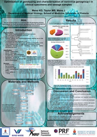
Figure 4: NoV GI diversity: GI.4 was the most
abundant strain and was detected in 63.2%
(12/19) of NoV GI positive samples. GI.2, GI.3 and
GI.5 were detected at similar levels of 10.5%
(2/19) whereas GI.6 was the least detected in 5%
(1/19) of samples.
Introduction
Results
Discussion and Conclusion
Five NoV GI genotypes circulated in Gauteng and the Free State between April 2015 to March 2016
Figure 3: Materials and Methods.
Samples were collected from the
Rotavirus Sentinel Surveillance
Programme (RSSP) and Randwater for
genotyping of NoV GI. Primers were
optimised for typing of Nov GI. NoV GI
samples were amplified, cloned and
sequenced to characterise genotypes.
Figure 3: Percentage of NoV GI positive sewage
samples from which the partial capsid region
could be amplified using semi-nested PCR.
TAN, M. & JIANG, X. 2005. The P domain of norovirus capsid protein forms a subviral particle that binds to
histo-blood group antigen receptors. Journal of virology, 79, 14017-14030.
CHAN, M. C et al. 2015. Rapid emergence and predominance of a broadly recognizing and fast-evolving
norovirus GII. 17 variant in late 2014. Nature communications, 6.
Figure 6: Neighbour-joining phylogenetic analysis of 35
capsid nucleotide sequences (323 bp, 5’-end) obtained from
sewage samples from Gauteng and the Free State. Reference
sequences, indicated by accession numbers, and the most
closely related sequences in GenBank are included. Bootstrap
values >70% are indicated.
Figure 1: Norovirus capsid. The capsid contains
180 proteins organised in an icosahedral shape.
Two major domains are the N-terminal shell (S) and
the C-terminal protruding (P) domains (Tan and
Jiang, 2005).
57%
43%
Amplification success rate
(n=35)
Positive
Negative
0
1
2
3
4
5
May
(n=2)
Jun
(n=2)
Jul
(n=1)
Aug
(n=1)
Sep
(n=2)
Oct
(n=2)
Nov
(n=2)
Dec
(n=3)
Jan
(n=2)
Feb
(n=1)
Mar
(n=1)
NumberofNoVGIclonessequenced
Months
NoV GI genotypes circulating from May 2015 to March 2016
G1.6
G1.5
G1.4
G1.3
G1.2
Figure 5: NoV GI circulation: GI.4 circulated in 8/11 months and was the predominant strain
overall. GI.2 appeared in December 2015. Between one and three genotypes co-circulated per
month.
Sample selection
Sewage samples
(Apr 2015-March 2016)
Stool specimens
(<5 yrs with diarrhoea
from RSSP)
Recovery + easyMAG
nucleic acid extraction
Manual nucleic acid
extraction
RNA
Primer optimisation
based on NoV GI
sequences in GenBank
Genotyping
cDNA
Semi-nested RT-PCR
(partial capsid)
Sewage Stool specimens
Cloning Direct sequencing
Colony PCR
Sanger Sequencing (at
least 3 clones/sample)
Phylogenetic Analysis
Acknowledgements
10.5%
10.5%
63.2%
10.5%
5.3%
Genotype distibution (n=19)
GI.2
GI.3
GI.4
G1.5
GI.6